SlideShare a Scribd company logo
1 of 57
Digestive system
Prepared by: Ahisha Kolapkar
Assistant Professor(Pharmaceutics)
VIVA Institute of Pharmacy
Digestive system
The digestive system contributes to homeostasis by breaking
down food into forms that can be absorbed and used by
body cells. It also absorbs water, vitamins, and minerals, and
it eliminates wastes from the body.
foods must be broken down into molecules that are small
enough to enter body cells, a process known as digestion.
The organs involved in the breakdown of food collectively
called the digestive system
Two groups of organs compose the digestive
system
1.Gastrointestinal (GI) tract, or alimentary canal (alimentary nourishment), is a continuous tube
that extends from the mouth to the anus through the thoracic and abdominopelvic cavities.
Organ -Mouth , Pharynx, Esophagus, Stomach, Small intestine, and Large intestine
length of the GI tract is about 5–7 meters (16.5–23 ft. ) in a living person when the muscles along
the wall of the GI tract organs are in a state of tonus (sustained contraction)
It is longer in a cadaver (about 7–9 meters or 23–29.5 ft. ) because of the loss of muscle tone after
death
2.The accessory digestive organs include the teeth, tongue, salivary glands, liver, gallbladder, and
pancreas
Digestive system performs six basic processes
• 1. Ingestion- taking foods and liquids into the mouth (eating)
• 2. Secretion- Accessory digestive organs secrete a total of about 7 liters of water, acid, buffers, and
enzymes into the lumen (interior space) of the tract.
• 3. Mixing and propulsion -Alternating contractions and relaxations of smooth muscle in the walls of
the GI tract mix food and secretions and propel them toward the anus.
• 4.Digestion -In mechanical digestion the teeth cut and grind food before it is swallowed, and then
smooth muscles of the stomach and small intestine churn the food. As a result, food molecules
become dissolved and thoroughly mixed with digestive enzymes. In chemical digestion the large
carbohydrate, lipid, protein, and nucleic acid molecules in food are split into smaller molecules by
hydrolysis.
• 5.Absorption.-The absorbed substances pass into blood or lymph and circulate to cells throughout the
bod
• 6. Defecation- Wastes, indigestible substances, bacteria, cells sloughed from the lining of the GI tract,
through the anus in a process called defecation. The eliminated material is termed feces or stool
Organ of Digestive System
Digestive system
When we eat food it is crushed by teeth and mixed by tongue and
saliva (saliva makes it soft) and provide lubrication to the food which
helps in swallowing.
• Saliva also contains lysozyme which kills the bacteria which is
present in food.
• Swallowed food ,enter into oesophagus (Food pipe) with the
help of peristalsis movement followed by food enter into
stomach.
• Food passes oesophagus in the form of bolus i.e. food +saliva.
• In stomach , concentrated HCL is present which helps kill the
foreign particles present in food and convert large particles of
food into small particles.
• Chewed food + HCL passes stomach in the form of Chyme
Digestive system
• There are 3 enzymes that acts on food in stomach
Amylase -Digestion of carbohydrates
Lipase - Digestion of fats and oil
Trypsin-Digestion of proteins.
• Liver also acts on chyme as the chyme passes through the stomach to intestine.
• Liver creates bile juice and bile salt .
• Bile juice converts acidic food converts to basic because food gets transferred into intestine
only in basic nature then food enters into small intestine ,Duodenum ,jejunum and ileum .
• Bile salts helps large particles of fats converts to small particles through emulsification
process.
Digestive system
• Small intestine
1. Main site of absorption
2. Portion between stomach and large intestine
3. Highly convoluted tube
4. Absorbs 90% nutrients from food we eat.
• Presence of villi and microvilli are present which result in better absorption.
• Length of small intestine 7 m , four to five times longer than large intestine.
• Brush border enzyme are digestive enzyme synthesised in small intestine.
Duodenum -
• Majority of chemical digestion takes place here.
• Initial and small part of small intestine.
• Hollow tube like structure.
Digestive system
Jejunum
• Part of small intestine.
• Also composed of muscle to help in movement of food.
• The jejunum helps to further digest food coming from the stomach.
Ileum
• Lower end of small intestine which continues into large intestine.
• Blood vessels are fewer in Ileum as compared to jejunum.
• Food enters into large intestine.
• Large intestine , absorbs water and vitamin. It is smaller than small intestine.
1. Cecum
• It is first part of large intestine.
• Inferior end of cecum (Appendix)
Digestive system
2.Colon
• Ascending ,Transverse and descending colon , where undigested food temporarily stored and
eliminated through anus.
• It helps the body absorb water and nutrients from food before excreting the waste.
• The colon’s job is to dehydrate what’s left of food and form it into stool.
3.Rectum
• Generally 12 cm ,connects to anus.
• Stores faeces temporarily.
• The primary function of the rectum is to collect and hold your faeces until it's time to release
it. Your rectum relaxes and stretches to accommodate the waste as it gradually comes in from
your colon.
Digestive system
4.Anus
• The last part of the digestive system.
• External opening of rectum
• The lining of the upper anus is able to detect rectal contents i.e. solid
liquid or gas.
• It's where faeces comes out from the body.
LAYERS OF THE GI TRACT
1. Mucosa - Inner lining of the GI tract, is a
mucous membrane. It is composed of (1) a
layer of epithelium in direct contact with the
contents of the GI tract, (2) a layer of
connective tissue called the lamina propria,
and (3) a thin layer of smooth muscle
(muscularis mucosa)
2. Submucosa - Areolar connective tissue that
binds the mucosa to the muscularis. It
contains many blood and lymphatic vessels
that receive absorbed food molecules.
LAYERS OF THE GI TRACT
3.Muscularis of the mouth, pharynx, and superior and middle parts of the esophagus contains skeletal
muscle that produces voluntary swallowing. Skeletal muscle also forms the external anal sphincter,
which permits voluntary control of defecation. , the muscularis consists of smooth muscle that is
generally found in two sheets: an inner sheet of circular fibers and an outer sheet of longitudinal
fibers. Involuntary contractions of the smooth muscle help break down food, mix it with digestive
secretions, and propel it along the tract.
4. serosa is a serous membrane composed of areolar connective tissue and simple squamous
epithelium. The serosa is also called the visceral peritoneum because it forms a portion of the
peritoneum.
Stomach
J-shaped enlargement of the GI tract directly inferior to
the diaphragm in the abdomen. The stomach connects
the esophagus to the duodenum, the first part of the
small intestine. Because a meal can be eaten much more
quickly than the intestines can digest and absorb it,
functions of the stomach is to serve as a mixing chamber
and holding reservoir. At appropriate intervals after food
is ingested, the stomach forces a small quantity of
material into the first portion of the small intestine. The
position and size of the stomach vary continually; the
diaphragm pushes it inferiorly with each inhalation and
pulls it superiorly with each exhalation.
Anatomy of the Stomach
• The stomach is continuous with the oesophagus at the cardiac sphincter and with the
duodenum at the pyloric sphincter.
• It has two curvatures, lesser curvature and greater curvature.
• Lesser curvature - short , lies on the posterior surface of the stomach and the downward
continuation of the posterior wall of the oesophagus.
• Greater curvature - The Oesophagus joins the stomach the anterior region angles acutely
upwards , curves downwards forming the greater curvature.
• The stomach is divided into three regions , the fundus , the body and the antrum.
• Fundus-The dome-shaped section at the top of the stomach .It does not generally stores
food unless stomach is full.
Anatomy of the Stomach
• Body- The body of the stomach stores food until it passes into small intestine .When empty the
volume of body is 50 millimeters but it expands to 1 litre when eating.
• Antrum - It is lower section of the stomach. Strong contractions mix food with stomach acid to
create substance called chyme. The wave like movements that the contractions produce push
chyme towards pyloric sphincter.
• At the distal end of pyloric antrum is pyloric sphincter , guarding the opening between the
stomach and the duodenum.
• When the stomach is inactive the pyloric sphincter is relaxed and open.
• When the stomach contains for the sphincter is closed
Function of Stomach
1. The major function of the stomach is to store and digest the food and liquid one consumes
during meals.
2. It produces hydrochloric acid and enzymes to help digest food and other foreign objects like
bacteria.
3. This acidic environment of the stomach kills unwanted microorganisms.
4. The stomach muscles contract at regular intervals to churn the food ingested and thus helps
in digestion.
5. Pepsin is a stomach enzyme that serves to digest proteins found in ingested food.
Histology of Stomach
Gastric juice
is the
combined
secretions
of mucous
cells,
parietal
cells, and
chief cells.
Acid Production
Secretion of HCL by
Parietal cell in the
stomach
Acid Production in Stomach
• Parietal cells of gastric glands produce HCL.
• water (H2O) and carbon dioxide (CO2) combine within the parietal cell to produce
carbonic acid (H2CO3), which is catalysed by carbonic anhydrase.
• Carbonic acid then dissociates into a hydrogen ion (H+) and a bicarbonate ion (HCO3–).
• The hydrogen ion that is formed is transported into the stomach lumen via the H+ – K+
ATPase ion pump. This pump uses ATP as an energy source to exchange potassium ions
into the parietal cells of the stomach with H+ ions
• The bicarbonate ion is transported out of the cell into the blood via a transporter
protein called anion exchanger which transports the bicarbonate ion out the cell
in exchange for a chloride ion (Cl–). This chloride ion is then transported into the
stomach lumen via a chloride channel.
• This results in both hydrogen and chloride ions being present within the stomach
lumen. Their opposing charges leads to them associating with each other to form
hydrochloric acid (HCl).
This “alkaline tide” of bicarbonate ions entering the bloodstream after a meal may be
large enough to elevate blood pH slightly and make urine more alkaline. HCl secretion by
parietal cells can be stimulated by several sources: acetylcholine (ACh) released by
parasympathetic neurons, gastrin secreted by G cells, and histamine, which is a paracrine
substance released by mast cells in the nearby lamina propria.
Acetylcholine and gastrin stimulate parietal cells to secrete more HCl in the presence of
histamine. Receptors for all three substances are present in the plasma membrane of
parietal cells. The histamine receptors on parietal cells are called H2 receptors; they
mediate different responses than do the H1 receptors involved in allergic responses.
• The strongly acidic fluid of the stomach kills many microbes in food.
• HCl partially denatures (unfolds) proteins in food and stimulates the secretion of
hormones that promote the flow of bile and pancreatic juice.
• Proteolytic (protein-digesting) enzyme in the stomach is pepsin, which is secreted by chief
cells. Pepsin severs certain peptide bonds between amino acids, breaking down a protein
chain of many amino acids into smaller peptide fragments.
• pepsin is secreted in an inactive form called pepsinogen; in this form, it cannot digest the
proteins in the chief cells that produce it. Pepsinogen is not converted into active pepsin
until it comes in contact with hydrochloric acid secreted by parietal cells or active pepsin
molecules.
• Another enzyme of the stomach is gastric lipase, which splits the short-chain triglycerides
(fats and oils) in fat molecules (such as those found in milk) into fatty acids and
monoglycerides.
Regulation of Acid production through
PSN system
• There is always a small quantity of gastric juice is present in the stomach , even when it
contains no food , this is known as fasting juice.
• Secretion reaches its maximum level about 1 hour after a meal then declines to the
fasting level after about 4 hours.
• There are 3 phases of secretion of gastric juices
1. Cephalic phase
2. Gastric phase
3. Intestinal phase
1. Cephalic phase
• This flow of juice occurs before food reaches the stomach ,due to reflex stimulation of
the vagus (parasympathetic ) nerves initiated by the sight , smell or taste of food.
• When the vagus nerves have been cut (vagotomy) this phase of gastric secretion stops.
• Sympathetic stimulation , e.g.during emotion states also inhibits gastric activity.
1. Gastric phase
• It is been stimulated by the presence of food the enteroendocrine cells in the pyloric
antrum and duodenum secrete gastrin , a hormone which passes directly into the
circulating blood.
• Gastrin circulating in the blood which supplies the stomach ,stimulates the gastric
glands to produce more gastric juice(HCL).
• In this way secretion of digestive juice is continued after completion of a meal and the end
of the cephalic phase.
• Gastrin secretion is suppressed when the pH in the pyloric antrum falls to about 1.5.
3.Intestinal phase
• When the partially digested contents of the stomach reach the small intestine , two
hormones , secretin and cholecystokinin are produced by endocrine cells in the intestinal
mucosa.
• They slow down the secretion of gastric juice and reduce gastric motility .
• By slowing the emptying rate of the stomach , the chyme in the duodenum becomes more
thoroughly mixed with bile and pancreatic juice.
• This phase of gastric secretion is most marked following a meal with a high
fat content.
• The rate at which the stomach empties depends largely on the type of food
eaten.
• A carbohydrate meal leaves the stomach in 2 to 3 hours , a protein meals
remains longer and a fatty meal remains in the stomach longest.
Pepsin role in Protein digestion
• In stomach Gastric gland is present which is made of chief cells which secrete
pepsin (Inactive from).
• Inactive form of pepsin is known pepsinogen or inactive zygomer.
• Stomach lining contains parietal cells ,which secrete HCl that lower the pH of
stomach (pH 1.5-2).
• Low pH causes the activation of pepsin from pepsinogen.
• Role pepsin-Protein is made of long chain of amino acid , pepsin breaks the long
chain of amino acid helps in digestion of protein .
Anatomy of Salivary glands
• These are exocrine gland i.e. a gland that makes substances such as
sweat, tears, saliva, milk, and digestive juices, and releases them
through a duct or opening to a body surface.
• These glands secrete saliva. The pH value of saliva before a meal is 7.4 ,
hence it is slightly basic to have a green colour. The pH value of saliva
after a meal is 5.8, hence it is acidic having a yellow colour.
• It is stimulated by Facial VII and Glossopharyngeal (IX) by cranial nerves.
• In Human being , there are three pair of salivary glands situated outside
of buccal cavity.
1.Parotid gland
2.Submandibular or Submaxillary gland
3.Sublingual gland
1.Parotid gland
• Largest salivary gland
• It is situated just below the auditory capsule (ear).
• Their ducts are called parotid duct ,this opens in the vestibule of the upper jaw i.e. the
buccal vestibule.
• Whenever this gland get infected by virus causes Mumps disease due to swelling of gland.
2.Submandibular or Submaxillary gland
• It is located at junction of upper and the lower jaw.
• Maximum saliva is secreted by this gland called wharton's duct
• It has largest salivary duct.
• It duct opens in the lower jaw just behind incision teeth.
3.Sublingual gland
• It is smallest salivary gland.
• It is found/located in lower jaw.
• Many ducts arises from this called the duct of Rivinus.
• This ducts opens into buccopharyngeal cavity on the ventral side of the
tongue.
Composition of Saliva
• It contains 99.5% water and other substance 0.5% like mucus ,starch,
lysozyme and few ions like sodium, potassium ,chlorine, urea(pH
balance),uric acid(anti-oxidant) ,IgA antibody are also present.
Role of Saliva
• Moistening or softening of Helps in making of food bolus so it can be swallowed
easily.
• It contains enzyme amylase which helps in breaking down of starch into maltose
and dextrin. Thus digestion food starts within mouth itself.
• Contain lysozyme helps to break down food and helps to kill the bacteria present
in food.
Pancrea
Pancrea
• Important organ in digestive system.
• Length of 15 cm (about 6 inches) and 2 inches wide
• It is located in the upper abdomen, directly behind the stomach and next to small intestine
• Comprised primarily of a network of tubes or pancreatic duct
• Pancreas is heterocrine gland (99%-exocrine and 1%-endocrine gland)
• These release liquid in upper portion of small intestine which is called duodenum
• Anatomically the pancreas is divided into 3 regions
• Head , body of pancreas and Tail.
• The head of the pancreas is on the right side of the abdomen and is connected to the duodenum
through the pancreatic duct. The tail of the pancreas extends to the left side of the body.
• Endocrine(1%)-The Pancreas produce chemical hormone that regulate blood sugar.
• It has three types of cells
• Alpha cells - Secrete glucagon, Beta cells-Secrete Insulin and Delta cells -Secrete
Somatostatin pancreatic polypeptide.
• Exocrine(99%) -The pancreas produce enzyme that helps in digestion of food.
• Amylase-Digestion of carbohydrate
• Lipase-Digestion of fats/oil
• Protease-Digestion of protein into amino acids wrapped in protective layer
• Digestive enzyme to reach GIT they travel through pancreatic duct and release into the
duodenum at major papilla.
• Once they completely out from the pancreas the protective layer removed and enzyme
become active.
• Bile from the gall bladder also enter the duodenum at the major papilla.
• Bile large fat molecule-small fat molecule ,hence easier for lipase to digest.
• When the pancreas is healthy it contributes to a healthy digestive system.
• However when our pancreas does not function correctly or is unwell we face
troubles is digesting food properly.
• Pancreas is made up of numerous acini i.e.-group of secretory cells surrounding a
cavity.
• It synthesize and secrete almost all the digestive enzymes active in the lumen of
the small intestine which are necessary for nutrient digestion.
LIVER
• The liver is located in the upper right portion of the abdomen. It is the largest gland in the
human body that performs several important functions.
• It is the only organ that has the ability to regenerate efficiently.
• Its weight between 1.44 to 1.66 kilograms.
• The liver is a triangular, bilobed structure consisting of a larger right lobe and a smaller left
lobe. The falciform ligament separates the two lobes.
• A layer of fibrous tissue called Glisson’s capsule covers the outside of the liver.
• This capsule is further covered by the peritoneum.
• This helps to hold the liver in place and protects it from physical damage
LIVER
It has two main sources of blood:
• Hepatic portal vein carries nutrient-rich blood from
the digestive system.
• Hepatic artery carries oxygenated blood from the
heart.
FUNCTION OF LIVER
1.Production of Bile
It is produced in liver and passed to gallbladder, their it becomes concentrated ,storage or
transport into first region of small intestine. Bile, which helps in the digestion and absorption
of fats, vitamins and cholesterol is produced in the liver.
2.Absorption of Bilirubin
Bilirubin is found in bile.
Bilirubin is formed by the breakdown of haemoglobin. The iron released is stored in the liver
to make next-generation blood cells.
3.Carbohydrate Metabolization
• The carbohydrates stored in the liver as glycogen are broken down into glucose and
released into the blood to maintain glucose levels.
• Glucose are stored as glycogen and released whenever energy is required.
4.Storage of Vitamins and Minerals
• The liver vitamins (A, D, E, K, and B12) are stored in the liver. It also stores iron from
hemoglobin in the form of ferritin to form new red blood cells.
5.Production of albumin (Protein in blood serum)
• It transports fatty acids and steroid hormones to help maintain the correct pressure and the
leaking of blood vessels.
6.Supporting Blood Clots
• Vitamin K is necessary for the creation of certain coagulants that helps clot the blood.
• Bile is responsible for the absorption of vitamin K. If bile is not produced, clotting factors will
not be produced.
7.Metabolization of Fats
• Bile helps in the breakdown and digestion of fats.
8.Filter of blood
• The liver filters and removes compounds from the body including
• Hormones(Estrogen and Aldosterone) and compound from outside the body (alcohol and
other drugs)
9.Immunological function
• It contains high number of kupffer cells that are involved in immune activity. These cells
destroy any disease causing agents that might enter liver through gut
Movements of GIT
1. Mobility in Mouth
2. Mobility in Esophagus
3. Mobility in stomach
4. Mobility in Small and Large intestine
1.Mobility in Mouth
• Chewing is reflex in nature.
• Breakdown of food into small pieces which results in swallowing.
• Helps in digestion of all type of food specifically cellulose containing food. e.g. vegetables.
• e.g. Starch digestion through salivary amylase.
• Swallowing -Transport of food from mouth to stomach.
a. Buccal phase-food moves from mouth to pharynx
b. Pharyngeal phase-food moves from pharynx to esophagus
c. Esophagus phase-food moves from esophagus to stomach by peristalsis
movement.
2.Mobility in Esophagus
• Esophageal peristalsis sweeps down from the esophagus
3.Mobility in stomach
Proximal (fundus and body)
• It is thin walled
• Weak contraction and infrequent
• To store food
Distal (Pylorus)
• Thick walled
• Strong contraction and frequent peristaltic contractions which results in mixing and propelling of
food.
• Distal is also responsible for gastric emptying into duodenum.
4.Mobility in Small intestine
Segmentation -
• Contraction of circular muscle for further movement of food.
Peristalsis-
• Involuntary contraction and relaxation of intestinal muscle.
Function-
• Mixing of food helps in digestion.
• Maximum exposure of food to membranes of intestinal cell which helps in digestion and
absorption.
5.Mobility of large intestine
Segmentation-
• Contraction of circular muscle for further movement of food.
Mass movement-
• Haustral churning is activated by the presence of chyme and serves to move food slowly to the
next hautra, along with mixing the chyme to help with water absorption. Mass movement
stronger and serve to move the chyme to the rectum quickly.
• peristalsis and mass perstalsis
Defecation-
• Voluntary contraction of the diaphragm and abdominal muscle and relaxation of the external anal
sphincter
Digestion and absorption of nutrients
• Small intestine is the main site of digestion and site for nutrient absorption.
Digestion in small intestine
Two types of digestion occurs in small intestine
a) Mechanical digestion
b) Chemical digestion
a)Mechanical digestion
• Digestion occurs by two types of movements of small intestine
1)Segmentation-mixing contractions
2)Peristalsis called migrating motility complexes.
• Segmentation mix chyme with the digestive juices and bring the particles of food
into contact with the mucosa for absorption
• After most of meal has been absorbed ,segmentation stops and peristalsis begin
and pushed the chyme forward.
Chemical digestion in small intestine
• The chyme entering the small intestine contains partially digested carbohydrate ,
proteins and lipids.
Digestion of carbohydrates
• Amylase cleaves interior alpha -1,4 glucose linkage .
• In large polymers of starch to form disaccharide ,trisaccharide and oligosaccharide .
• Oligosaccharide and disaccharide in the bush border of enterocytes digest small
fragments to the monosaccharides , glucose ,galactose and fructose.
Digestion of proteins
• Protein digestion of starts in the stomach , where protein are fragmented into
peptides by the action of pepsin.
• The enzyme in pancreatic juice -trypsin, chymotrypsin , carboxypeptidase and
elastase , continues to breakdown of proteins into peptides.
• Aminopeptidase and dipeptidase completes the protein digestion in the brush
border.
• Aminopeptidase cleaves off the amino acids at the end of a peptide and
dipeptidase splits dipeptidase into single amino acids.
Digestion of lipids
• Triglycerides constitute about 90% of dietary lipid ,cholesterol ,phospholipid
,sphingolipids ,fatty acids and fat soluble vitamins.
• Dietary lipids are first emulsified by mechanical digestion (chewing,antral
contraction and segmentation) which produces fine droplets that are suspended in
aqueous liquid.
• Digestion of lipids begins in the stomach by the combined action of swallowed
lingual lipase from salivary gland and chief cells in the fundus.
• These lipase contents triglycerides to fatty acids and diglycerides.
Digestion of Nucleic acid
• Pancreatic juice contains nuclease , ribonuclease which digest RNA and
deoxyribonuclease which digest DNA
• Nucleotide that result from the action of two nuclease are further digested by
brush-border enzyme called nucleosides' and phosphatase into pentoses
phosphate and nitrogenous base
Absorption of nutrients
• Passage of the digested nutrients from GI tract into blood or lymph is called absorption
• Absorption occurs via , diffusion , facilitated diffusion ,osmosis and active transport
• Almost 90% of all absorption of nutrients occur in small intestine and other 10% in
stomach and large intestine
Absorption of monosaccharides
• All carbohydrate are absorbed as monosaccharides , capacity is 120 gms/hour
• Monosaccharides pass from the lumen through the apical membrane facilitated
diffusion or active transport.
• Fructose is transported via facilitated diffusion while glucose and galactose are
transported into absorptive cells of villi via secondary active transport.
• Monosaccharides then move out of the absorptive cells through their basolateral
surface via facilitated diffusion and enter the capillaries of villi
Absorption of Amino acids , dipeptides and tripeptides
• Most protein are absorbed as amino acids via active transport process that occur
mainly in the duodenum and jejunum
• Normally 95-98% of the protein present in small intestine digested and absorbed.
• Some amino acids enter absorptive cells of the villi via Na+ dependent secondary
active transport processes other amino acids are actively transported themselves
• One symporter brings in dipeptides and tripeptides together H+ the peptides then
are hydrolysed to single amino acids.
• Amino acids move out of the absorptive cells via diffusion and enter capillaries of the villus
• Both monosaccharides and amino acids are transported in the blood to the liver by way of
hepatic portal system
Absorption of lipids
• All dietary lipids are absorbed via simple diffusion
• Adults absorbs about 95% of the lipids present in small intestine and newborn infants
absorbs only 85% of lipids
• As a result of emulsification and digestion triglycerides are broken into monosaccharides
and fatty acids which can be either short chain fatty acids or long chain fatty acids
• Short chain fatty acids dissolve in the watery intestinal chyme and pass through the
absorptive cells via simple diffusion
• Long chains fatty acids and monoglycerides are surrounded by bile salts in
the intestinal chyme forming tiny sphere called micelles
• Micelles once formed moved from the inferior of the small intestinal lumen
to the brush border of the absorptive cells.
Absorptive of Electrolysis
• Many of the electrolytes absorbed by the small intestines come from GIT
secretions and some are part of ingested food and liquids
• Na+ ions are actively transported out of the absorptive cells by basolateral
sodium-potassium pumps after they have moved into the absorption cells
via diffusion and secondary active transported
• Calcium ions are absorbed actively
Absorption of Vitamins
• Fat soluble vitamins A,D ,E and K included with ingested dietary lipids in
micelles and are absorbed via simple diffusion
• Water soluble vitamins , B vitamins and vitamin C are also absorbed via
simple diffusion
Absorption of water
• Total volume of fluid that enters the small intestine each day comes from
ingestion of liquids and from various GIT secretions
• Small intestine absorbs about 8.3 liters of the liquid and remainder passes
into large intestine .Absorption of water from the small intestine depends
on the absorption of electrolytes and nutrients to maintain an osmotic
balance with the blood

More Related Content

Similar to Digestive system.pptx

GASTROINTESTINAL AND BILLIARY SYSTEM MLS-PH-OPTOMETRY.pptx
GASTROINTESTINAL AND BILLIARY SYSTEM MLS-PH-OPTOMETRY.pptxGASTROINTESTINAL AND BILLIARY SYSTEM MLS-PH-OPTOMETRY.pptx
GASTROINTESTINAL AND BILLIARY SYSTEM MLS-PH-OPTOMETRY.pptxRuthCincinEgun
 
Digestive system
Digestive systemDigestive system
Digestive systemLermaPendon
 
Nutrition & digestive system
Nutrition & digestive systemNutrition & digestive system
Nutrition & digestive systemitutor
 
Basic ap chapter 23 powerpoint 2017
Basic ap chapter 23 powerpoint   2017Basic ap chapter 23 powerpoint   2017
Basic ap chapter 23 powerpoint 2017Kathy Richards
 
Digestive system notes
Digestive system notesDigestive system notes
Digestive system notesTia Hohler
 
DIGESTIVE SYSTEM_CLASS 10th.pptx
DIGESTIVE SYSTEM_CLASS 10th.pptxDIGESTIVE SYSTEM_CLASS 10th.pptx
DIGESTIVE SYSTEM_CLASS 10th.pptxALKANANDA7
 
Digestive system part 2 ( pharynx, esophagus and stomach) english
Digestive system  part 2 ( pharynx, esophagus and stomach)  englishDigestive system  part 2 ( pharynx, esophagus and stomach)  english
Digestive system part 2 ( pharynx, esophagus and stomach) englishMY STUDENT SUPPORT SYSTEM .
 
Digestive system powerpoint.pptx
Digestive system powerpoint.pptxDigestive system powerpoint.pptx
Digestive system powerpoint.pptxLelaniePalomar1
 
HAP 4 SEMESTER 2 BPHARMACY AS PER PCI SYLLABUS
HAP 4 SEMESTER 2 BPHARMACY AS PER PCI SYLLABUSHAP 4 SEMESTER 2 BPHARMACY AS PER PCI SYLLABUS
HAP 4 SEMESTER 2 BPHARMACY AS PER PCI SYLLABUSchristinajohn24
 
The Digestive System.pptx
The Digestive System.pptxThe Digestive System.pptx
The Digestive System.pptxRupaSingh83
 
The Digestive System.pptx
The Digestive System.pptxThe Digestive System.pptx
The Digestive System.pptxRupaSingh83
 
Introduction to Digestive System
Introduction to Digestive System Introduction to Digestive System
Introduction to Digestive System Pharmacy Universe
 
Grade 7 chapter1 lesson1 - the digestive system
Grade 7 chapter1 lesson1 - the digestive systemGrade 7 chapter1 lesson1 - the digestive system
Grade 7 chapter1 lesson1 - the digestive systemLermaPendon1
 
Digestive features
Digestive featuresDigestive features
Digestive featuresDinDin Horneja
 
Digestive System notes.pptx
Digestive System notes.pptxDigestive System notes.pptx
Digestive System notes.pptxMeghanaMeghu11
 
Nutrition in mammals
Nutrition in mammalsNutrition in mammals
Nutrition in mammalsSaad Adil
 

Similar to Digestive system.pptx (20)

Snsd world
Snsd worldSnsd world
Snsd world
 
GASTROINTESTINAL AND BILLIARY SYSTEM MLS-PH-OPTOMETRY.pptx
GASTROINTESTINAL AND BILLIARY SYSTEM MLS-PH-OPTOMETRY.pptxGASTROINTESTINAL AND BILLIARY SYSTEM MLS-PH-OPTOMETRY.pptx
GASTROINTESTINAL AND BILLIARY SYSTEM MLS-PH-OPTOMETRY.pptx
 
Digestive system
Digestive systemDigestive system
Digestive system
 
Nutrition & digestive system
Nutrition & digestive systemNutrition & digestive system
Nutrition & digestive system
 
Lek 13-14.pptx
Lek 13-14.pptxLek 13-14.pptx
Lek 13-14.pptx
 
Basic ap chapter 23 powerpoint 2017
Basic ap chapter 23 powerpoint   2017Basic ap chapter 23 powerpoint   2017
Basic ap chapter 23 powerpoint 2017
 
Digestive system notes
Digestive system notesDigestive system notes
Digestive system notes
 
DIGESTIVE SYSTEM_CLASS 10th.pptx
DIGESTIVE SYSTEM_CLASS 10th.pptxDIGESTIVE SYSTEM_CLASS 10th.pptx
DIGESTIVE SYSTEM_CLASS 10th.pptx
 
Digestive system part 2 ( pharynx, esophagus and stomach) english
Digestive system  part 2 ( pharynx, esophagus and stomach)  englishDigestive system  part 2 ( pharynx, esophagus and stomach)  english
Digestive system part 2 ( pharynx, esophagus and stomach) english
 
Digestive system powerpoint.pptx
Digestive system powerpoint.pptxDigestive system powerpoint.pptx
Digestive system powerpoint.pptx
 
HAP 4 SEMESTER 2 BPHARMACY AS PER PCI SYLLABUS
HAP 4 SEMESTER 2 BPHARMACY AS PER PCI SYLLABUSHAP 4 SEMESTER 2 BPHARMACY AS PER PCI SYLLABUS
HAP 4 SEMESTER 2 BPHARMACY AS PER PCI SYLLABUS
 
The Digestive System.pptx
The Digestive System.pptxThe Digestive System.pptx
The Digestive System.pptx
 
The Digestive System.pptx
The Digestive System.pptxThe Digestive System.pptx
The Digestive System.pptx
 
Introduction to Digestive System
Introduction to Digestive System Introduction to Digestive System
Introduction to Digestive System
 
Digestive system
Digestive systemDigestive system
Digestive system
 
Grade 7 chapter1 lesson1 - the digestive system
Grade 7 chapter1 lesson1 - the digestive systemGrade 7 chapter1 lesson1 - the digestive system
Grade 7 chapter1 lesson1 - the digestive system
 
Digestive features
Digestive featuresDigestive features
Digestive features
 
Digestive System notes.pptx
Digestive System notes.pptxDigestive System notes.pptx
Digestive System notes.pptx
 
Life processes
Life processesLife processes
Life processes
 
Nutrition in mammals
Nutrition in mammalsNutrition in mammals
Nutrition in mammals
 

Recently uploaded

Crayon Activity Handout For the Crayon A
Crayon Activity Handout For the Crayon ACrayon Activity Handout For the Crayon A
Crayon Activity Handout For the Crayon AUnboundStockton
 
Employee wellbeing at the workplace.pptx
Employee wellbeing at the workplace.pptxEmployee wellbeing at the workplace.pptx
Employee wellbeing at the workplace.pptxNirmalaLoungPoorunde1
 
AMERICAN LANGUAGE HUB_Level2_Student'sBook_Answerkey.pdf
AMERICAN LANGUAGE HUB_Level2_Student'sBook_Answerkey.pdfAMERICAN LANGUAGE HUB_Level2_Student'sBook_Answerkey.pdf
AMERICAN LANGUAGE HUB_Level2_Student'sBook_Answerkey.pdfphamnguyenenglishnb
 
Like-prefer-love -hate+verb+ing & silent letters & citizenship text.pdf
Like-prefer-love -hate+verb+ing & silent letters & citizenship text.pdfLike-prefer-love -hate+verb+ing & silent letters & citizenship text.pdf
Like-prefer-love -hate+verb+ing & silent letters & citizenship text.pdfMr Bounab Samir
 
Earth Day Presentation wow hello nice great
Earth Day Presentation wow hello nice greatEarth Day Presentation wow hello nice great
Earth Day Presentation wow hello nice greatYousafMalik24
 
AmericanHighSchoolsprezentacijaoskolama.
AmericanHighSchoolsprezentacijaoskolama.AmericanHighSchoolsprezentacijaoskolama.
AmericanHighSchoolsprezentacijaoskolama.arsicmarija21
 
Framing an Appropriate Research Question 6b9b26d93da94caf993c038d9efcdedb.pdf
Framing an Appropriate Research Question 6b9b26d93da94caf993c038d9efcdedb.pdfFraming an Appropriate Research Question 6b9b26d93da94caf993c038d9efcdedb.pdf
Framing an Appropriate Research Question 6b9b26d93da94caf993c038d9efcdedb.pdfUjwalaBharambe
 
Introduction to AI in Higher Education_draft.pptx
Introduction to AI in Higher Education_draft.pptxIntroduction to AI in Higher Education_draft.pptx
Introduction to AI in Higher Education_draft.pptxpboyjonauth
 
call girls in Kamla Market (DELHI) 🔝 >༒9953330565🔝 genuine Escort Service 🔝✔️✔️
call girls in Kamla Market (DELHI) 🔝 >༒9953330565🔝 genuine Escort Service 🔝✔️✔️call girls in Kamla Market (DELHI) 🔝 >༒9953330565🔝 genuine Escort Service 🔝✔️✔️
call girls in Kamla Market (DELHI) 🔝 >༒9953330565🔝 genuine Escort Service 🔝✔️✔️9953056974 Low Rate Call Girls In Saket, Delhi NCR
 
ROOT CAUSE ANALYSIS PowerPoint Presentation
ROOT CAUSE ANALYSIS PowerPoint PresentationROOT CAUSE ANALYSIS PowerPoint Presentation
ROOT CAUSE ANALYSIS PowerPoint PresentationAadityaSharma884161
 
What is Model Inheritance in Odoo 17 ERP
What is Model Inheritance in Odoo 17 ERPWhat is Model Inheritance in Odoo 17 ERP
What is Model Inheritance in Odoo 17 ERPCeline George
 
Influencing policy (training slides from Fast Track Impact)
Influencing policy (training slides from Fast Track Impact)Influencing policy (training slides from Fast Track Impact)
Influencing policy (training slides from Fast Track Impact)Mark Reed
 
Introduction to ArtificiaI Intelligence in Higher Education
Introduction to ArtificiaI Intelligence in Higher EducationIntroduction to ArtificiaI Intelligence in Higher Education
Introduction to ArtificiaI Intelligence in Higher Educationpboyjonauth
 
Proudly South Africa powerpoint Thorisha.pptx
Proudly South Africa powerpoint Thorisha.pptxProudly South Africa powerpoint Thorisha.pptx
Proudly South Africa powerpoint Thorisha.pptxthorishapillay1
 
Full Stack Web Development Course for Beginners
Full Stack Web Development Course  for BeginnersFull Stack Web Development Course  for Beginners
Full Stack Web Development Course for BeginnersSabitha Banu
 
Types of Journalistic Writing Grade 8.pptx
Types of Journalistic Writing Grade 8.pptxTypes of Journalistic Writing Grade 8.pptx
Types of Journalistic Writing Grade 8.pptxEyham Joco
 
Romantic Opera MUSIC FOR GRADE NINE pptx
Romantic Opera MUSIC FOR GRADE NINE pptxRomantic Opera MUSIC FOR GRADE NINE pptx
Romantic Opera MUSIC FOR GRADE NINE pptxsqpmdrvczh
 

Recently uploaded (20)

Crayon Activity Handout For the Crayon A
Crayon Activity Handout For the Crayon ACrayon Activity Handout For the Crayon A
Crayon Activity Handout For the Crayon A
 
Model Call Girl in Bikash Puri Delhi reach out to us at 🔝9953056974🔝
Model Call Girl in Bikash Puri  Delhi reach out to us at 🔝9953056974🔝Model Call Girl in Bikash Puri  Delhi reach out to us at 🔝9953056974🔝
Model Call Girl in Bikash Puri Delhi reach out to us at 🔝9953056974🔝
 
Employee wellbeing at the workplace.pptx
Employee wellbeing at the workplace.pptxEmployee wellbeing at the workplace.pptx
Employee wellbeing at the workplace.pptx
 
AMERICAN LANGUAGE HUB_Level2_Student'sBook_Answerkey.pdf
AMERICAN LANGUAGE HUB_Level2_Student'sBook_Answerkey.pdfAMERICAN LANGUAGE HUB_Level2_Student'sBook_Answerkey.pdf
AMERICAN LANGUAGE HUB_Level2_Student'sBook_Answerkey.pdf
 
Like-prefer-love -hate+verb+ing & silent letters & citizenship text.pdf
Like-prefer-love -hate+verb+ing & silent letters & citizenship text.pdfLike-prefer-love -hate+verb+ing & silent letters & citizenship text.pdf
Like-prefer-love -hate+verb+ing & silent letters & citizenship text.pdf
 
Earth Day Presentation wow hello nice great
Earth Day Presentation wow hello nice greatEarth Day Presentation wow hello nice great
Earth Day Presentation wow hello nice great
 
AmericanHighSchoolsprezentacijaoskolama.
AmericanHighSchoolsprezentacijaoskolama.AmericanHighSchoolsprezentacijaoskolama.
AmericanHighSchoolsprezentacijaoskolama.
 
Framing an Appropriate Research Question 6b9b26d93da94caf993c038d9efcdedb.pdf
Framing an Appropriate Research Question 6b9b26d93da94caf993c038d9efcdedb.pdfFraming an Appropriate Research Question 6b9b26d93da94caf993c038d9efcdedb.pdf
Framing an Appropriate Research Question 6b9b26d93da94caf993c038d9efcdedb.pdf
 
Introduction to AI in Higher Education_draft.pptx
Introduction to AI in Higher Education_draft.pptxIntroduction to AI in Higher Education_draft.pptx
Introduction to AI in Higher Education_draft.pptx
 
call girls in Kamla Market (DELHI) 🔝 >༒9953330565🔝 genuine Escort Service 🔝✔️✔️
call girls in Kamla Market (DELHI) 🔝 >༒9953330565🔝 genuine Escort Service 🔝✔️✔️call girls in Kamla Market (DELHI) 🔝 >༒9953330565🔝 genuine Escort Service 🔝✔️✔️
call girls in Kamla Market (DELHI) 🔝 >༒9953330565🔝 genuine Escort Service 🔝✔️✔️
 
ROOT CAUSE ANALYSIS PowerPoint Presentation
ROOT CAUSE ANALYSIS PowerPoint PresentationROOT CAUSE ANALYSIS PowerPoint Presentation
ROOT CAUSE ANALYSIS PowerPoint Presentation
 
TataKelola dan KamSiber Kecerdasan Buatan v022.pdf
TataKelola dan KamSiber Kecerdasan Buatan v022.pdfTataKelola dan KamSiber Kecerdasan Buatan v022.pdf
TataKelola dan KamSiber Kecerdasan Buatan v022.pdf
 
What is Model Inheritance in Odoo 17 ERP
What is Model Inheritance in Odoo 17 ERPWhat is Model Inheritance in Odoo 17 ERP
What is Model Inheritance in Odoo 17 ERP
 
Influencing policy (training slides from Fast Track Impact)
Influencing policy (training slides from Fast Track Impact)Influencing policy (training slides from Fast Track Impact)
Influencing policy (training slides from Fast Track Impact)
 
Introduction to ArtificiaI Intelligence in Higher Education
Introduction to ArtificiaI Intelligence in Higher EducationIntroduction to ArtificiaI Intelligence in Higher Education
Introduction to ArtificiaI Intelligence in Higher Education
 
Proudly South Africa powerpoint Thorisha.pptx
Proudly South Africa powerpoint Thorisha.pptxProudly South Africa powerpoint Thorisha.pptx
Proudly South Africa powerpoint Thorisha.pptx
 
Full Stack Web Development Course for Beginners
Full Stack Web Development Course  for BeginnersFull Stack Web Development Course  for Beginners
Full Stack Web Development Course for Beginners
 
Types of Journalistic Writing Grade 8.pptx
Types of Journalistic Writing Grade 8.pptxTypes of Journalistic Writing Grade 8.pptx
Types of Journalistic Writing Grade 8.pptx
 
Rapple "Scholarly Communications and the Sustainable Development Goals"
Rapple "Scholarly Communications and the Sustainable Development Goals"Rapple "Scholarly Communications and the Sustainable Development Goals"
Rapple "Scholarly Communications and the Sustainable Development Goals"
 
Romantic Opera MUSIC FOR GRADE NINE pptx
Romantic Opera MUSIC FOR GRADE NINE pptxRomantic Opera MUSIC FOR GRADE NINE pptx
Romantic Opera MUSIC FOR GRADE NINE pptx
 

Digestive system.pptx

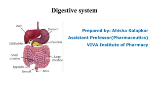
  • 1. Digestive system Prepared by: Ahisha Kolapkar Assistant Professor(Pharmaceutics) VIVA Institute of Pharmacy
  • 2. Digestive system The digestive system contributes to homeostasis by breaking down food into forms that can be absorbed and used by body cells. It also absorbs water, vitamins, and minerals, and it eliminates wastes from the body. foods must be broken down into molecules that are small enough to enter body cells, a process known as digestion. The organs involved in the breakdown of food collectively called the digestive system
  • 3. Two groups of organs compose the digestive system 1.Gastrointestinal (GI) tract, or alimentary canal (alimentary nourishment), is a continuous tube that extends from the mouth to the anus through the thoracic and abdominopelvic cavities. Organ -Mouth , Pharynx, Esophagus, Stomach, Small intestine, and Large intestine length of the GI tract is about 5–7 meters (16.5–23 ft. ) in a living person when the muscles along the wall of the GI tract organs are in a state of tonus (sustained contraction) It is longer in a cadaver (about 7–9 meters or 23–29.5 ft. ) because of the loss of muscle tone after death 2.The accessory digestive organs include the teeth, tongue, salivary glands, liver, gallbladder, and pancreas
  • 4. Digestive system performs six basic processes • 1. Ingestion- taking foods and liquids into the mouth (eating) • 2. Secretion- Accessory digestive organs secrete a total of about 7 liters of water, acid, buffers, and enzymes into the lumen (interior space) of the tract. • 3. Mixing and propulsion -Alternating contractions and relaxations of smooth muscle in the walls of the GI tract mix food and secretions and propel them toward the anus. • 4.Digestion -In mechanical digestion the teeth cut and grind food before it is swallowed, and then smooth muscles of the stomach and small intestine churn the food. As a result, food molecules become dissolved and thoroughly mixed with digestive enzymes. In chemical digestion the large carbohydrate, lipid, protein, and nucleic acid molecules in food are split into smaller molecules by hydrolysis. • 5.Absorption.-The absorbed substances pass into blood or lymph and circulate to cells throughout the bod • 6. Defecation- Wastes, indigestible substances, bacteria, cells sloughed from the lining of the GI tract, through the anus in a process called defecation. The eliminated material is termed feces or stool
  • 6. Digestive system When we eat food it is crushed by teeth and mixed by tongue and saliva (saliva makes it soft) and provide lubrication to the food which helps in swallowing. • Saliva also contains lysozyme which kills the bacteria which is present in food. • Swallowed food ,enter into oesophagus (Food pipe) with the help of peristalsis movement followed by food enter into stomach. • Food passes oesophagus in the form of bolus i.e. food +saliva. • In stomach , concentrated HCL is present which helps kill the foreign particles present in food and convert large particles of food into small particles. • Chewed food + HCL passes stomach in the form of Chyme
  • 7. Digestive system • There are 3 enzymes that acts on food in stomach Amylase -Digestion of carbohydrates Lipase - Digestion of fats and oil Trypsin-Digestion of proteins. • Liver also acts on chyme as the chyme passes through the stomach to intestine. • Liver creates bile juice and bile salt . • Bile juice converts acidic food converts to basic because food gets transferred into intestine only in basic nature then food enters into small intestine ,Duodenum ,jejunum and ileum . • Bile salts helps large particles of fats converts to small particles through emulsification process.
  • 8. Digestive system • Small intestine 1. Main site of absorption 2. Portion between stomach and large intestine 3. Highly convoluted tube 4. Absorbs 90% nutrients from food we eat. • Presence of villi and microvilli are present which result in better absorption. • Length of small intestine 7 m , four to five times longer than large intestine. • Brush border enzyme are digestive enzyme synthesised in small intestine. Duodenum - • Majority of chemical digestion takes place here. • Initial and small part of small intestine. • Hollow tube like structure.
  • 9. Digestive system Jejunum • Part of small intestine. • Also composed of muscle to help in movement of food. • The jejunum helps to further digest food coming from the stomach. Ileum • Lower end of small intestine which continues into large intestine. • Blood vessels are fewer in Ileum as compared to jejunum. • Food enters into large intestine. • Large intestine , absorbs water and vitamin. It is smaller than small intestine. 1. Cecum • It is first part of large intestine. • Inferior end of cecum (Appendix)
  • 10. Digestive system 2.Colon • Ascending ,Transverse and descending colon , where undigested food temporarily stored and eliminated through anus. • It helps the body absorb water and nutrients from food before excreting the waste. • The colon’s job is to dehydrate what’s left of food and form it into stool. 3.Rectum • Generally 12 cm ,connects to anus. • Stores faeces temporarily. • The primary function of the rectum is to collect and hold your faeces until it's time to release it. Your rectum relaxes and stretches to accommodate the waste as it gradually comes in from your colon.
  • 11. Digestive system 4.Anus • The last part of the digestive system. • External opening of rectum • The lining of the upper anus is able to detect rectal contents i.e. solid liquid or gas. • It's where faeces comes out from the body.
  • 12. LAYERS OF THE GI TRACT 1. Mucosa - Inner lining of the GI tract, is a mucous membrane. It is composed of (1) a layer of epithelium in direct contact with the contents of the GI tract, (2) a layer of connective tissue called the lamina propria, and (3) a thin layer of smooth muscle (muscularis mucosa) 2. Submucosa - Areolar connective tissue that binds the mucosa to the muscularis. It contains many blood and lymphatic vessels that receive absorbed food molecules.
  • 13. LAYERS OF THE GI TRACT 3.Muscularis of the mouth, pharynx, and superior and middle parts of the esophagus contains skeletal muscle that produces voluntary swallowing. Skeletal muscle also forms the external anal sphincter, which permits voluntary control of defecation. , the muscularis consists of smooth muscle that is generally found in two sheets: an inner sheet of circular fibers and an outer sheet of longitudinal fibers. Involuntary contractions of the smooth muscle help break down food, mix it with digestive secretions, and propel it along the tract. 4. serosa is a serous membrane composed of areolar connective tissue and simple squamous epithelium. The serosa is also called the visceral peritoneum because it forms a portion of the peritoneum.
  • 14. Stomach J-shaped enlargement of the GI tract directly inferior to the diaphragm in the abdomen. The stomach connects the esophagus to the duodenum, the first part of the small intestine. Because a meal can be eaten much more quickly than the intestines can digest and absorb it, functions of the stomach is to serve as a mixing chamber and holding reservoir. At appropriate intervals after food is ingested, the stomach forces a small quantity of material into the first portion of the small intestine. The position and size of the stomach vary continually; the diaphragm pushes it inferiorly with each inhalation and pulls it superiorly with each exhalation.
  • 15. Anatomy of the Stomach • The stomach is continuous with the oesophagus at the cardiac sphincter and with the duodenum at the pyloric sphincter. • It has two curvatures, lesser curvature and greater curvature. • Lesser curvature - short , lies on the posterior surface of the stomach and the downward continuation of the posterior wall of the oesophagus. • Greater curvature - The Oesophagus joins the stomach the anterior region angles acutely upwards , curves downwards forming the greater curvature. • The stomach is divided into three regions , the fundus , the body and the antrum. • Fundus-The dome-shaped section at the top of the stomach .It does not generally stores food unless stomach is full.
  • 16. Anatomy of the Stomach • Body- The body of the stomach stores food until it passes into small intestine .When empty the volume of body is 50 millimeters but it expands to 1 litre when eating. • Antrum - It is lower section of the stomach. Strong contractions mix food with stomach acid to create substance called chyme. The wave like movements that the contractions produce push chyme towards pyloric sphincter. • At the distal end of pyloric antrum is pyloric sphincter , guarding the opening between the stomach and the duodenum. • When the stomach is inactive the pyloric sphincter is relaxed and open. • When the stomach contains for the sphincter is closed
  • 17. Function of Stomach 1. The major function of the stomach is to store and digest the food and liquid one consumes during meals. 2. It produces hydrochloric acid and enzymes to help digest food and other foreign objects like bacteria. 3. This acidic environment of the stomach kills unwanted microorganisms. 4. The stomach muscles contract at regular intervals to churn the food ingested and thus helps in digestion. 5. Pepsin is a stomach enzyme that serves to digest proteins found in ingested food.
  • 18. Histology of Stomach Gastric juice is the combined secretions of mucous cells, parietal cells, and chief cells.
  • 19. Acid Production Secretion of HCL by Parietal cell in the stomach
  • 20. Acid Production in Stomach • Parietal cells of gastric glands produce HCL. • water (H2O) and carbon dioxide (CO2) combine within the parietal cell to produce carbonic acid (H2CO3), which is catalysed by carbonic anhydrase. • Carbonic acid then dissociates into a hydrogen ion (H+) and a bicarbonate ion (HCO3–). • The hydrogen ion that is formed is transported into the stomach lumen via the H+ – K+ ATPase ion pump. This pump uses ATP as an energy source to exchange potassium ions into the parietal cells of the stomach with H+ ions
  • 21. • The bicarbonate ion is transported out of the cell into the blood via a transporter protein called anion exchanger which transports the bicarbonate ion out the cell in exchange for a chloride ion (Cl–). This chloride ion is then transported into the stomach lumen via a chloride channel. • This results in both hydrogen and chloride ions being present within the stomach lumen. Their opposing charges leads to them associating with each other to form hydrochloric acid (HCl).
  • 22. This “alkaline tide” of bicarbonate ions entering the bloodstream after a meal may be large enough to elevate blood pH slightly and make urine more alkaline. HCl secretion by parietal cells can be stimulated by several sources: acetylcholine (ACh) released by parasympathetic neurons, gastrin secreted by G cells, and histamine, which is a paracrine substance released by mast cells in the nearby lamina propria. Acetylcholine and gastrin stimulate parietal cells to secrete more HCl in the presence of histamine. Receptors for all three substances are present in the plasma membrane of parietal cells. The histamine receptors on parietal cells are called H2 receptors; they mediate different responses than do the H1 receptors involved in allergic responses.
  • 23. • The strongly acidic fluid of the stomach kills many microbes in food. • HCl partially denatures (unfolds) proteins in food and stimulates the secretion of hormones that promote the flow of bile and pancreatic juice. • Proteolytic (protein-digesting) enzyme in the stomach is pepsin, which is secreted by chief cells. Pepsin severs certain peptide bonds between amino acids, breaking down a protein chain of many amino acids into smaller peptide fragments. • pepsin is secreted in an inactive form called pepsinogen; in this form, it cannot digest the proteins in the chief cells that produce it. Pepsinogen is not converted into active pepsin until it comes in contact with hydrochloric acid secreted by parietal cells or active pepsin molecules. • Another enzyme of the stomach is gastric lipase, which splits the short-chain triglycerides (fats and oils) in fat molecules (such as those found in milk) into fatty acids and monoglycerides.
  • 24. Regulation of Acid production through PSN system • There is always a small quantity of gastric juice is present in the stomach , even when it contains no food , this is known as fasting juice. • Secretion reaches its maximum level about 1 hour after a meal then declines to the fasting level after about 4 hours. • There are 3 phases of secretion of gastric juices 1. Cephalic phase 2. Gastric phase 3. Intestinal phase
  • 25. 1. Cephalic phase • This flow of juice occurs before food reaches the stomach ,due to reflex stimulation of the vagus (parasympathetic ) nerves initiated by the sight , smell or taste of food. • When the vagus nerves have been cut (vagotomy) this phase of gastric secretion stops. • Sympathetic stimulation , e.g.during emotion states also inhibits gastric activity. 1. Gastric phase • It is been stimulated by the presence of food the enteroendocrine cells in the pyloric antrum and duodenum secrete gastrin , a hormone which passes directly into the circulating blood. • Gastrin circulating in the blood which supplies the stomach ,stimulates the gastric glands to produce more gastric juice(HCL).
  • 26. • In this way secretion of digestive juice is continued after completion of a meal and the end of the cephalic phase. • Gastrin secretion is suppressed when the pH in the pyloric antrum falls to about 1.5. 3.Intestinal phase • When the partially digested contents of the stomach reach the small intestine , two hormones , secretin and cholecystokinin are produced by endocrine cells in the intestinal mucosa. • They slow down the secretion of gastric juice and reduce gastric motility . • By slowing the emptying rate of the stomach , the chyme in the duodenum becomes more thoroughly mixed with bile and pancreatic juice.
  • 27. • This phase of gastric secretion is most marked following a meal with a high fat content. • The rate at which the stomach empties depends largely on the type of food eaten. • A carbohydrate meal leaves the stomach in 2 to 3 hours , a protein meals remains longer and a fatty meal remains in the stomach longest.
  • 28. Pepsin role in Protein digestion • In stomach Gastric gland is present which is made of chief cells which secrete pepsin (Inactive from). • Inactive form of pepsin is known pepsinogen or inactive zygomer. • Stomach lining contains parietal cells ,which secrete HCl that lower the pH of stomach (pH 1.5-2). • Low pH causes the activation of pepsin from pepsinogen. • Role pepsin-Protein is made of long chain of amino acid , pepsin breaks the long chain of amino acid helps in digestion of protein .
  • 29. Anatomy of Salivary glands • These are exocrine gland i.e. a gland that makes substances such as sweat, tears, saliva, milk, and digestive juices, and releases them through a duct or opening to a body surface. • These glands secrete saliva. The pH value of saliva before a meal is 7.4 , hence it is slightly basic to have a green colour. The pH value of saliva after a meal is 5.8, hence it is acidic having a yellow colour. • It is stimulated by Facial VII and Glossopharyngeal (IX) by cranial nerves. • In Human being , there are three pair of salivary glands situated outside of buccal cavity. 1.Parotid gland 2.Submandibular or Submaxillary gland 3.Sublingual gland
  • 30. 1.Parotid gland • Largest salivary gland • It is situated just below the auditory capsule (ear). • Their ducts are called parotid duct ,this opens in the vestibule of the upper jaw i.e. the buccal vestibule. • Whenever this gland get infected by virus causes Mumps disease due to swelling of gland. 2.Submandibular or Submaxillary gland • It is located at junction of upper and the lower jaw. • Maximum saliva is secreted by this gland called wharton's duct • It has largest salivary duct. • It duct opens in the lower jaw just behind incision teeth.
  • 31. 3.Sublingual gland • It is smallest salivary gland. • It is found/located in lower jaw. • Many ducts arises from this called the duct of Rivinus. • This ducts opens into buccopharyngeal cavity on the ventral side of the tongue. Composition of Saliva • It contains 99.5% water and other substance 0.5% like mucus ,starch, lysozyme and few ions like sodium, potassium ,chlorine, urea(pH balance),uric acid(anti-oxidant) ,IgA antibody are also present.
  • 32. Role of Saliva • Moistening or softening of Helps in making of food bolus so it can be swallowed easily. • It contains enzyme amylase which helps in breaking down of starch into maltose and dextrin. Thus digestion food starts within mouth itself. • Contain lysozyme helps to break down food and helps to kill the bacteria present in food.
  • 34. Pancrea • Important organ in digestive system. • Length of 15 cm (about 6 inches) and 2 inches wide • It is located in the upper abdomen, directly behind the stomach and next to small intestine • Comprised primarily of a network of tubes or pancreatic duct • Pancreas is heterocrine gland (99%-exocrine and 1%-endocrine gland) • These release liquid in upper portion of small intestine which is called duodenum • Anatomically the pancreas is divided into 3 regions • Head , body of pancreas and Tail. • The head of the pancreas is on the right side of the abdomen and is connected to the duodenum through the pancreatic duct. The tail of the pancreas extends to the left side of the body.
  • 35. • Endocrine(1%)-The Pancreas produce chemical hormone that regulate blood sugar. • It has three types of cells • Alpha cells - Secrete glucagon, Beta cells-Secrete Insulin and Delta cells -Secrete Somatostatin pancreatic polypeptide. • Exocrine(99%) -The pancreas produce enzyme that helps in digestion of food. • Amylase-Digestion of carbohydrate • Lipase-Digestion of fats/oil • Protease-Digestion of protein into amino acids wrapped in protective layer • Digestive enzyme to reach GIT they travel through pancreatic duct and release into the duodenum at major papilla. • Once they completely out from the pancreas the protective layer removed and enzyme become active.
  • 36. • Bile from the gall bladder also enter the duodenum at the major papilla. • Bile large fat molecule-small fat molecule ,hence easier for lipase to digest. • When the pancreas is healthy it contributes to a healthy digestive system. • However when our pancreas does not function correctly or is unwell we face troubles is digesting food properly. • Pancreas is made up of numerous acini i.e.-group of secretory cells surrounding a cavity. • It synthesize and secrete almost all the digestive enzymes active in the lumen of the small intestine which are necessary for nutrient digestion.
  • 37. LIVER • The liver is located in the upper right portion of the abdomen. It is the largest gland in the human body that performs several important functions. • It is the only organ that has the ability to regenerate efficiently. • Its weight between 1.44 to 1.66 kilograms. • The liver is a triangular, bilobed structure consisting of a larger right lobe and a smaller left lobe. The falciform ligament separates the two lobes. • A layer of fibrous tissue called Glisson’s capsule covers the outside of the liver. • This capsule is further covered by the peritoneum. • This helps to hold the liver in place and protects it from physical damage
  • 38. LIVER It has two main sources of blood: • Hepatic portal vein carries nutrient-rich blood from the digestive system. • Hepatic artery carries oxygenated blood from the heart.
  • 39. FUNCTION OF LIVER 1.Production of Bile It is produced in liver and passed to gallbladder, their it becomes concentrated ,storage or transport into first region of small intestine. Bile, which helps in the digestion and absorption of fats, vitamins and cholesterol is produced in the liver. 2.Absorption of Bilirubin Bilirubin is found in bile. Bilirubin is formed by the breakdown of haemoglobin. The iron released is stored in the liver to make next-generation blood cells.
  • 40. 3.Carbohydrate Metabolization • The carbohydrates stored in the liver as glycogen are broken down into glucose and released into the blood to maintain glucose levels. • Glucose are stored as glycogen and released whenever energy is required. 4.Storage of Vitamins and Minerals • The liver vitamins (A, D, E, K, and B12) are stored in the liver. It also stores iron from hemoglobin in the form of ferritin to form new red blood cells. 5.Production of albumin (Protein in blood serum) • It transports fatty acids and steroid hormones to help maintain the correct pressure and the leaking of blood vessels.
  • 41. 6.Supporting Blood Clots • Vitamin K is necessary for the creation of certain coagulants that helps clot the blood. • Bile is responsible for the absorption of vitamin K. If bile is not produced, clotting factors will not be produced. 7.Metabolization of Fats • Bile helps in the breakdown and digestion of fats. 8.Filter of blood • The liver filters and removes compounds from the body including • Hormones(Estrogen and Aldosterone) and compound from outside the body (alcohol and other drugs) 9.Immunological function • It contains high number of kupffer cells that are involved in immune activity. These cells destroy any disease causing agents that might enter liver through gut
  • 42. Movements of GIT 1. Mobility in Mouth 2. Mobility in Esophagus 3. Mobility in stomach 4. Mobility in Small and Large intestine 1.Mobility in Mouth • Chewing is reflex in nature. • Breakdown of food into small pieces which results in swallowing. • Helps in digestion of all type of food specifically cellulose containing food. e.g. vegetables. • e.g. Starch digestion through salivary amylase.
  • 43. • Swallowing -Transport of food from mouth to stomach. a. Buccal phase-food moves from mouth to pharynx b. Pharyngeal phase-food moves from pharynx to esophagus c. Esophagus phase-food moves from esophagus to stomach by peristalsis movement. 2.Mobility in Esophagus • Esophageal peristalsis sweeps down from the esophagus
  • 44. 3.Mobility in stomach Proximal (fundus and body) • It is thin walled • Weak contraction and infrequent • To store food Distal (Pylorus) • Thick walled • Strong contraction and frequent peristaltic contractions which results in mixing and propelling of food. • Distal is also responsible for gastric emptying into duodenum.
  • 45. 4.Mobility in Small intestine Segmentation - • Contraction of circular muscle for further movement of food. Peristalsis- • Involuntary contraction and relaxation of intestinal muscle. Function- • Mixing of food helps in digestion. • Maximum exposure of food to membranes of intestinal cell which helps in digestion and absorption.
  • 46. 5.Mobility of large intestine Segmentation- • Contraction of circular muscle for further movement of food. Mass movement- • Haustral churning is activated by the presence of chyme and serves to move food slowly to the next hautra, along with mixing the chyme to help with water absorption. Mass movement stronger and serve to move the chyme to the rectum quickly. • peristalsis and mass perstalsis Defecation- • Voluntary contraction of the diaphragm and abdominal muscle and relaxation of the external anal sphincter
  • 47. Digestion and absorption of nutrients • Small intestine is the main site of digestion and site for nutrient absorption. Digestion in small intestine Two types of digestion occurs in small intestine a) Mechanical digestion b) Chemical digestion
  • 48. a)Mechanical digestion • Digestion occurs by two types of movements of small intestine 1)Segmentation-mixing contractions 2)Peristalsis called migrating motility complexes. • Segmentation mix chyme with the digestive juices and bring the particles of food into contact with the mucosa for absorption • After most of meal has been absorbed ,segmentation stops and peristalsis begin and pushed the chyme forward.
  • 49. Chemical digestion in small intestine • The chyme entering the small intestine contains partially digested carbohydrate , proteins and lipids. Digestion of carbohydrates • Amylase cleaves interior alpha -1,4 glucose linkage . • In large polymers of starch to form disaccharide ,trisaccharide and oligosaccharide . • Oligosaccharide and disaccharide in the bush border of enterocytes digest small fragments to the monosaccharides , glucose ,galactose and fructose.
  • 50. Digestion of proteins • Protein digestion of starts in the stomach , where protein are fragmented into peptides by the action of pepsin. • The enzyme in pancreatic juice -trypsin, chymotrypsin , carboxypeptidase and elastase , continues to breakdown of proteins into peptides. • Aminopeptidase and dipeptidase completes the protein digestion in the brush border. • Aminopeptidase cleaves off the amino acids at the end of a peptide and dipeptidase splits dipeptidase into single amino acids.
  • 51. Digestion of lipids • Triglycerides constitute about 90% of dietary lipid ,cholesterol ,phospholipid ,sphingolipids ,fatty acids and fat soluble vitamins. • Dietary lipids are first emulsified by mechanical digestion (chewing,antral contraction and segmentation) which produces fine droplets that are suspended in aqueous liquid. • Digestion of lipids begins in the stomach by the combined action of swallowed lingual lipase from salivary gland and chief cells in the fundus. • These lipase contents triglycerides to fatty acids and diglycerides.
  • 52. Digestion of Nucleic acid • Pancreatic juice contains nuclease , ribonuclease which digest RNA and deoxyribonuclease which digest DNA • Nucleotide that result from the action of two nuclease are further digested by brush-border enzyme called nucleosides' and phosphatase into pentoses phosphate and nitrogenous base
  • 53. Absorption of nutrients • Passage of the digested nutrients from GI tract into blood or lymph is called absorption • Absorption occurs via , diffusion , facilitated diffusion ,osmosis and active transport • Almost 90% of all absorption of nutrients occur in small intestine and other 10% in stomach and large intestine Absorption of monosaccharides • All carbohydrate are absorbed as monosaccharides , capacity is 120 gms/hour • Monosaccharides pass from the lumen through the apical membrane facilitated diffusion or active transport. • Fructose is transported via facilitated diffusion while glucose and galactose are transported into absorptive cells of villi via secondary active transport.
  • 54. • Monosaccharides then move out of the absorptive cells through their basolateral surface via facilitated diffusion and enter the capillaries of villi Absorption of Amino acids , dipeptides and tripeptides • Most protein are absorbed as amino acids via active transport process that occur mainly in the duodenum and jejunum • Normally 95-98% of the protein present in small intestine digested and absorbed. • Some amino acids enter absorptive cells of the villi via Na+ dependent secondary active transport processes other amino acids are actively transported themselves • One symporter brings in dipeptides and tripeptides together H+ the peptides then are hydrolysed to single amino acids.
  • 55. • Amino acids move out of the absorptive cells via diffusion and enter capillaries of the villus • Both monosaccharides and amino acids are transported in the blood to the liver by way of hepatic portal system Absorption of lipids • All dietary lipids are absorbed via simple diffusion • Adults absorbs about 95% of the lipids present in small intestine and newborn infants absorbs only 85% of lipids • As a result of emulsification and digestion triglycerides are broken into monosaccharides and fatty acids which can be either short chain fatty acids or long chain fatty acids • Short chain fatty acids dissolve in the watery intestinal chyme and pass through the absorptive cells via simple diffusion
  • 56. • Long chains fatty acids and monoglycerides are surrounded by bile salts in the intestinal chyme forming tiny sphere called micelles • Micelles once formed moved from the inferior of the small intestinal lumen to the brush border of the absorptive cells. Absorptive of Electrolysis • Many of the electrolytes absorbed by the small intestines come from GIT secretions and some are part of ingested food and liquids • Na+ ions are actively transported out of the absorptive cells by basolateral sodium-potassium pumps after they have moved into the absorption cells via diffusion and secondary active transported • Calcium ions are absorbed actively
  • 57. Absorption of Vitamins • Fat soluble vitamins A,D ,E and K included with ingested dietary lipids in micelles and are absorbed via simple diffusion • Water soluble vitamins , B vitamins and vitamin C are also absorbed via simple diffusion Absorption of water • Total volume of fluid that enters the small intestine each day comes from ingestion of liquids and from various GIT secretions • Small intestine absorbs about 8.3 liters of the liquid and remainder passes into large intestine .Absorption of water from the small intestine depends on the absorption of electrolytes and nutrients to maintain an osmotic balance with the blood