1. 1. Introduction
Many studies have found that cells take up
nanoparticles mainly through endocytosis
pathway other than passive route. From the
studies we did before, we observed that
1)SLNs enter MCF-7/ADR cells by
endocytosis; 2) multidrug resistant (MDR)
cancer cells MCF-7/ADR expressed caveolin,
which is a regulator of caveolin-mediated
endocytosis (CvME), but not its parental
MCF-7, suggesting solid lipid nanoparticles
can enter MCF-7/ADR by CvME.
To get more clear evidences for endocytosis
uptake of SLNs in MCF-7/ADR, we observed
the entry of fluorescence marker for
endocytosis pathway and dye incorporated in
SLNs into cells using confocal microscopy
under the treatment with endocytosis inhibitors.
2. Materials and methods
• Confocal laser scanning microscope
• 3 x 10⁵ cells/well were seeded on glass cover
slips in 6-well plates and incubated overnight.
• Cell were pretreated with different kinds of
inhibitors.
• Add 0.4 μM Rhodamine123/TM or dyes and
incubate for 2 hours
• Remove the medium and wash 5 times by
PBS.
• Cells were fixed in 4% paraformaldehyde
• After washing cells 3 times with PBS cell
nuclei were stained with DAPI.
• Use mounting solution and prepared for
confocal microscopy measurements.
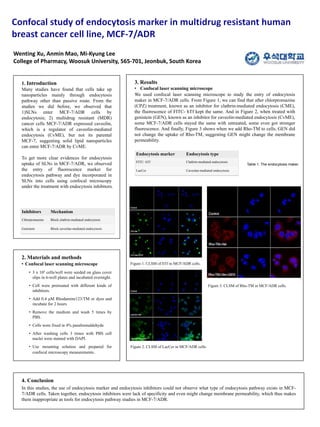
3. Results
• Confocal laser scanning microscope
We used confocal laser scanning microscope to study the entry of endocytosis
maker in MCF-7/ADR cells. From Figure 1, we can find that after chlorpromazine
(CPZ) treatment, known as an inhibitor for clathrin-mediated endocytosis (CME),
the fluorescence of FITC- hTf kept the same. And in Figure 2, when treated with
genistein (GEN), known as an inhibitor for caveolin-mediated endocytosis (CvME),
some MCF-7/ADR cells stayed the same with untreated, some even got stronger
fluorescence. And finally, Figure 3 shows when we add Rho-TM to cells, GEN did
not change the uptake of Rho-TM, suggesting GEN might change the membrane
permeability.
Wenting Xu, Anmin Mao, Mi-Kyung Lee
College of Pharmacy, Woosuk University, 565-701, Jeonbuk, South Korea
4. Conclusion
In this studies, the use of endocytosis marker and endocytosis inhibitors could not observe what type of endocytosis pathway exists in MCF-
7/ADR cells. Taken together, endocytosis inhibitors were lack of specificity and even might change membrane permeability, which thus makes
them inappropriate as tools for endocytosis pathway studies in MCF-7/ADR.
Inhibitors Mechanism
Chlorpromazine Block clathrin-mediated endocytosis
Genistein Block caveolae-mediated endocytosis
Endocytosis marker Endocytosis type
FITC- hTf Clathrin-mediated endocytosis
LacCer Caveolae-mediated endocytosis
Figure 1. CLSM of hTf in MCF/ADR cells.
Figure 2. CLSM of LacCer in MCF/ADR cells.
Figure 3. CLSM of Rho-TM in MCF/ADR cells.
Table 1. The endocytosis maker.
Editor's Notes
Copyright Colin Purrington (http://colinpurrington.com/tips/academic/posterdesign).