Downloaded 70 times


![CHAPTER SEVEN: Pediatric Burn Injuries 69
III. INITIAL EVALUATION
A. History
The events leading to the thermal injury and the past medical history are extremely
important in the initial evaluation of an infant and child. The potential for child
abuse must always be considered, particularly in children under 4 years of age. A
careful and detailed history is essential and should include a record of past ill-nesses,
immunizations, and allergies.
B. Physical (Extent of Injury)
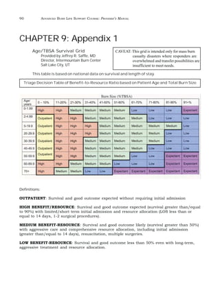
The severity of injury depends on age, body surface involvement, and depth of burn.
Measurement of body surface can be related to the Rules of Nines when one consid-ers
that the head and neck represent 18 percent of body surface rather than nine
percent, and each lower extremity represents 14 percent body surface area as com-pared
to the adult’s 18 percent. A Lund & Browder Chart is helpful in detailing the
extent of burn and in calculating the percentage of body at different stages.
IV. IMMEDIATE RESUSCITATIVE MEASURES
A. Airway
Fundamental considerations of airway thermal injuries have been discussed in
Chapter 2. Children may have few physical or radiographic signs of pulmonary
injury in the first 24 hours post burn. All pediatric patients with suspected inhala-tion
injury should be prepared for immediate transfer to a burn center. Endotracheal
intubation is indicated in infants and children with significant respiratory distress
or compromise of the airway by edema involving the glottis and upper airway.
Younger children and those with larger burns are more likely to require intubation.
For those children being transported to a burn center, the airway should be as-sessed
and secured prior to transport. Endotracheal intubation may also be indi-cated
for those children requiring large volume I.V. fluid resuscitation during
transport.
Intubation should be undertaken by one experienced in managing the child’s
airway. The infant’s larynx is located more cephalad than the adult’s. Angulation of
the infant glottis is more acute in an infant and the glottis is found more anteriorly
than in the adult. These anatomical differences make intubation by the inexperi-enced
more difficult. The diameter of the child’s external nares or small finger
should be used to gauge the size of the endotracheal tube (or the equation [16 + age
in years]/4). An uncuffed endotracheal tube should be used in infants and small
children. Remember that the narrowest point of the pediatric airway is at the cricoid
and not at the glottis.](https://image.slidesharecdn.com/ablsprovidermanual201010181-141106092420-conversion-gate01/85/ABLS-Course-Manual-69-320.jpg)


This document provides information on initial assessment and management of burn patients, including: - Components of primary and secondary surveys to evaluate burn patients - Using the "Rule of Nines" to estimate burn extent - Distinguishing between partial-thickness and full-thickness burns - Stating the American Burn Association referral criteria for transfer to burn centers


