
Recommended
More Related Content
Similar to Enfermedad diverticular
Similar to Enfermedad diverticular (20)
Recently uploaded
Recently uploaded (20)
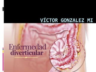
Enfermedad diverticular
- 2. Enfermedad Diverticular Generalidades Incidencia y Epidemiología Etiopatogenia Clínica Diagnóstico por Imágenes Manejo Médico Complicaciones Indicaciones Quirúrgicas y generalidades de cirugía NOTENGO CONFLICTOS DE INTERESES CON ESTA PRESENTACIÓN, SOLO INTERESES ACADÉMICOS
- 3. Generalidades Sigmoides Pocos o muchos Asintomático o sintomática [1]D.E. Beck et al. (eds.), The ASCRS Textbook of Colon and Rectal Surgery: Second Edition, DOI 10.1007/978-1-4419-1584- 9_22, © Springer Science+Business Media, LLC 2011
- 4. INCIDENCIA Prevalencia según la Edad 40- 5% , 60- 30% , 80- 65% a 80% Prevalencia según el género < 50 Más común en el sexo masculino 50–70 Leve preponderancia femenina > 70 Más común en el sexo femenino Perforación Hombres vs Mujeres [1]D.E. Beck et al. (eds.), The ASCRS Textbook of Colon and Rectal Surgery: Second Edition, DOI 10.1007/978-1-4419-1584- 9_22, © Springer Science+Business Media, LLC 2011
- 5. Etiopatogenia Presión elevada Segmentación Vasa Teoría colinérgicca [1] D.E. Beck et al. (eds.), The ASCRS Textbook of Colon and Rectal Surgery: Second Edition, DOI 10.1007/978-1-4419-1584- 9_22, © Springer Science+Business Media, LLC 2011
- 6. [2]World Gastroenterology Organisation Practice Guidelines. Enfermedad Diverticular. 2014
- 7. Otros Factores de Riesgo AINES ESTEROIDES OPIÁCEOS Tabaquismo [3] [3] Aldoori WH, Giovannucci EL, Rimm EB, et al. A prospective study of alcohol, smoking, caffeine, and the risk of symptomatic diverticular disease in men. Ann Epidemiol. 1995;5:221-8. [1] D.E. Beck et al. (eds.), The ASCRS Textbook of Colon and Rectal Surgery: Second Edition, DOI 10.1007/978-1-4419-1584- 9_22, © Springer Science+Business Media, LLC 2011
- 8. Clínica [1] D.E. Beck et al. (eds.), The ASCRS Textbook of Colon and Rectal Surgery: Second Edition, DOI 10.1007/978-1-4419-1584- 9_22, © Springer Science+Business Media, LLC 2011
- 9. Clínica Dolor Abdominal Edad Náuseas y Vómitos?? Disuria Peritonitis Abscesos [1] D.E. Beck et al. (eds.), The ASCRS Textbook of Colon and Rectal Surgery: Second Edition, DOI 10.1007/978-1-4419-1584- 9_22, © Springer Science+Business Media, LLC 2011
- 10. Imágenes USG:TAUS+TRUS MRI S = 69–98% E= 75–100%. [1] D.E. Beck et al. (eds.), The ASCRS Textbook of Colon and Rectal Surgery: Second Edition, DOI 10.1007/978-1-4419-1584-9_22, © Springer Science+Business Media, LLC 2011 [2]World Gastroenterology Organisation Practice Guidelines. Enfermedad Diverticular. 2014
- 11. Diagnóstico Diferencial IBS, Carcinoma appendicitis, bowel obstruction, ischemic colitis, gynecologic disease, and urologic disease. [1] D.E. Beck et al. (eds.), The ASCRS Textbook of Colon and Rectal Surgery: Second Edition, DOI 10.1007/978-1-4419-1584- 9_22, © Springer Science+Business Media, LLC 2011
- 12. Manejo Médico Dieta con fibra 20-30 g diarios Descanso intestinal Antibióticos G- Anticolinérgicos
- 13. Manejo Quirúrgico INDICACIONES (1) failure of abscess to respond to nonoperative management with clinical deterioration (2) free perforation with or whithout Peritonitis (3) obstruction.
- 14. Complicaciones Sangrados Fístulas Abscesos Obstrucción [1]D.E. Beck et al. (eds.), The ASCRS Textbook of Colon and Rectal Surgery: Second Edition, DOI 10.1007/978-1-4419-1584- 9_22, © Springer Science+Business Media, LLC 2011
- 15. Bibliografía [1]D.E. Beck et al. (eds.), The ASCRS Textbook of Colon and Rectal Surgery: Second Edition, DOI 10.1007/978-1-4419-1584-9_22, © Springer Science+Business Media, LLC 2011 [2]World Gastroenterology Organisation Practice Guidelines. Enfermedad Diverticular. 2014 [3] Aldoori WH, Giovannucci EL, Rimm EB, et al. A prospective study of alcohol, smoking, caffeine, and the risk of symptomatic diverticular disease in men. Ann Epidemiol. 1995;5:221-8.
- 16. GRACIAS POR LA ATENCIÓN Y RECOMENDACIONES
Editor's Notes
- These changes most commonly occur in the sigmoid colon but may involve the entire colon. The continuum can range from the presence of a single diverticulum (a sac or pouch in the wall of an organ) to many diverticula (which may be too numerous to count). It can refer to an asymptomatic state (diverticulosis) or to any one of a number of mbinations of inflammatory symptoms, changes, and complications (diverticulitis). Symptoms may result from: simple physiologic changes in colonic motility related to altered neuromuscular activity in the sigmoid colon, varying degrees of localized inflammatory response, or complex inflammatory interactions leading to diffuse peritonitis and septic shock. These more complex symptoms and resulting complications arise from breaches in the integrity of the wall of one or more diverticula.
- Although the exact incidence is not well established, numerous autopsy, radiographic and endoscopic series have shown that the incidence has increased dramatically over the past 75 years,1-4 from around 5% near the turn of the century to 50% or more by 1975.23 It is now estimated that the risk of developing diverticular disease in the USA approximates 5% by age 40 and may rise to over 80% by age 80. There is some evidence that males are more frequently affected at a younger age compared to females; however, significant bias may influence this impression. Young females may frequently be under diagnosed due to confusion with gynecologic diseases in women who are of child-bearing age. Older females may be over diagnosed due to confusion with irritable bowel syndrome (IBS). There also appears to be a dichotomy in age and sex with regard to complications of diverticular disease, particularly perforation. The incidence of perforation is higher in males under age 50. In contrast, the incidence of perforation is higher in females over age 50. Men have a higher incidence of bleeding than women; however, women have a higher incidence of fistula formation compared to men. Younger men present with fistula more frequently, while older men present more frequently with bleeding.
- Diverticulosis is associated with high intraluminal pressures. Pressures in patients with diverticular disease have been found to be as high as 90 mmHg during peak contraction. This represents a value nearly nine times higher than seen in patients with normal colons.12 It has been theorized that abnormally high pressures lead to segmentation. Segmentation refers to a process whereby the colon effectively functions as a series of separate compartments rather than as one continuous tube. These pressures predispose to herniation of mucosa through the muscular defects that occur where blood vessels penetrate to reach the submucosa and mucosa (vasa recta brevia). Most of these penetrations occur between the mesenteric and anti-mesenteric tinea where, coincidentally, most diverticula are found. As the mucosa herniates, it does so without dragging the muscular layer along, leaving the diverticula denuded of muscle, which is consistent with the definition of an acquired process. Diverticula may be true, containing all layers of the bowel wall (congenital), or false, lacking the muscular layer (acquired or pulsion diverticula). Thus, the most common diverticula are acquired or pulsion diverticula. Pain associated with diverticular disease may be related to muscle spasm as well as inflammation. Perforation can occur in the absence of inflammation and may be secondary to the extremely high intraluminal pressure.
- The incidence increases with age and with the adoption of a diet high in red meat, refined sugars, and milled flour but low in whole grains, fruits, and vegetables. La baja ingesta de fibras fue descrita por primera vez como un agente etiológico posible para el desarrollo de ED por Painter y Burkitt a fines de los 60 [5, 6]. Si bien inicialmente la teoría fue recibida con resistencia, el seguimiento confirmó su papel en la afección, siendo demostrado por publicaciones como el Estudio de Seguimiento de los Profesionales de la Salud [7]. • El riesgo relativo de presentar ED es 0.58 para los hombres que ingieren poca fibra en su dieta • La ED es menos común en los vegetarianos [8] La actual teoría que plantea a la fibra como un agente protector contra los divertículos y posteriormente contra la diverticulitis sostiene que: La fibra insoluble provoca la formación de heces más voluminosas, disminuyendo así la efectividad en la segmentación colónica. El resultado general es que la presión intracolónica se mantiene próxima al rango normal durante la peristalsis colónica
- Nonsteroidal anti-inflammatory drugs (NSAIDs) have been linked to increased rates of complications related to diverticular disease. The plausible mechanism of action I The use of opiate pain medications has been shown to raise intracolonic pressure and slow intestinal transit, The use of corticosteroids is associated with a higher risk of perforation and more severe inflammatory complications. The postulated mechanism is immunosuppressive and antiinflammatory effects hinder confinement of perforation in its early stages, resulting in more serious sequelae A recent large case-control study demonstrated that smokers had three times the risk of developing complications from diverticular disease than did nonsmokers.
- Patients with acute diverticulitis typically complain of left lower quadrant abdominal pain. However, in a patient with a redundant sigmoid colon an inflamed segment might present with pain in the right lower quadrant, thus complicating the differential diagnosis with appendicitis. The pain is generally constant in nature, not colicky. Radiation may occur to the back, ipsilateral flank, groin, and even the leg. The pain may be preceded or accompanied by episodes of constipation or diarrhea. It commonly is progressive in nature if appropriate treatment is not instituted. Patients presenting with acute diverticulitis will be tender to palpation in the left lower quadrant and left iliac region. There may be limited rigidity or localized guarding to deeper palpation. With resolution of the acute phase, palpation may reveal a mass in the left lower quadrant. Classically, there is no prodromal epigastric pain with diverticulitis as one might expect to see with appendicitis.
- The primary value of abdominal X-rays is to rule out pneumoperitoneum or to assess for a possible obstruction, therefore plain films of the abdomen should include supine upright or left lateral decubitus views. A water soluble contrast study can evaluate the lumen of the bowel if there is concern about distal bowel obstruction. It may be an important part of the assessment for the possible use of a colonic stent if malignant disease is suspected. Contrast studies have been shown to identify fistulas, most commonly colovaginal or coloenteric. • La diverticulitis a menudo es considerada como un trastorno predominantemente extraluminal. La TAC ofrece el beneficio de evaluar tanto el intestino como el mesenterio con una sensibilidad = 69–98% y una especificidad = 75–100%. • Los hallazgos tomográficos más comúnmente observados en la diverticulitis aguda incluyen: 1. engrosamiento de la pared intestinal 2. grasa mesentérica en franjas 3. abscesos asociados An important advantage of a CT scan is the ability to document diverticulitis, even if uncomplicated, when the diagnosis is in doubt. It has been demonstrated that CT can recognize and stratify patients according to the severity of their disease. It can distinguish uncomplicated disease with a predictably short length of hospital stay from complicated disease as defined by abscess, fistula, peritonitis or obstruction and a predictably long length of stay. It also provides information about extracolonic pathology and anatomic variation which is useful for surgical planning. MRI = CAT Transrectal ultrasound (TRUS) has been utilized in the evaluation of diverticular disease in conjunction with transabdominal ultrasound (TAUS). Combining TRUS with TAUS reveals complications not visualized on TAUS alone including inflamed diverticula. • Endoscopía - procto sigmoidoscopía / sigmoidoscopía flexible. El uso de endoscopía con la insuflación inherente de aire está relativamente contraindicado en agudo porque aumenta las posibilidades de perforación. Endoscopy in the face of acute diverticulitis must be undertaken with extreme caution due to risk of perforation and decreased chance of successful cecal intubation. It can provide important information prior to operation but will change acute management in less than 1% of cases.51 Generally, in the absence of an urgent indication, colonoscopy should
- In many ways, the distinction between chronic diverticulitis and noninflammatory diverticular disease relies upon the pathologist while the distinction between noninflammatory diverticular disease and IBS relies on the diagnostic acumen of the clinician and the long-term outcomes of resection. Due to the prevalence of diverticular disease many patients with IBS will have concomitant diverticular disease. However, due to the fact that diverticular disease is most commonly asymptomatic, the presence of diverticulosis in these patients will often not be the source of their symptoms but rather just a source of confusion in the differential. It is helpful to be familiar with the Rome II criteria (Table 22-3) for the diagnosis of IBS in order to sort through this differential.
- The primary management of asymptomatic diverticular disease is diet. The goal of dietary manipulation is to increase the bulkiness of stool thus increasing lumen size, decreasing transit time, and decreasing intraluminal pressures. This decreases segmentation which has been described as a significant factor in the development of diverticular disease. The ideal amount of fiber is not known; however, the recommended daily amount is 20-30 gm. What is less clear is whether a high fiber diet can prevent diverticulitis and its complications in patients who already have diverticulosis. Appropriate antibiotics should be instituted. The most predominant organisms cultured from acute diverticular abscess and peritonitis include the aerobic and facultative bacteria Escherichia coli and Streptococcus spp. The most frequently isolated anaerobes include Bacteroides spp. (B.fragilis group), Peptostreptococcus, Clostridium, and Fusobacterium spp.91 The use of anticholinergics as adjunctive therapy is based on theoretically reducing pain related to spasm and hypermotility in the sigmoid colon. Efficacy has not been proven
- The indications for surgery of acute disease include (1) failure of phlegmon or abscess to respond to nonoperative management with clinical deterioration (increasing fever, leukocytosis, tachycardia, hypotension, signs of sepsis, or a worsening physical examination), (2) free perforation with peritonitis, and (3) obstruction. Perforation without peritonitis may not require operation ( Surgical options include primary resection with anastomosis with or without proximal diversion, resection with proximal colostomy and oversewing of the rectal remnant (Hartmann’s procedure) or mucous fistula (Mikulicz operation), simple diversion with drainage of the affected segment, diversion with oversewing of the perforation site and, rarely, subtotal colectomy. The historical discussion of these options would include the use of a three-stage approach with diversion and drainage followed by a second operation for resection and a third operation for reestablishment of intestinal continuity.
- It is postulated that perforation then occurs leading to a characteristic response which results in varying degrees of inflammation. The perforation might cause microabscess, phlegmon, large abscess, fistulas, or even free perforation. Free perforations occur rarely, while fistulas are more likely, with the bladder being the most common site of fistula formation. Saint’s Triad Saint’s triad is a described association of diverticulosis, cholelithiasis and hiatal hernia. Although it has been suggested that the triad occurs in 3-6% of the general population, Polycystic Kidney Disease There is a such a high incidence of diverticulosis among patients with autosomal dominant polycystic kidney disease that some consider it an extra-renal manifestation. ABSCESO • La formación de un absceso diverticular complicado depende de la capacidad de los tejidos pericólicos de controlar (localizar) la diseminación del proceso inflamatorio. • En general, los abscesos intra-abdominales se forman por: o Fuga anastomótica = 35% o Enfermedad diverticular = 23% La diseminación limitada de la perforación da lugar a un flemón, mientras que al seguir avanzando (aunque manteniéndose localizado) se crea un absceso. • Signos/Síntomas o fiebre+/- leucocitosis a pesar de antibióticos adecuados, tumoración dolorosa • Tratamiento o Absceso pericólico pequeño - 90% responde a los antibióticos y manejo conservador. o Drenaje percutáneo de los abscesos (DPA) es el tratamiento de elección para las colecciones simples, bien definidas. Un grupo de la Universidad de Minnesota publicó tasas generales de éxito de 76% para DPA. o 100% de los abscesos uniloculares simples se resolvieron con DPA y antibióticoterapia. Entre los factores identificados como limitantes del éxito de esta estrategia de manejo se incluyen: 1. colección multilocular 2. abscesos acompañados de fístulas entéricas 3. abscesos que contienen material sólido o semisólido PERFORACIÓN (Perforación libre) • Afortunadamente la perforación libre es infrecuente. Ocurre más frecuentemente en el paciente inmunocomprometido. La perforación libre está asociada a una alta tasa de mortalidad, presentándose en hasta 35% de los casos. En la mayoría de los casos se require una intervención quirúrgica urgente. FISTULAS Las fístulas ocurren en 2% de los pacientes con enfermedad diverticular complicada. La fístula se forma a partir de un proceso inflamatorio local que produce un absceso que se descomprime espontáneamente, perforándose hacia una víscera adyacente o a través de la piel. Habitualmente hay un único tracto fistuloso, pero se pueden encontrar tractos múltiples en 8% de los pacientes. • Un proceso inflamatorio local asociado con un absceso que se descomprime espontáneamente, perforándose a las vísceras adyacentes o a través de la piel. Habitualmente hay un único episodio, pero puede ocurrir en más de una oportunidad en 8% de los pacientes. • Las fístulas son más frecuentes: o en los hombres que en las mujeres (2:1) o en los pacientes con antecedentes de cirugía abdominal o en pacientes inmunocomprometidos Tipos de fistulas relacionadas con Enfermedad Diverticular: • Colovesical: 65% • Colovaginal: 25% • Colocutánea: (no disponible) • Coloentérica: (no disponible) Diagnóstico: • El diagnóstico puede requerir múltiples exámenes, pero lo más frecuente es que se vea en la TAC, en el enema baritado, la vaginoscopía, cistoscopía, o fistulografía. Tendencias: SANGRADO A La Enfermedad Diverticular sigue siendo la causa más común de sangrado digestivo bajo masivo, responsable de 30– 50% de los casos. Se estima que 15% de los pacientes con diverticulosis sangrará en algún momento de la vida. El sangrado habitualmente es abrupto, indoloro y de gran volumen, siendo 33% masivo, requiriendo una transfusión de emergencia. A pesar de esto, el sangrado se detiene espontáneamente en 70–80% de los casos. Se ha demostrado que los AINEs aumentan el riesgo de enfermedad diverticular, habiéndose tratado más de 50% de los casos de sangrado diverticular con AINEs. La enfermedad diverticular es responsable de sangrado colónico porque a medida que el divertículo se hernia, los vasos que penetran, responsables de la debilidad de la pared intestinal, se extienden sobre la cúpula del divertículo. Con esta configuración, estos vasos quedan separados de la luz intestinal sólo por un recubrimiento mucoso fino.