2. W
hen members of the surgical team at Boston Children’s
Hospital, MA, opened the skull of 18-month-old Violet
Pietrok in October 2014 to correct her congenital facial
malformation, they referred to a three-dimensional (3-D) mold
created from computerized tomography (CT) images of Violet’s
head to perform the delicate procedure.1
3-D printing tech-
nology, also known as “additive manufacturing” because its
production method adds very thin layers of material upon one
another to create 3-D objects, allows surgeons to plan where to
cut down to the millimeter and to practice complex operations
on patient-specific rubber or plastic molds.2
Although 3-D printers have been used in the last couple of
decades to create surgical tools, laboratory equipment, and pros-
thetic limbs, only recently has software been developed to translate
a patient’s magnetic resonance imaging (MRI) or CT scan images
into a replica of a patient’s organ. Estimates suggest that as of 2015,
75 U.S. hospitals and 200 worldwide have access to a high-level
3-D printer, and that number is expected to grow.3
Fellows of the American College of Surgeons (ACS) are actively
using this technology, not only at Boston Children’s Hospital but
at other institutions around the country, including Washington
University School of Medicine, St. Louis, MO, where surgeons
used a 3-D model of Myah McWilliams’ skull to fix the five-year-
old’s severe facial asymmetry in December 2015.4
In this article, members of the College describe their expe-
riences with 3-D printing models, identify its benefits and
limitations, and consider future applications for this technology.
Preoperative planning with 3-D printing
Adnan Siddiqui, MD, PhD, FACS, FAHA, neurovascular surgeon
and vice-chair, neurosurgery, Jacobs School of Medicine and Bio-
medical Sciences, University of Buffalo, NY, has been using 3-D
printers as an aid in operations for complex aneurysms for the
last few years.5
He uses the technology to help him gain a deeper
HIGHLIGHTS
• Describes the benefits of 3-D printing
as a preoperative planning tool
• Outlines 3-D printing relevancy across
the spectrum of subspecialties
• Explains how 3-D modeling
enhances resident education
• Defines 3-D modeling challenges,
including slow build times, a
lack of autonomy, and expensive
equipment and software costs
• Considers future applications for 3-D
printing, including bioprinting
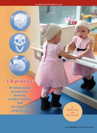
Above: Dr. Meara and
colleagues mark Violet’s
calvarium to designate the
osteotomy pattern for the
craniectomy and removal
of the frontal bones
Overleaf: Violet after surgery
(photos courtesy of Boston
Children’s Hospital)
V101 No 7 BULLETIN American College of Surgeons
10 |
3-D PRINTING AND SURGICAL CARE
3. understanding of a patient’s unique vasculature in a
way that traditional imaging alone cannot provide, as
well as to develop a preoperative surgical plan.5
“When you do an angiogram and you take a blood
vessel picture or snapshot, a CT angiogram, an MRI
angiogram, it looks like, ‘There’s the problem, there’s
where I need to go,’” said Dr. Siddiqui, chief medical
officer, The Jacobs Institute, Buffalo. “The fact of the
matter is, when you try to perform the procedure in
reality, the impediments are such that you can’t make
your way up there with the tools you have or there is
less space in the artery than you originally thought,
and you need to make adjustments on the fly.” For
example, the surgeon may need to abandon the first
approach and try something else, such as entering
through the neck rather than the groin to reach an
aneurysm.
“What this translates into is multiple procedures,
multiple variations, longer procedures, [and] more
possibilities for complications,” Dr. Siddiqui said.
Employing a patient-specific 3-D model preoperatively
may be able to alleviate some of those problems.
Dr. Siddiqui and his colleagues at The Gates Vas-
cular Institute and University at Buffalo, including
scientists, clinicians, and trainees, meet weekly to dis-
cuss what cases would be best served by the technology.
“[Then] we say, hold on, this looks like a complicated
case. Let’s 3-D print this entire vascular anatomy, put
it in the lab, attach it to a flow pump, and let’s do the
whole procedure, from groin to vertex, artificially on
the 3-D printed model and work out the kinks,” he
said. The advantage of practicing on a 3-D printout
is to shift any trial-and-error element for a procedure
from a patient to a replaceable model, with the goal of
achieving improved outcomes and safer patient care.
“With 3-D printing, we are now able to take some
of the most complicated cases—specifically those with
lots of nuances to their anatomy that really aren’t appre-
ciable on a computer screen—and generate a model
that you can turn around in your hand,” said Albert
S. Woo, MD, an Associate Member of the ACS and
chief, pediatric plastic surgery, and director, craniofa-
cial program, division of plastic surgery, Warren Alpert
Medical School of Brown University, Providence, RI.
Dr. Woo and Steven Couch, MD, FACS, oculofacial
plastic surgeon and assistant professor of ophthalmol-
ogy, Washington University, used 3-D modeling to
carefully reconstruct Myah McWilliams’ orbital defor-
mity while protecting the young patient’s tear ducts.4
(At the time of Myah’s procedure, Dr. Woo was asso-
ciate professor, Washington University, and chief of
pediatric plastic surgery, St. Louis Children’s Hospital.)
Dr. Couch noted that a 3-D printed model made
preoperative visualization a more tangible experi-
ence. “The standard is, you look at a 2-D screen and
look at multiple views on the area—so in my case, you
look at coronal, axial, sagittal images—and you try to
develop a 3-D model in your mind,” he said, and the
repositioning or canting of the bone were done men-
tally. Dr. Couch acknowledges that while surgeons
have been doing this conceptualization for a long time,
“3-D printing potentially improves our ability to accu-
rately visualize in the preoperative setting.”
A 3-D-printed model was especially useful in
Myah’s case, in which a multidisciplinary team
needed to work together. “A model allows us to say,
‘My goal is to change this portion, this portion, and
this portion. How will that affect your portion of the
surgery? Is there a better way to expose this area?’”
Dr. Couch explained.
Dr. Siddiqui (in
background, right)
showing a medical device
executive how to coil an
aneurysm on a 3-D-printed
brain artery model, at the
State University of New
York (SUNY) at Buffalo’s
Toshiba Stroke and
Vascular Research Center
JUL 2016 BULLETIN American College of Surgeons
| 11
3-D PRINTING AND SURGICAL CARE
4. Dr. Siddiqui (right)
and Liza Pope, MS,
research associate,
SUNY at Buffalo, study
a 3-D printed bifurcated
aneurysm model to
determine optimal
coil placement
Use across subspecialties
More than a year ago, Washington University obtained a grant to pur-
chase a professional grade 3-D printer, and since then Dr. Woo and
colleagues have used the printing lab to optimize the results of several
of their patients.
“Initially, I had anticipated that the people who would be the most
interested in 3-D modeling would be those who deal with bone defor-
mities—plastic surgeons like myself, or oral maxillofacial surgeons,
neurosurgeons, and orthopaedic surgeons. The interesting thing is that
the greatest enthusiasm for this technology has actually been from sur-
geons who don’t work on bones, so to speak, but actually on the soft
tissues,” said Dr. Woo.
“Not too long ago, a cardiac surgery colleague was working on a case
with an aortic deformity and we were able to model-out not only the
heart but the aorta for this young infant who was less than a year old,”
said Dr. Woo. “When the cardiac surgery team was trying to decide
whether the child needed open-heart surgery versus a cardiac catheter-
ization, we were able to not only print a 3-D model for them, we were
able to give the interventional cardiologist the opportunity to choose
the texture and the softness of the printed materials so that the model
was as close to a normal infant’s [heart] as possible. They could actu-
ally practice the surgery on a model that was much more true to life.”
“For orthopaedic and craniofacial surgery, the hard tissue simula-
tion is excellent…because the plastic is roughly the same consistency as
bone, so you have a fairly realistic simulation,” said John G. Meara, MD,
DMD, MBA, FACS, plastic surgeon-in-chief, Boston Children’s Hospi-
tal, MA. In fact, Dr. Meara and his team used several 3-D models to
plan the operation on Violet Pietrok. “We had a couple of 3-D-printed
models made because I wanted to have the ability to do the proce-
dure on those heads and to develop a couple of osteotomy patterns
as backups. We revised the pattern three times before we got to the
point where I could see exactly where the cuts needed to be.”
This exacting preparation was a key element in correcting the
young girl’s rare facial deformity. “Violet had a complex Tessier cleft,
and we did a facial bipartition—we moved the two halves of her face
Dr. Woo
Dr. Couch
V101 No 7 BULLETIN American College of Surgeons
12 |
3-D PRINTING AND SURGICAL CARE
5. Neurological residents
learn and practice stroke
intervention surgery
with Dr. Siddiqui (at far
right) on 3-D-printed
in the Jacobs Institute’s
Training Center
closer together,” Dr. Meara said. “The preoperative
planning was very helpful because we actually did
the surgery on the 3-D printed models to give us
an idea of how the orbits would come together,” he
added, explaining that he and his surgical team made
subtle changes in the way they had initially planned
to cut the bones based on potential interference in
the affected area. Similar to Dr. Siddiqui’s experience,
Dr. Meara found that he “didn’t have to revise things
in the OR [operating room] because we were able to
make those adjustments on the model beforehand.”
A tactile understanding of the procedure translates
into increased confidence for the surgeon going into
that nonstandard operation, and, according to Dr.
Meara, the 3-D-printed models also are useful inside
the OR.1,6
“Even in Violet’s case, there were times I
would have someone bring the models over to me
while operating just for me to take a look from differ-
ent angles, to get my bearings in terms of where the
orbit was, where the optic nerve was,” he said. “You
might be making a bone cut that is five millimeters
away from a fairly important anatomic structure, so
it’s nice to have someone be able to spin that around
in front of you to get a much clearer 3-D image in
your head of what you need to do on the patient.”
As general surgery and the surgical subspecial-
ties continue to move toward minimally invasive
techniques and as these procedures become more
sought after by patients, surgeons are facing new
challenges that can be alleviated, in part, through
3-D modeling. “With the rising demand for mini-
mally invasive procedures, surgeons are no longer
able to fillet open the anatomy and see everything
they need to,” said Dr. Woo. “Frequently, you are
looking at the anatomy through a very tiny incision or
through an unusual angle and with a limited view of
what exactly is going on. With a 3-D model on hand,
it really helps the surgeon get his or her bearings
by having a millimeter-to-millimeter exact correla-
tion from model to patient.” These models can also
be sterilized so that the surgeon can manipulate the
model on the operative field while performing the
procedure.
Surgical education and training benefits
For surgeons—whether the user is an experienced
surgeon, a resident, or a medical student—there is no
replacement for the knowledge afforded by holding a
solid object and understanding its nuances.
“We are a major teaching center, and education and
training are typically at the core of what we do,” said
Amar Singh, MD, FACS, a urologist with Erlanger
Health System, Chattanooga, TN. “A very useful part
of resident education is having [trainees] hold a model
in their hands and look at the 3-D contour of the tumor
and consider how they can approach it surgically.” Sur-
geons who have done thousands of organ-preserving
kidney operations, for example, know the pitfalls of the
procedure, “and communicating that knowledge base
to a resident is so much easier when you have some-
thing tangible,” Dr. Singh said.
In a panel session titled Emerging Technologies in
Simulation presented at the ninth annual meeting of
the Consortium of ACS-Accredited Education Insti-
tutes (ACS-AEI) in March 2016, Robert Sweet, MD,
FACS, executive director of the Institute for Simula-
tion Healthcare (formerly the Institute for Simulation
and Interprofessional Studies), University of Washing-
ton, Seattle, underscored the pedagogic benefits of 3-D
JUL 2016 BULLETIN American College of Surgeons
| 13
3-D PRINTING AND SURGICAL CARE
6. printing technology, particularly for resident education. “You can do
these cases that you might not normally see during residency [with
3-D printed models],” said Dr. Sweet, a professor of urology. “If we’re
going to credential people to do things that they’ve never seen, that’s
problematic. 3-D printing offers us the ability to essentially immortal-
ize these rare cases and create a library of opportunities for students for
things that they might see during their residency or training program.”
Katherine A. Barsness, MD, MSCi, FACS, pediatric surgeon; direc-
tor, surgical simulation at Ann and Robert H. Lurie Children’s Hospital
of Chicago; and associate professor of surgery and medical education,
Northwestern University Feinberg School of Medicine, Chicago, IL,
said she has been using 3-D printers at Northwestern Simulation since
2011 to create new tools to train surgeons in her specialty.7
Dr. Bars-
ness also spoke at the ACS-AEI Emerging Technologies in Simulation
panel session on the topic of hybrid simulation—the use of surgically
modified real tissue placed into 3-D printed thoracic and abdominal
cavities. In an interview, Dr. Barsness said, “Specifically, it started in
pediatric surgery. It was used to work with the size limitations inher-
ent to neonatal surgery—when you’re trying to simulate that small
space, 3-D printing is the most accurate way we’re able to do that. It
has allowed us to create size-appropriate, anatomically correct teach-
ing aids, so that no longer is a newborn infant exposed to the risk of
the learning curve; rather, the learning curve is borne on the back of
the simulation,” which is especially meaningful with the fragile tissue
at play in neonates.
The educational and training benefits of the technology also extend
to experienced surgeons. “There’s a technology called flow diversion,
which is used to treat complex brain aneurysms. There are physicians
who have done a few cases as a part of their training, but the condition
is relatively rare,” Dr. Siddiqui said. To enhance their understanding of
the condition, he and his colleagues at The Jacobs Institute have devel-
oped an advanced users course, in which physicians from all over the
world fly into Buffalo and spend a couple of days at the institute watch-
ing surgeons perform live demonstrations of complex cases. “Then we
3-D print some of those cases and have the physicians practice on the
Violet before her
operation
3-D models used for planning and
performing Violet’s operation
V101 No 7 BULLETIN American College of Surgeons
14 |
3-D PRINTING AND SURGICAL CARE
BOSTONCHILDREN’SHOSPITALBOSTONCHILDREN’SHOSPITALBOSTONCHILDREN’SHOSPITAL
7. BOSTONCHILDREN’SHOSPITAL
3-D models to gain a stronger familiarity with what
they will have to do to be successful with those proce-
dures on patients,” he said.
The customizability of 3-D printed models also is a
useful feature, Dr. Siddiqui noted. “If you have a patient
with a perfect aneurysm that would be great for train-
ing, you print out that model, and then it can be easily
modified in the future to include different complica-
tions,” he said. “A bend here, an additional aneurysm
there—you can represent any additional problem you
want to a student, fellow, or even yourself, in order to
work in a variety of training scenarios. That’s a unique
advantage of how this all gets done.”
Quality metrics as a key to reimbursement
While 3-D modeling is beneficial in the areas of
education and training, the measurable value of 3-D
modeling on improved surgical outcomes is yet to be
determined. However, anecdotal evidence suggests
this technology can reduce the risk for complica-
tions and lessen the time the patient spends under
anesthesia.
“We’ve done some early work showing a decrease
in OR times as a result of the models,” Dr. Meara said.
“If you can cut an hour off a 10-hour case, each hour
in the OR is extremely expensive, so maybe a $500 or
$800 model is not terribly expensive if you are cut-
ting an hour or an hour-and-a-half of OR time,” he
said, adding that he was able to save approximately
one to two hours on Violet’s case because he had a
clearer idea what he and the surgical team were going
to do in the OR. So while the infrastructure costs in
acquiring an on-site 3-D printer are considerable—the
high-quality model at Boston Children’s Hospital
costs $400,000, and printers in use at other hospi-
tals can run upward of $100,000—with efficient use,
the technology has the potential to save money over
time.1
A pilot study—reportedly the first in the world to
review 3-D modeling in a hospitalwide setting—was
launched by the Erlanger Health System in Octo-
ber 2015, to examine how 3-D printing can be used
to improve surgical outcomes and the challenges—
including costs—involved in using this technology
in a large public hospital system.8,9
The study was
conducted in partnership with the University of
Tennessee College of Medicine-Chattanooga and
3D Operations, Inc. (3D Ops), a provider of patient-
specific 3-D printed models.
“During the first six months [of the study] we
are trying to figure out how to create these models
accurately and what is the most cost-effective way
to do this so that every surgeon has access [to this
technology],” said Christopher Keel, DO, a urologic
surgeon, Associate Member of the ACS, and the first
surgeon at Erlanger to use a 3-D model for surgi-
cal planning.10
To prepare for that operation, which
involved a kidney with multiple large tumors, a model
was created using a 3-D printer that costs approxi-
mately $250,000, including software.
“We are doing this in a research setting in col-
laboration with industry because there is no
reimbursement currently for 3-D printed models,”
explained Dr. Singh. “We do believe this technology
helps us provide better outcomes, but there is no way
to quantitate that in dollars and cents right now. If I
am a hospital or health system or university, unless
there is a grant or philanthropic money, it might not
make sense to purchase a high-end printer. This is an
Peter Weinstock, MD,
PhD (left), and Dr. Meara
examine a 3-D printed
model of Violet’s skull
JUL 2016 BULLETIN American College of Surgeons
| 15
3-D PRINTING AND SURGICAL CARE
8. Dr. Singh
Dr. Keel
extremely dynamic technology—the printers that are out there could
be outdated within the next two years. However, you do see situa-
tions where organizations are leasing the technology and equipment
or partnering with another entity,” said Dr. Singh.
Drs. Singh and Keel emphasized the importance of quality metrics
as a key to reimbursement, particularly if 3-D printing technology can
be linked to better outcomes and enhanced efficiency.
Hurdles to adoption
At least three primary limitations are associated with 3-D printing
technology for use in surgical models—slow build times, a lack of
autonomy, and expensive equipment and software costs. Build times
for a single model can last from a few hours to an entire day and can
vary depending on whether the 3-D printer is housed on-site or with
an outside company. “Speed is one of the things that developers are
currently working on,” Dr. Keel said. “It depends on the type of organ
you are printing. They’ve printed a whole brain before, and obviously,
that takes more time than it does to print a kidney, particularly if it’s
a smaller kidney.”
Another challenge to wide adoption of 3-D printing is the training
involved in mastering the scan conversion software; as a result, sur-
geons must typically rely on experts in printer technology to generate
models.11
“A lot of these printers need babysitters to sit and watch as
they are printing because there can be errors [or because] you have
to switch out the materials,” Dr. Sweet said.
“The reality is that there is a huge amount of infrastructure that
needs to be developed [by a hospital] before the first case,” Dr. Woo
added. “Not only do you need to have a 3-D printer available, which
is a significant cost in and of itself, you also need the technical exper-
tise to be able to manipulate the data in order to create the model
that you want, and generally that’s not something that surgeons can
do themselves.”
Furthermore, medical-grade printers are expensive and cover a
range of price points. According to Dr. Sweet, high-end 3-D printers
can cost up to $850,000 depending on the scale, number of inks, and
performance capabilities of the device. At the low end of the market,
namely the consumer market, 3-D printers sell for as low as $140.
“Most of the market right now is either on the low end or the high
end, but what’s interesting is the merging [of the two markets] right in
the middle,” said Dr. Sweet. “This emerging middle market is where
you are getting some of the capabilities of the high-end machines, but
with lower costs driven by scale and so on, and this is what we need.”
V101 No 7 BULLETIN American College of Surgeons
16 |
3-D PRINTING AND SURGICAL CARE
9. “You don’t necessarily need the most expensive
printer to do what we want to do, although the higher
grade printers have much higher resolution and have
more flexibility and allow you to use not just one
material but multiple materials,” said Dr. Woo. “I can
mix a plastic type of material with a rubber material
and find the perfect mix so that we are able to get the
texture or the flexibility in the material that we want
to allow us to achieve our goals.”
Future applications
Although 3-D printing already has affected surgical
practice and training, the technology has even greater
potential going forward. The future of 3-D printing
in health care will likely revolve around bio-printing,
which entails creating biological structures through
a layered manufacturing method, similar to the one
used today, but instead of using a resin polymer, bio-
printing uses a stem cell base—tissue grown in a lab.
“That is the next step in evolution. We might even see
both those types of models merge together,” accord-
ing to Dr. Singh.
Work already is under way to merge the field of
artificial materials and biologics in 3-D printing, Dr.
Sweet said, through the use of advanced inks and
embedded sensor technology. “When you’re dealing
with electronics, they’re two dimensional, they’re hard,
rigid, and brittle, and they have very high processing
temperatures from a manufacturing standpoint, where
biological structures are three dimensional, soft, flex-
ible, stretchable, and are temperature-sensitive,” he
said. “So what is nice about 3-D printing is that it can
actually solve both those problems and you can start
embedding electronics with biological structures.”
Intertwining organic and inorganic materials also
may create new possibilities in functional human tissue
and organ generation. “There is a core printing and a
shell around it, and what you can actually do is implant
drugs, small growth factors, and those can be activated
and released when you want them to be released using
laser or mechanical energy,” Dr. Sweet added. “So imag-
ine that—imagine being able to print an organ with
little beads of growth factors embedded in it that may
be released over time slowly or when you activate them
for release. This opens a whole new world for us, not
only in training, but for organ replacement and heal-
ing as well.”
The usability of 3-D printed materials inside patients
is still in its early stages, but surgeons already are con-
sidering the potential. “I guarantee you that someday,
you will be able to print 3-D pieces of, for example,
orbital bones, which you’ll be able to feed stem cells
into and have it turn into bone,” Dr. Meara said.
Dr. Siddiqui believes that the ability to print func-
tional organic components would be a benefit to
vascular surgery. “In vascular surgery, we’re always
looking for the best grafts, whether that is a radial
Sample 3-D heart model
used in the Erlanger
Health System study
JUL 2016 BULLETIN American College of Surgeons
| 17
3-D PRINTING AND SURGICAL CARE
10. REFERENCES
1. Weintraub K. Off the 3-D printer, practice parts for the surgeon.
The New York Times. January 26, 2015. Available at: www.nytimes.
com/2015/01/27/science/off-the-3-d-printer-practice-parts-for-the-
surgeon.html?_r=0. Accessed April 26, 2016.
2. Osawa J. Next to use 3-D printing: Your surgeon. The Wall Street
Journal. April 8, 2013. Available at: www.wsj.com/articles/SB10001
424127887324504704578410764264855512. Accessed April 26, 2016.
3. Storrs C. How a 3-D printer changed a 4-year-old’s heart and life.
CNN. October 6, 2015. Available at: www.cnn.com/2015/10/06/
health/3d-printed-heart-simulated-organs/. Accessed April 26,
2016.
4. Bernhard B. 3-D printing comes to the hospital. St. Louis Post-
Dispatch. January 17, 2016. Available at: www.stltoday.com/
lifestyles/health-med-fit/health/d-printing-comes-to-the-hospital/
article_bd0ac87b-2f02-5d37-ae75-c761e10f7e45.html. Accessed April
26, 2016.
5. Mangan D. New brain surgery innovation: Practice on a 3-D
model. CNBC. November 24, 2015. Available at: www.cnbc.
com/2015/11/23/new-brain-surgery-innovation-practice-on-a-3-d-
model.html. Accessed April 1, 2016.
6. Boston Children’s Hospital. Violet’s Journey—Part three: Inside
the operating room. January 19, 2015. Available at: www.youtube.
com/watch?v=zcmZeTqhLyI. Accessed April 26, 2016.
7. Dunne N. 3-D printed models provide lifelike simulations for
training pediatric surgeons. Northwestern Medicine News Center.
October 15, 2014. Available at: http://news.feinberg.northwestern.
edu/2014/10/barsness-3d-printing/. Accessed April 20, 2016.
8. Pulford M. Erlanger, 3D Ops announce 6-month pilot of pre-
surgery 3-D printing. Nooga.com. October 20, 2015. Available at:
http://nooga.com/171376/erlanger-3d-ops-announce-6-month-
pilot-of-pre-surgery-3-d-printing/. Accessed April 26, 2016.
9. Bailey R. Blue is for tumor, clear is for kidney. The Pulse.
November 4, 2015. Available at: www.chattanoogapulse.com/
columns/tech/blue-is-for-tumor-clear-is-for-kidney/. Accessed
April 26, 2016.
10. Johnson S. A model operation: Local startup says it can cut
surgical costs by 3D printing patient’s organs. Chattanooga Times
Free Press. November 1, 2015. Available at: www.timesfreepress.
com/news/edge/story/2015/nov/01/model-operation-local-
startsays-it-ccut-surgi/332231/. Accessed April 26, 2015.
11. Lewis C. 3-D printing and surgery a ‘paradigm shift.’ Crain’s New
York Business. January 11, 2016. Available at: www.plasticsnews.
com/article/20160111/NEWS/160119985/3-d-printing-and-surgery-
a-paradigm-shift. Accessed April 21, 2016.
12. Gilpin L. 3D ‘bioprinting’: 10 things you should know about
how it works. TechRepublic. April 23, 2014. Available at: www.
techrepublic.com/article/3d-bioprinting-10-things-you-should-
know-about-how-it-works. Accessed April 26, 2016.
artery graft, or a saphenous vein graft, or a
cephalic vein, or an internal mammary artery—
it’s a perpetual issue with trying to the find the
right graft, the right diameter, with properties
that will allow it to serve as an effective conduit
and not cause spasms,” he said. “Being able to
print artificial vessels would be a major step for-
ward in any vascular surgery.”
Ultimately, the goal is to one day be able to
print entire functional organs for use in human
transplantation; these organs, created from a
patient’s stem cells, could avert the need for
immunosuppressive drugs and alleviate the
ever-growing need for donor organs.12
Achiev-
ing that aim is years away. The complexity of
replicating a biologically viable liver or heart
via 3-D printing is beyond the current scope of
the technology, but the science is continuously
improving.
As the capabilities of 3-D technology advance
and lead to improved patient safety and out-
comes, the surgeons interviewed for this article
predict that 3-D printers will be as common in
hospitals as CT scans within a decade. “It’s the
same thing I told a medical student last week,”
said Dr. Singh. “I’ve been in practice for nine
years, and I don’t do a single operation the same
way I did nine years ago. The ultimate goal is
to provide equivalent or superior care and out-
comes for your patients and minimize their
complications, and if you have a technology that
is affordable or that will become affordable as it
continues to be developed, you can resist it all
you want, but the wave is going to sweep over
you, and we have seen that with every sort of
minimally invasive approach.”
“There are some incredible benefits of this
technology for the surgeons who are willing to
get out of their comfort zone,” added Dr. Woo.
“I firmly believe that 3-D printing is here to stay
and it’s actually going to revolutionize not just
the practice of surgery, but really the practice
of medicine.” ♦
V101 No 7 BULLETIN American College of Surgeons
18 |
3-D PRINTING AND SURGICAL CARE