Kridsada Sirisabhabhorn, Supaporn Pumpa, Surapong Pornprasitseang and Palakor...
Final_Presentation_7-27-11[2]
1. CHARACTERIZATION OF INFLAMMATION IN A MOUSE MODEL OF ASTHMA
CONCLUSIONS
Asthmatic airways display a significant increase in total white blood cells
compared to normal airways, suggesting that the ovalbumin model elicits an
inflammatory response in the lungs.
The increase in eosinophils in our asthma model and their concordant presence
in the airways of asthma patients suggests that these cells are instrumental to
the pathology of the asthmatic response.
Clinical significance: This mouse model may be useful because it can assist
us in understanding more about the causes of human asthma. Furthermore,
this model may provide us with a route by which to test the safety and
effectiveness of new asthma therapies prior to human administration.
ABSTRACT
Background: Asthma is a chronic disorder of the lungs in which the bronchi
(airways) constrict in response to the triggering allergen, thus affecting one’s
ability to breathe. Although this disorder affects millions and takes thousands
of lives each year, there is still little in the way of a treatment for the disease
itself. Therefore, good animal models of asthma are needed to investigate the
specific cause of this disease and possibly help researchers find a cure. The
purpose of this study was to generate a relevant mouse model of asthma in
order to understand the lungs’ inflammatory response to the model allergen
ovalbumin (OVA).
Hypothesis: Compared to normal (non-sick) mice, asthmatic mice will
demonstrate an enhanced inflammatory response in the lung, as noted by an
increase in total white blood cell counts and an increase in eosinophils.
Results: Ovalbumin-challenged mice showed an enhanced lung immune
response compared to normal mice as noted by a significant increase in airway
white blood cells, specifically eosinophils. Eosinophils are most
characteristically found in asthmatic airways and their presence is thought to
be indicative of disease severity.
Tianna Edwards, Linda Guernsey, Sonali Bracken, Alexander Adami, Prabitha Natarajan, Steven Szczepanek,
Mary Cearley, Sonali Shah, and Roger S. Thrall
Department of Health Career Opportunity Programs, Summer Research Fellowship Program
Department of Immunology, University of Connecticut Health Center, Farmington, CT 06032
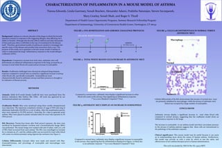
FIGURE 1: OVA SENSITIZATION AND AEROSOL CHALLENGE PROTOCOL
METHODS
Animals: Adult (6–8 week) female C57BL/6J mice were purchased from the
Jackson Laboratory (Bar Harbor, ME, USA). All work was approved by our
institutional animal care committee.
Ovalbumin Model: Mice were sensitized using three weekly intraperitoneal
(i.p.) injections. The injections contained a mixture of 25µg of OVA and 2mg of
aluminum hydroxide (ALUM) in 0.5 ml of saline. After the sensitization period,
mice were exposed to 1% OVA aerosol, 1 hour/day, for 7 days to generate acute
asthma. Mice were placed in plastic restraint tubes for nose-only exposure to 1%
OVA aerosol.
BAL Recovery: Twenty-four hours after final aerosol exposure, the mice were
sacrificed by ketamine/xylazine overdose and a bronchoalveolar lavage (BAL)
was performed. The mouse lungs were washed with 5ml of saline, with 3 to 4 ml
of BAL fluid recovered from each animal. The BAL was centrifuged at 1700rpm
for 10 minutes at 4°C, and the cellular pellet was recovered for total white blood
cell counts on a hemocytometer using nigrosin dye exclusion (see Figure 1).
Cellular Differentials: Cytospin slides were made and stained with May-
Grunwald/Giemsa, and percentage of eosinophils and macrophages were
determined.
FIGURE 2: TOTAL WHITE BLOOD CELLS INCREASE IN ASTHMATIC MICE
Compared to normal mice, asthmatic mice display a significant increase in white
blood cell counts in the airways, thus signifying an inflammatory response.
***p<0.0001 (Student’s Unpaired T Test)
FIGURE 3: ASTHMATIC MICE DISPLAY AN INCREASE IN EOSINOPHILS
Compared to normal mice, asthmatic mice display a significant increase in eosinophils
in the airways, thus signifying that the inflammation displayed in Figure 2 is likely due
to an asthmatic response. ***p<0.0001 (Student’s Unpaired T Test)
Normal
Asthmatic
Macrophage
Eosinophil
FIGURE 4: CELLULAR DIFFERENTIALS IN NORMAL VERSUS
ASTHMATIC MICE
Cellular differentials of the BAL demonstrate that airways of normal mice (top)
are primarily inhabited by macrophages, while the airways of asthmatic mice
(below) are occupied by a large number of eosinophils.
This work was funded by: NIH/AI R01 HL-43573 (RST)