2. improve the molecular orbital overlap between the organic
ligand and the SiQD.40
To date we have found very few reports that link an aromatic
ligand to the SiQD using a vinyl linkage and none combining
computation and experimental results.4,41
However, the styryl
ligand considered was not designed to form a Type-II energy
alignment with SiQDs and only a slightly broadened PL was
observed; both Type-II energy alignment and conjugated
bridges are essential for effective tuning of SiQDs optical
properties by peripheral ligands.40
Reported here is the design and synthesis of 4-ethynyl-N,N-
bis(4-methoxyphenyl)aniline (MeOTPA) as the organic
molecule to functionalize SiQDs. The triphenylamine moiety
is a typical electron rich material that has a comparatively high-
lying HOMO level compared with many other organic
materials used for organic electronics devices. A para-
substitution of electron donating methoxy groups can further
raise the HOMO level of MeOTPA and make it high enough
(−4.46 eV, 278 nm) to form the Type-II energy alignment with
SiQDs. Furthermore, the hole transport properties of the
triphenylamine-based ligand may facilitate hole transport in the
double superexchange system previously reported by our
group.40,42
The terminal alkyne functional group was added
to the triphenylamine so as to form vinyl connective bridges
with the Si−H sites of the SiQD upon hydrosilylation
chemistry.
A theoretical study of the molecular orbital energy of
MeOTPA functionalized SiQDs was carried out based on the
time-dependent density functional theory (TDDFT). The
computational methodology is the same as that detailed in
our earlier study.40
A SiQD (Si849H344) with a diameter of 3.1
nm (Figure 2) was chosen as a model system. In the
computational study, only four MeOTPA molecules were
considered because minimal changes were observed in the
optoelectronic analysis of smaller dots using more than four
MeOTPA attachments. Specifically, changing the number of
MeOTPA ligands from 4 to 8, 12, and 16 did not significantly
change the absorption peaks although it does influence the
shape of the spectra at the low energy edge because of the
localization of low-energy orbitals. In addition, a comparison of
the optoelectronic properties of small dots with decyl and
hydride passivation gave almost identical results. This implies
that a direct comparison can be made between our computa-
tionally generated dots with hydrogen passivation and our
experimental setting in which decyl passivation is employed.
The computationally predicted influence of MeOTPA
functionalization is shown in Figure 2, where frontier orbitals
are shown for hydrogen (left) versus MeOTPA (right)
termination. The latter shows a significantly lower optical gap
of 1292 nm (0.96 eV) as compared with those having only
hydrogen termination (1033 nm, 1.20 eV). This 0.24 eV (259
nm) red shift is consistent with our earlier prediction of a Type-
II aligned organic/SiQD hybrid system.40
The results are
summarized in Table 1.
To experimentally verify our theoretical predictions, the
MeOTPA ligand was synthesized via a Sonogashira reaction of
trimethylsilylacetylene with 4-iodo-N,N-bis(4-methoxyphenyl)-
aniline followed by a potassium hydroxide removal of the
trimethylsilyl group to obtain the terminal alkyne (Figure 3a).
The decyl and MeOTPA functionalized SiQDs were prepared
using thermal hydrosilylation, (Figure 3b).6
After passivation by
MeOTPA or 1-decene, the resultant SiQDs became very
soluble in common solvents such as dichloromethane,
qualitatively indicating that surface functionalization had
occurred (Figure 3b).
FT-IR was used to follow the MeOTPA reaction on the
SiQD surface. In Supporting Information Figure S1a, the
successful preparation of MeOTPA functionalized SiQD is
suggested by the formation of -Si-CC- stretching bands at
1599 cm−1
, although this also overlaps with the aromatic −C
C− bonds from MeOTPA.4,41
Additional evidence comes from
the presence of aromatic C−H stretching bands around 3000
cm−1
and MeOTPA feature absorption at about 1500, 1240,
1035, and 825 cm−1
. Evidence of unreacted Si−H is shown in
the 2100 cm−1
region, which is not surprising as total ligand
coverage of the SiQD surface is unlikely due to steric issues and
is commonly seen in other SiQD hydrosilylation systems.
Furthermore, no CC−H stretch at 3283 cm−1
and CC at
2100 cm−1
(Supporting Information Figure S1b) are observed
when compared with MeOTPA starting materials as shown in
Supporting Information Figure S1b. The absence of MeOTPA
starting material also indicates the MeOTPA SiQDs have been
well purified. Supporting Information Figure S1c shows the FT-
IR of decyl-passivated SiQDs. The absorption bands at 2920,
1465, and 1378 cm−1
are features from the alkyl stretching and
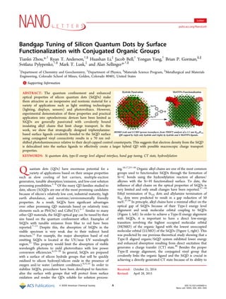
Figure 1. Electron transition in Type-I (left) and Type-II (right)
molecular orbital energy level aligned organic functionalized SiQDs
hybrid systems.
Figure 2. HOMO (red) and LUMO (green) isosurfaces from TDDFT
analysis of a 3.1 nm Si849H344 QD capped by (left) only hydride and
(right) by hydride and four MeOTPA ligands. Isosurfaces are for a
fixed value of the absolute value of electron orbitals, 0.009 Å−3/2
.
Table 1. Theoretical Calculation of Molecular Orbital
Energy of Hydride Passivated, Decyl Passivated, MeOTPA
Functionalized SiQDs (3.1 nm) and MeOTPA Molecule
Based on Time Dependent Density Functional Theory
(TDDFT)
energy (eV) Si849H348 Si849H344 (MeOTPA)4 MeOTPA
HOMO −5.19 −4.89 −4.46
LUMO −4.00 −3.93 −2.10
optical gap 1.19 (1042 nm) 0.96 (1291 nm) 2.36 (525 nm)
Nano Letters Letter
DOI: 10.1021/nl504051x
Nano Lett. XXXX, XXX, XXX−XXX
B
3. deformation demonstrating surface alkylation of SiQDs. The
absence of −CC− stretch at 1641 cm−1
(Supporting
Information Figure S1d) also indicates that 1-decene has
been thoroughly removed during the purification process
despite also being used in large excess. As is the case for the
MeOTPA SiQDs, a minor and broad peak at 2088 cm−1
results
from unreacted Si−H on the SiQD surface. In the FT-IR of
many reported SiQDs passivated with alkyl chains, the intense
and structureless band around 1080−1090 cm−1
is commonly
ascribed to Si−O−Si from surface oxidation.43,44
A similar
relatively broad peak was also observed in our decyl-passivated
SiQDs at 1031 cm−1
that is quite different from the reported
values (>50 cm−1
). It has been reported that partial oxidation of
SiHx surfaces could shift the hydride stretching band to higher
energy with a characteristic stretch at 2250 cm−1
for O3Si−H,
while formation of Si−C bonds has been predicted to shift the
frequency to lower energy.41
It was also reported that the
vibration of Si−O not only appears at 1090 cm−1
, but also at
∼460 cm−1
with an intense but smaller band of 60% less in
absorption intensity.43
This could be very useful and
straightforward for the evaluation of the oxidation degree of
SiQDs. To make sure that the 460 cm−1
band can be used as
reference for our hybrid SiQDs, our hydride SiQDs were
intentionally oxidized by heating them in open air for 24 h, and
Figure 3. (a) The synthesis of MeOTPA ligand. (b) The preparation of hydride terminated SiQDs and MeOTPA functionalized SiQDs.
Nano Letters Letter
DOI: 10.1021/nl504051x
Nano Lett. XXXX, XXX, XXX−XXX
C
4. the FT-IR spectrum was subsequently investigated. As seen in
Supporting Information Figure S1e, the similar intense and
broad absorption band at 1081 cm−1
and the smaller band at
458 cm−1
were observed in such oxidized SiQDs, indicating the
reference absorption band around 460 cm−1
should be
applicable to our SiQDs as well. Thus, the absence of an
observable 460 cm−1
absorption band in the FT-IR spectra of
decyl and MeOTPA passivated SiQDs indicates there was no
significant oxidation in our materials. It also further confirms
that the 1031 cm−1
absorption band that we observed in our
SiQD materials is not from exhaustive oxidation but rather
suboxide formation.36
FT-IR of our starting hydride terminated
SiQDs are shown in Supporting Information Figure S1f
showing the characteristic Si−H at 2098 cm−1
. A small peak
around 1030 cm−1
is observed indicating surface suboxides.
Some C−H absorption bands are seen in the 3000 cm−1
region
from residual solvent when depositing the sample on the ATR
crystal. In both of our SiQD systems, we are not so concerned
with some oxidation on the surface as this will be inevitable in
practical applications. As shown in the next sections, the surface
oxidation does not seem to affect the new optoelectronic
properties associated with attaching our conjugated ligands.
Further evaluation of functionalized SiQDs was performed
using X-ray photoelectron spectroscopy (XPS). The size of the
functionalized SiQDs is smaller than the sampling depth of
XPS, therefore XPS analysis provides bulk rather than surface
composition. SiQDs functionalized with MeOTPA show ∼72%
carbon, 14% silicon, 11% oxygen, and 3% nitrogen (Supporting
Information Table S1). XPS provides solid confirmation of
surface functionalization demonstrating a carbon to nitrogen
ratio of 25 that is only slightly higher than the value expected
based on stoichiometry of MeOTPA of 22. On the basis of the
stoichiometry of MeOTPA, at least 6% of oxygen should be
associated with ligand, thus 5% from surface oxidation. The
XPS ratio of C/Si is slightly higher than expected for a 2.8 nm
SiQD with 80% coverage (65% carbon, 19% silicon, 7.8%
oxygen, and 3.4% nitrogen), which can be explained by the
presence of adventitious carbon and sampling multiple layers of
quantum dots rather than monolayer. Deconvolution of high-
resolution C 1s spectra shown in Figure 4a1 provides
information about the relative amount of CC, C−N, C−O,
CO, and O−CO, and along with the shakeup feature
located at ∼291 eV further confirms a match between expected
and observed structures of the MeOTPA-SiQD. In comparison,
C 1s spectra acquired from decyl functionalized SiQDs shows
C−C and various C−O species and no shakeup feature,
confirming lack of conjugation in the structure of the ligand
(Figure 4a2). Both samples show peak binding energy lower
than those of C−C and CC groups, suggesting formation of
bonds between C and Si. High-resolution Si 2p spectra
representative of MeOTPA-SiQDs is shown in Figure 4b. The
spectrum is fit with four peaks, each consisting of two
components, 2p3/2 and 2p1/2, separated by 0.6 eV. The 2p3/2
component of the first doublet located at 99.6 eV is attributed
to Si(0).45
The second 2p3/2 component located at 100.4 eV is
due to Si−C species, further corroborating formation of
bonding between Si and C atoms of the ligand.42,46,47
Doublet
with 2p3/2 component located at 102.4 eV is due to Si−Ox
species, that is, suboxides.42,48,49
A small component at 103.0
eV could also be attributed to Si−Ox species, which are more
similar to those observed in SiO2. On the basis of these
assignments, the amounts of Si(0), Si−C, and Si−Ox species
are estimated at 58 ± 3.3, 18 ± 1.0, and 23.9 ± 4.3% (which
normalized to the total amount of measured silicon
corresponds to 7.9, 2.5 and 3.25%, respectively). Considering
the total amount of oxygen of 11% and the fact that at least 6%
of oxygen is associated with ligand, the amount of oxygen that
could be associated with SiQD is ∼5%. On the basis of the
amount of oxygen bonded to Si (5%) and amount of Si bonded
to oxygen (3.25%), the average ratio of oxygen atoms bonded
to Si is 1.5, which is more typical of suboxides as oppose to fully
oxidized Si in SiO2. The spectrum of Si 2p acquired from decyl
functionalized SiQDs is fairly similar to that of MeOTPA-
SiQDs with small differences related to distribution of Si−Ox
bonds (Figure 4b2). This difference is not surprising
considering that some heterogeneity in the Si-Ox species is
observed even when comparing several analysis areas for the
same sample/batch.
Absorption and photoluminescence (PL) spectra of
MeOTPA- and decyl-passivated SiQD dichloromethane sol-
utions were measured and presented in Figure 5. Photo-
luminescence excitation (PLE) spectra for MeOTPA ligand,
MeOTPA- and decyl-passivated SiQDs are provided in
Supporting Information Figure S2. The MeOTPA-SiQDs
exhibit an enhanced absorption in the range from 290−450
nm compared with the decyl-passivated dots. Some of this
enhanced absorption results from the newly emerged shoulder
absorption at 295 nm that is predicted to be the direct
absorption transition from the MeOTPA ligand attached to the
SiQD.40
In the region from 350 to 450 nm, the absorption
intensity of MeOTPA-SiQDs has a slower decay than that of
decyl-passivated dots, implying the optical gap of MeOTPA
dots has been reduced. While the absorption bands of the
SiQDs are relatively structureless and do not show an abrupt
edge, it is clear from Figure 5a that the band edge for
MeOTPA-SiQD has been red shifted by at least 75 nm over the
decyl-SiQD analog.
In the PL spectra (Figure 5b), the decyl-SiQD emission peak
at 679 nm (1.83 eV) suggests that the mean dot diameter is
Figure 4. High-resolution XPS spectra of C 1s (a) and Si 2p for SiQDs
modified with (1) MeOTPA and (2) decyl.
Nano Letters Letter
DOI: 10.1021/nl504051x
Nano Lett. XXXX, XXX, XXX−XXX
D
5. approximately 3.1 nm.50,51
This agrees well with the trans-
mission electron microscopy (TEM) results of MeOTPA-
SiQDs as shown in Figure 6. A size count histogram (Figure 6a
and Supporting Information Figure S8) for 329 dots reveals an
average size of 5.0 nm with a mode of 2.8 nm, both consistent
with the 3.1 nm size suggested from the decyl-SiQD PL results
(same hydride terminated SiQD precursor used for both
materials). Consistent with the reduced optical gap observed in
the absorption spectra, the normalized PL spectrum of
MeOTPA-SiQDs (peak at 749 nm (1.66 eV)) shows a
significant red shift of 70 nm versus the decyl-SiQDs. This
0.17 eV change in the emission peak is in qualitative agreement
with our computational prediction of 0.24 eV for 3.1 nm
SiQDs. The 72 meV difference is smaller than the uncertainty
associated with the TDDFT methodology as applied to
SiQDs.52
These experimental results strongly suggest that our
computational predictions of the reduced optical gap in such
Type-II energy level aligned hybrid SiQDs material are valid.
Hydride-terminated SiQDs that are significantly larger than
what is studied here are expected to have HOMO levels that
result in a Type-I interface between dot and MeOTPA. The
direct excitation from the MeOTPA HOMO to SiQD LUMO
loses dominance with the independent excitation from the
SiQDs and organic ligand becoming stronger. This is consistent
with the 295 nm shoulder in the absorption spectrum of
MeOTPA-SiQDs, indicative of a large portion of independent
excitation transitions associated only with the ligand.
It should also be noted that, in contrast to the broadened and
slightly red-shifted PL observed in styrene functionalized
SiQDs,4
the PL of MeOTPA-SiQDs has completely shifted
away from the PL of decyl-passivated SiQDs. This indicates the
CT state dominates the excited state of MeOTPA-SiQDs. In
other words, in addition to indirect excitons, direct excitons
generated from the independent absorption transition of SiQDs
dissociate into CT states. This is evidenced by red-shifted PL
that comes primarily from direct relaxation of the CT state. We
conclude from this that a Type-II aligned hybrid material
Figure 5. (a) Absorption and (b) PL spectra of MeOTPA and decyl-
passivated SiQDs in dichloromethane solutions.
Figure 6. TEM characterization of MeOTPA-SiQDs. (a) Histogram
showing the particle size distribution. (b) Bright-field TEM micro-
graph illustrating a representative SiQD size and morphology. (c) The
selected area diffraction pattern of many SiQDs illustrating excellent
crystallinity and primarily Si phase to be present.
Nano Letters Letter
DOI: 10.1021/nl504051x
Nano Lett. XXXX, XXX, XXX−XXX
E
6. system will lead to efficient exciton dissociation, one of the
most important steps in solar cell photoconversion.
In order to further explore the contribution of the MeOTPA
ligand on the SiQD systems, we performed cyclic voltammetry
(CV) on MeOTPA-SiQD (Supporting Information Figure
S5a), a model compound of MeOTPA attached to triethylsilane
with a vinyl group (Supporting Information Figure S5b), the
starting MeOTPA ligands (Supporting Information Figure S6c)
and ferrocene as a control (Supporting Information Figure
S5d). The HOMO level for the MeOTPA-SiQD of −4.46 eV
differs substantially from the model compound (−4.66 eV) and
starting ligand (−4.76 eV) suggesting that the oxidized
MeOTPA ligand is being influenced by the SiQD, hence the
MeOTPA and SiQD are electronically communicating through
the vinyl linkage. We do not see any CV response on the decyl
SiQD material.
In summary, we demonstrate by using both computation and
experimentation, that strategically designed aromatic amine
ligands, linked by conjugation to SiQD surfaces, participate in
charge transport in such hybrid systems. This has led to an
unprecedented 70 nm red-shifted photoluminescence versus
their decyl terminated analogues and a 300 meV shift in the
HOMO level toward vacuum from cyclic voltammetry. We
believe this result can lead to new opportunities for SiQD
application in the optoelectronic community. In addition, it is
further evidence of the efficacy in using computation to help
direct the design and synthesis of new materials.
■ ASSOCIATED CONTENT
*S Supporting Information
Synthetic procedures, experimental details, FT-IR, CV, and
PLE spectra, and other supporting results. The Supporting
Information is available free of charge on the ACS Publications
website at DOI: 10.1021/nl504051x.
■ AUTHOR INFORMATION
Corresponding Authors
*E-mail: (T.Z.) tianleizhou@ymail.com.
*E-mail: (A.S.) aselli@mines.edu.
Notes
The authors declare no competing financial interest.
■ ACKNOWLEDGMENTS
This research is supported by the Renewable Energy Materials
Research Science and Engineering Center (REMRSEC) under
Award Number DMR-0820518, and by startup funds (A.S.)
from Colorado School of Mines (C.S.M.). The authors
acknowledge the Golden Energy Computing Organization at
the Colorado School of Mines for the use of resources acquired
with financial assistance from the National Science Foundation
and the National Renewable Energy Laboratory (NREL). The
authors also acknowledge the surface analysis facilities at
NREL.
■ REFERENCES
(1) Beard, M. C.; Knutsen, K. P.; Yu, P.; Luther, J. M.; Song, Q.;
Metzger, W. K.; Ellingson, R. J.; Nozik, A. J. Nano Lett. 2007, 7 (8),
2506−12.
(2) Pandey, A.; Guyot-Sionnest, P. Science 2008, 322 (5903), 929−
32.
(3) Timmerman, D.; Valenta, J.; Dohnalova, K.; de Boer, W. D.;
Gregorkiewicz, T. Nat. Nanotechnol. 2011, 6 (11), 710−3.
(4) Dung, M. X.; Tung, D. D.; Jeong, S.; Jeong, H. D. Chem.Asian
J. 2013, 8 (3), 653−64.
(5) Liu, C. Y.; Holman, Z. C.; Kortshagen, U. R. Nano Lett. 2009, 9
(1), 449−52.
(6) Cheng, X.; Lowe, S. B.; Reece, P. J.; Gooding, J. J. Chem. Soc. Rev.
2014, 43, 2680−2700.
(7) Sato, K.; Tsuji, H.; Hirakuri, K.; Fukata, N.; Yamauchi, Y. Chem.
Commun. 2009, 25, 3759−61.
(8) Sato, K.; Hirakuri, K. J. Appl. Phys. 2006, 100 (11), 11303.
(9) Sato, K.; Kishimoto, N.; Hirakuri, K. J. Appl. Phys. 2007, 102
(10), 104305.
(10) Kang, Z.; Liu, Y.; Tsang, C. H. A.; Ma, D. D. D.; Fan, X.; Wong,
N.-B.; Lee, S.-T. Adv. Mater. 2009, 21 (6), 661−664.
(11) Dohnalova, K.; Poddubny, A. N.; Prokofiev, A. A.; de Boer, W.
D. A. M.; Umesh, C. P.; Paulusse, J. M. J.; Zuilhof, H.; Gregorkiewicz,
T. Light: Sci. Appl. 2013, 2, e47.
(12) Wilcoxon, J. P.; Samara, G. A.; Provencio, P. N. Phys. Rev. B
1999, 60 (4), 2704−2714.
(13) Dasog, M.; Yang, Z.; Veinot, J. G. C. CrystEngComm 2012, 14
(22), 7576−7578.
(14) Heath, J. R. Science 1992, 258 (5085), 1131−3.
(15) Warner, J. H.; Hoshino, A.; Yamamoto, K.; Tilley, R. D. Angew.
Chem., Int. Ed. 2005, 44 (29), 4550−4554.
(16) Tilley, R. D.; Yamamoto, K. Adv. Mater. 2006, 18 (15), 2053−
2056.
(17) Shiohara, A.; Hanada, S.; Prabakar, S.; Fujioka, K.; Lim, T. H.;
Yamamoto, K.; Northcote, P. T.; Tilley, R. D. J. Am. Chem. Soc. 2010,
132 (1), 248−53.
(18) Shiohara, A.; Prabakar, S.; Faramus, A.; Hsu, C.-Y.; Lai, P.-S.;
Northcote, P. T.; Tilley, R. D. Nanoscale 2011, 3 (8), 3364−3370.
(19) Wang, J.; Sun, S.; Peng, F.; Cao, L.; Sun, L. Chem. Commun.
2011, 47 (17), 4941−4943.
(20) Cheng, X.; Gondosiswanto, R.; Ciampi, S.; Reece, P. J.;
Gooding, J. J. Chem. Commun. 2012, 48 (97), 11874−11876.
(21) Dohnalová, K.; Gregorkiewicz, T.; Kůsová, K. J. Phys.: Condens.
Matter 2014, 26 (17), 173201.
(22) Ghosh, B.; Shirahata, N. Sci. Technol. Adv. Mater. 2014, 15 (1),
014207.
(23) Kelly, J. A.; Shukaliak, A. M.; Fleischauer, M. D.; Veinot, J. G. J.
Am. Chem. Soc. 2011, 133 (24), 9564−71.
(24) Yu, Y.; Hessel, C. M.; Bogart, T. D.; Panthani, M. G.; Rasch, M.
R.; Korgel, B. A. Langmuir 2013, 29 (5), 1533−40.
(25) Kelly, J. A.; Henderson, E. J.; Veinot, J. G. C. Chem. Commun.
2010, 46 (46), 8704−8718.
(26) Hua, F.; Swihart, M. T.; Ruckenstein, E. Langmuir 2005, 21
(13), 6054−62.
(27) Tilley, R. D.; Warner, J. H.; Yamamoto, K.; Matsui, I.; Fujimori,
H. Chem. Commun. 2005, 14, 1833−1835.
(28) Ciampi, S.; Harper, J. B.; Gooding, J. J. Chem. Soc. Rev. 2010, 39
(6), 2158−2183.
(29) Rosso-Vasic, M.; Spruijt, E.; Popovic, Z.; Overgaag, K.; van
Lagen, B.; Grandidier, B.; Vanmaekelbergh, D.; Dominguez-Gutierrez,
D.; De Cola, L.; Zuilhof, H. J. Mater. Chem. 2009, 19 (33), 5926−
5933.
(30) Siekierzycka, J. R.; Rosso-Vasic, M.; Zuilhof, H.; Brouwer, A. M.
J. Phys. Chem. C 2011, 115 (43), 20888−20895.
(31) Ruizendaal, L.; Pujari, S. P.; Gevaerts, V.; Paulusse, J. M. J.;
Zuilhof, H. Chem.Asian J. 2011, 6 (10), 2776−2786.
(32) Yang, C.-S.; Bley, R. A.; Kauzlarich, S. M.; Lee, H. W. H.;
Delgado, G. R. J. Am. Chem. Soc. 1999, 121 (22), 5191−5195.
(33) Mayeri, D.; Phillips, B. L.; Augustine, M. P.; Kauzlarich, S. M.
Chem. Mater. 2001, 13 (3), 765−770.
(34) Rogozhina, E.; Belomoin, G.; Smith, A.; Abuhassan, L.; Barry,
N.; Akcakir, O.; Braun, P. V.; Nayfeh, M. H. Appl. Phys. Lett. 2001, 78
(23), 3711−3713.
(35) Zou, J.; Baldwin, R. K.; Pettigrew, K. A.; Kauzlarich, S. M. Nano
Lett. 2004, 4 (7), 1181−1186.
Nano Letters Letter
DOI: 10.1021/nl504051x
Nano Lett. XXXX, XXX, XXX−XXX
F
7. (36) Purkait, T. K.; Iqbal, M.; Wahl, M. H.; Gottschling, K.;
Gonzalez, C. M.; Islam, M. A.; Veinot, J. G. C. J. Am. Chem. Soc. 2014,
136 (52), 17914−17917.
(37) Reboredo, F. A.; Galli, G. J. Phys. Chem. B 2004, 109 (3), 1072−
1078.
(38) Gali, A.; Vörös, M.; Rocca, D.; Zimanyi, G. T.; Galli, G. Nano
Lett. 2009, 9 (11), 3780−3785.
(39) Wang, R.; Pi, X.; Yang, D. J. Phys. Chem. C 2012, 116 (36),
19434−19443.
(40) Li, H.; Wu, Z.; Zhou, T.; Sellinger, A.; Lusk, M. T. Phys. Chem.
Chem. Phys. 2014, 16 (36), 19275−19281.
(41) Kelly, J. A.; Veinot, J. G. C. ACS Nano 2010, 4 (8), 4645−4656.
(42) Chaukulkar, R. P.; de Peuter, K.; Stradins, P.; Pylypenko, S.;
Bell, J. P.; Yang, Y. A.; Agarwal, S. ACS Appl. Mater. Interface 2014, 6
(21), 19026−19034.
(43) Vincent, J.; Maurice, V.; Paquez, X.; Sublemontier, O.; Leconte,
Y.; Guillois, O.; Reynaud, C.; Herlin-Boime, N.; Raccurt, O.; Tardif, F.
J. Nanopart. Res. 2010, 12 (1), 39−46.
(44) Lie, L. H.; Duerdin, M.; Tuite, E. M.; Houlton, A.; Horrocks, B.
R. J. Electroanal. Chem. 2002, 538−539 (0), 183−190.
(45) Chang, W. H.; Bello, I.; Lau, W. M. J. Vac. Sci. Technol., A 1993,
11 (4), 1221−1225.
(46) Delplancke, M. P.; Powers, J. M.; Vandentop, G. J.; Salmeron,
M.; Somorjai, G. A. J. Vac. Sci. Technol., A 1991, 9 (3), 450−455.
(47) Smith, K. L.; Black, K. M. J. Vac. Sci. Technol., A 1984, 2 (2),
744−747.
(48) Hollinger, G. Appl. Surf. Sci. 1981, 8 (3), 318−336.
(49) O’Hare, L.-A.; Parbhoo, B.; Leadley, S. R. Surf. Interface Anal.
2004, 36 (10), 1427−1434.
(50) Wolkin, M. V.; Jorne, J.; Fauchet, P. M.; Allan, G.; Delerue, C.
Phys. Rev. Lett. 1999, 82 (1), 197−200.
(51) Luo, J.-W.; Stradins, P.; Zunger, A. Energy Environ. Sci. 2011, 4
(7), 2546−2557.
(52) Tiago, M. L.; Chelikowsky, J. R. Phys. Rev. B 2006, 73 (20),
205334.
Nano Letters Letter
DOI: 10.1021/nl504051x
Nano Lett. XXXX, XXX, XXX−XXX
G