3. Host Susceptibility( Risk Factor) to Infection
• Local factors ; trauma, scar tissue, poor circulation, diminished
sensibility, chronic bone or joint disease and the presence of foreign
bodies (e.g implants)
• Systemic factors ; malnutrition, general illness, debility, diabetes,
rheumatoid disease, corticosteroid administration and
immunosuppression person.
4. Type of Osteomyelitis
1) Acute Haematogenous Osteomyelitis
2) Sub-acute Haematogenous Osteomyelitis
3) Post- Traumatic Osteomyelitis
4) Chronic Osteomyelitis
5. Acute vs Chronic
Acute:
Children
Certain adults (immunocompromised ,
local trauma, drugs addicts((heroin)))
Common pathogen : Staphylococcus
aureus, H. influenza (in children)
The infection develops two weeks of an
injury
Chronic:
Children (chronic may follow on acute)
Adults (open fracture , operation)
Common pathogen:
S. aureus ,E. coli, S. pyogenes, Proteus,
Pseudomonas
S. epidermidis (surgical implants)
The infection starts at least two months of an
injury
SUBACUTE
OSTEOMYLISTIS
7. 1. Inflammation
• Earliest change in the metaphysis is acute
inflammatory reaction with vascular congestion,
exudation of fluid and infiltration by
polymorphonuclear leucocyte.
• The intraosseous pressure rises rapidly, causing
intense pain, obstruction to blood flow and
intravascular thrombosis.
8. 2. Suppuration
• By the second or third day, pus forms
within the bone and forces its way along the
Volkmann canals to the surface where it
produces a subperiosteal abscess.
• From the subperiosteal abscess, pus can
spread along the shaft, to re-enter the bone
at another level or burst into the
surrounding soft tissues.
9. 3.Bone necrosis
• By the end of a week there is
usually microscopic evidence of bone
death.
• With the gradual in-growth of
granulation tissue, the boundary
between living and devitalized bone
becomes defined.
• Pieces of dead bone may separate
as sequestra.
10. 4.Reactive new bone formation
• if the pus is not released, new bone
starts forming on viable surfaces in
the bone and from the deep layers of
the stripped periosteum.
• With time, this new bone thickens to
form involucrum, enclosing the
sequestrum and infected tissue.
• If the infection persists, pus and tiny
sequestrated spicules of bone may
discharge through cloacae in the
involucrum and track by sinuses to
the skin surface.
11. 5. Resolution & healing
• If the infection is controlled and intraosseous pressure released at an early
stage, this dire progress can be halted.
• • The bone around the zone of infection becomes increasingly dense; this,
together with the periosteal reaction, results in thickening of the bone
• • If healing does not occur, a nidus of infection may remain locked inside
the bone, causing pus and sometimes bone debris to be discharged
intermittently through a persistent sinus.
12. Chronic OM
• This used to be the dreaded sequel
to acute haematogenous
osteomyelitis; nowadays, it more
frequently follows an open fracture or
an operation.
• The causative organisms:
– Staphylococcus aureus, Escherichia
coli, Streptococcus pyogenes, Proteus
mirabilis and Pseudomonas aeruginosa
– Staphylococcus epidermidis -
presence of foreign implants
13. Predisposing factors
• Untreated acute osteomyelitis
• Dead and dying bone around the focus of infection
• Poor penetration of new blood vessels
14. Clinical features
• Pain
• Fever
• Redness
• Tenderness
• Discharging sinus (seropurulent) with excoriation of surrounding skin
• Tissue become thick and folded inward at the site of scar or sinus
that adhere to underlying bone (long standing case)
• In post-traumatic OM will lead to deformed or ununited bone
15.
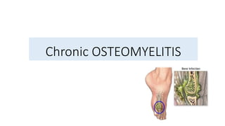
16. CHRONIC OSTEOMYELITIS
XRAY
Bone resorption
either as a patchy loss
of density or as frank
excavation around an
implant
Thickening and
sclerosis of the
surrounding bone.
CT and MRI
Extent of bone
destruction,
reactive oedema,
hidden
abscesses and
sequestra.
IMAGING
Acute flares ESR and
WBC levels may be
increased.
Organisms cultured from
discharging sinuses
should be tested
repeatedly for antibiotic
sensitivity.
LABORATORY
INVESTIGATIONS
19. TREATMENT
• ANTIBIOTIC
Chronic infection is seldom eradicated by antibiotics alone.
Bactericidal drugs Stop the spread of infection to healthy bone and control acute flares.
Choice of antibiotic Capable of penetrating sclerotic bone and non-toxic with long-term
use.
Example: fusidic acid, clindamycin and cephalosporins.
MRSA Vancomycin
administered for 4–6 weeks (starting from the beginning of treatment or the last
debridement) before considering operative treatment.
If surgical clearance fails, antibiotics should be continued for another 4 weeks before
considering another attempt at full debridement.
20. • LOCAL TREATMENT
A sinus may be painless and need dressing simply to protect the clothing.
An acute abscess may need urgent incision and drainage but this is only a
temporary measure.
TREATMENT
21. SURGERY
INDICATION FOR RADICAL SURGERY
Chronic haematogenous infections
Intrusive symptoms, failure of adequate antibiotic treatment, clear evidence of a
sequestrum or dead bone.
Post-traumatic infections
An intractable wound and/or an infected ununited fracture.
Postoperative infection
Presence of a foreign implant
prompt surgical intervention to remove the implant, whether in case of internal
fixation or substitution.
TREATMENT
22. • Debridement
All infected and dead soft tissue, devitalized bone or any infected
implant must be excised.
The wound is inspected after 3 or 4 days and if there are renewed
signs of tissue death, the debridement may have to be repeated.
Antibiotic cover is continued for at least 4 weeks after the last
debridement.
TREATMENT
23. • Grafting
Porous
antibiotic
Porous gentamicin impregnated beads laid in the cavity and left
for 2 or 3 weeks replaced with cancellous bone grafts.
Papineau
technique
The entire cavity is packed with small cancellous chips, mixed
with an antibiotic and a fibrin sealant.
Muscle flap
transfer
In suitable large wad of muscle with its blood supply intact can
be mobilized and laid into the cavity the surface is later
covered with a split-skin graft.
Lautenbach
approach
Radical excision of all avascular and infected tissue closed
irrigation and suction drainage appropriate antibiotic
solution in high concentration to allow the ‘dead space’ to be
filled by vascular granulation tissue.
In refractory cases, it may be possible to excise the infected
or devitalized segment of bone completely then close the gap
by the Ilizarov method.
This is especially useful if infection is associated with non-
union after fracture
24.
25. • Soft tissue cover
Small defects split thickness skin grafts.
Larger wounds local musculocutaneous flaps or free vascularized flaps.
Vacuum-assisted closure (VAC) may help when the deep infection is solved,
not before.
After care
Local trauma must be avoided
Any recurrence of symptoms however slight, should be taken seriously and
investigated.
TREATMENT