
Recommended
More Related Content
Similar to Porifera-good.ppt
Similar to Porifera-good.ppt (20)
More from TaniaPalChoudhury1
Recently uploaded
Recently uploaded (20)
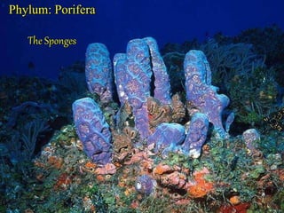
Porifera-good.ppt
- 2. I. General Ecological Characteristics Sponges are: • Sessile, benthic • Filter feeders • Competitors for space • Fed upon by specialist predators • Grow in many forms, solitary, colonial, branching, as thin sheets over substrates • From few cm to over 1 m in size • Estimated in some cases to be several hundred years old
- 3. The classification of sponges is based on skeletal morphology II. General Characteristics of the Porifera Body Plan
- 6. D. Class Calcarea (Calcispongiae) 1. These are calcareous sponges with spicules of calcium carbonate. 2. The spicules are straight or have three or four rays. E. Class Hexactinellida (Hyalospongiae) 1. These are glass sponges with six-rayed spicules of silica. 2. Nearly all are deep-sea forms; most are radially symmetrical. F. Class Demospongiae 1. This class contains 95% of living sponge species. 2. Spicules are siliceous but not six rayed; they may be absent or bound together by spongin. 3. All are leuconoid and all are marine except for Spongillidae, the freshwater sponges.
- 7. 12.1. Advent of Multicellularity A. Advantages 1. Nature’s experiments with larger organisms without cellular differentiation are limited. 2. Increasing the size of a cell causes problems of exchange; multicellularity avoids surface-to- mass problems. 3. Cell assemblages in sponges are distinct from other animals; but molecular evidence shows common ancestry.
- 8. 12.5. Phylum Porifera A. General Features 1. Porifera means "pore-bearing"; their sac-like bodies are perforated by many pores. 2. They are sessile and depend on water currents to bring in food and oxygen and carry away wastes. 3. Their body is a mass of cells embedded in gelatinous matrix and stiffened by spicules of calcium carbonate or silica and collagen. 4. They have no organs or tissues; cells are somewhat independent. 5. Being sessile, they have no nervous or sense organs and have simplest of contractile elements. 6. They are not from the mainstream of animal evolution; thus they are often called Parazoa. 7. Most of the 5000 species are marine; about 150 are freshwater. 8. Morphology changes with substrate, calmness of water, etc. 9. Sponges are ancient; fossils extend to Cambrian or earlier.
- 9. B. Form and Function 1. Body openings consist of small incurrent pores or ostia and a few excurrent oscula. 2. Openings are connected by a system of canals; water passes from ostia to osculum. 3. Choanocytes or flagellated collar cells line some of the canals. a. They keep the current flowing by beating of flagella. b. They trap and phagocytize food particles passing by. 4. The framework of the sponge is composed of needle-like calcareous or siliceous spicules or organic spongin fibers.
- 10. Overview Diversity: three major groups 1. Calcarea: Calcareous sponges Calcium carbonate (calcite) spicules Primarily shallow water and tropical (some exceptions) 2. Hexactinellida: Glass Sponges Siliceous, 6-rayed spicules Marine, primarily deep water Taxonomic Detail Desmospongia All other classes
- 11. Overview Diversity: three major groups 3. Demospongiae: Demosponges Siliceous spicules (never 6-rayed) and/or spongin for support
- 12. 5. There are three types of canal systems. a. Asconoids: Flagellated Spongocoels 1) Asconoids are simplest; they are small and tube- shaped. 2) Water enters a large cavity, the spongocoel, lined with choanocytes. 3) Choanocyte flagella pull water through. 4) All Calcarea are asconoids: Leucosolenia and Clathrina are examples.
- 13. b. Syconoids: Flagellated Canals 1) They resemble asconoids but are bigger with a thicker body wall. 2) The wall contains choanocyte- lined radial canals that empty into the spongocoel. (Fig.12-7) 3) Water entering filters through tiny openings. 4) Food is digested by choanocytes. 5) Flagella force the water through internal pores into the spongocoel and out the osculum. 6) The flagellated canals form by evagination of the body wall; this is developmental evidence of being derived from asconoid ancestors. 7) Classes Calcarea and Hexactinellida have species that are syconoid.
- 14. c. Leuconoids: Flagellated Chambers 1) These are most complex and are larger with many oscula. (Fig. 12-8) 2) Clusters of flagellated chambers are filled from incurrent canals, discharge to excurrent canals. 3) Most sponges are leuconoid; it is seen in most Calcarea and in all other classes. 4) This system increases flagellated surfaces compared to volume; more collar cells can meet food demands. Its all about surface-to- volume ratios and energy extraction!!!
- 15. 6. Types of Cells a. Sponge cells are arranged in a gelatinous matrix called mesohyl. b. Pinacocytes 1) These cells form the pinacoderm; they are flat epithelial-like cells. 2) Pinacocytes are somewhat contractile.
- 16. c. Choanocytes 1) These are oval cells with one end embedded in mesohyl. 2) The exposed end has a flagellum surrounded by a collar. 3) A collar is made of a row of microvilli forming a fine filtering device to strain food. (Fig. 12-10) 4) Particles too large to enter the collar are trapped in mucous and moved to the choanocyte where they are phagocytized. 5) Food engulfed by choanocytes is passed to neighboring archaeocytes for digestion.
- 17. 3) A collar is made of a row of microvilli forming a fine filtering device to strain food. (Fig. 12-10) 4) Particles too large to enter the collar are trapped in mucous and moved to the choanocyte where they are phagocytized. 5) Food engulfed by choanocytes is passed to neighboring archaeocytes for digestion.
- 18. d. Amoebocytes (Archaeocyte) 1) These cells move about in the mesohyl. 2) They phagocytize particles in the pinacoderm. 3) They can differentiate into any other type of cell. 4) Those called sclerocytes secrete spicules. 5) Spongocytes secrete spongin. 6) Collencytes secrete fibrillar collagen. 7) Lophocytes secrete lots of collagen but may look like collencytes.
- 19. e. Types of Skeletons 1) Collagen fibrils are found throughout intercellular matrix of sponges. 2) Various Demospongiae secrete a form of collagen called spongin. 3) Demospongiae also secrete some siliceous spicules. 4) Calcareous sponges secrete spicules of crystalline calcium carbonate. 5) Glass sponges have siliceous spicules with six rays. 6) Spicule patterns are important classification features.
- 20. Spicule formation: The sponge is a sessile (permanently attached; non-motile) animal. It utilizes a proteoglycan-like molecule termed the aggregation factor 31-32 for its adhesive properties.4 Structural support is based upon the formulation of a endoskeleton. Made up of collagen, spongin, and mineral sclera(spicules),the skeleton stabilizes and protects the sponge. 5 Skeletal formation is based on mineral secretions of calcite, aragonite, and/or silica. This formation also functions by: Precise control of intracellular calcium carbonate precipitation releases protons, which contribute to the acid pH maintenance necessary for sponge biological processes. HCO3+ Ca2 →CaCO3+H+ 4
- 21. 7. Sponge Physiology a. Filtration Rates 1) Leuconia, a small sponge, has 81,000 incurrent canals. 2) It would have more than two million flagellated chambers. 3.)Expulsion is strong enough to disperse wastes. 4) Some large sponges can filter 1500 liters of water a day. b. Particles are filtered nonselectively; choanocytes phagocytize 80%. c. Digestion is completely intracellular, primarily by amoebocytes. d. There are no excretory or respiratory organs; diffusion suffices. e. The only movements are very slow opening and closing of pores; nerve cells are not present.
- 22. C. Reproduction 1. Asexual Reproduction a. External buds are small individuals that break off after attaining a certain size. b. Internal buds or gemmules are formed by amoebocytes that collect in mesohyl and are coated with tough spongin and spicules; they survive drought, freezing, etc, and contain amoebocytes. 2. Sexual Reproduction a. Most are monoecious with both male and female sex cells in one individual. b. Sperm arise from transformed choanocytes. c. In some Demospongiae and Calcarea, oocytes develop from choanocytes; others derive them from archaeocytes. d. Sponges provide nourishment to the zygote until it is released as a ciliated larva. e. In some species, sperm are released out into the water where they enter the pores of other sponges. f. Choanocytes phagocytize the sperm and transfer them to carrier cells that carry sperm through mesohyl to oocytes. g. Some release both sperm and oocytes into water.
- 25. Vase Sponges
- 26. Barrel Sponges
- 27. Ball Sponges
- 28. Rope Sponges