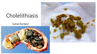
Cholelithiasis: Causes, Types, Diagnosis and Treatment
•Download as PPTX, PDF•
3 likes•633 views

General overview about gall stones, types, clinical features, diagnosis and treatment perspectives

Recommended
More Related Content
What's hot
What's hot (20)
Similar to Cholelithiasis: Causes, Types, Diagnosis and Treatment
Similar to Cholelithiasis: Causes, Types, Diagnosis and Treatment (20)
More from Suman Baral
Recently uploaded
Recently uploaded (20)
Cholelithiasis: Causes, Types, Diagnosis and Treatment
- 2. Incidence • In USA or Europe, 80% are cholesterol or mixed stones • Cholesterol or mixed stones contain 51-99 % pure cholesterol plus admixture of calcium salts, bile acids, bile pigments and phospholipids • Pigment stones contain less than 30% cholesterol
- 3. Pigment Stones Brown Contains calicium bilirubinate , palmitate or stearate as well as cholesterol Rare in Gall bladder Formed in Bile Duct Related to bile stasis and infection: Gram Negative bacteria(E.coli and Klebsiella) secretes beta-glucuronidase which deconjugates the soluble conjugated bilirubin Free indirect bilirubin precipitates and combines with cacium and bile to form brown stones Black Black stones are composed of insoluble bilirubin pigment polymer mixed with calcium phosphate and calcium bilirubinate Predisposing factors: Hemolytic Disorders, Mechanical Prosthetic valves, Cirrhosis, Gilbert’s Syndrome, Cystic Fibrosis, Ileal disease or resection
- 4. Factors forming Gall Stones Lithogenic Bile : • Increased biliary cholesterol: Obesity, Cholesterol rich diet, Clofibrate therapy • Decreased Bile acids: Primary Biliary Cirrhosis, Impaired EHC of bile acids- Ileal resection , cholestyramine therapy or colestipol(bile acid sequestrants) • Decreased biliary lecithin- MDR-3 gene mutation leads to defective lecithin secretion in bile Nucleation : Pronucleating Vs Anti-nucleating factors- excess pronucleation(mucin, non mucin glycoprotein,Infection) or defective anti nucleation (Apo A-I and A-II)
- 5. Risk Factors • Obesity • Rapid weight loss • First degree relatives • Childbearing • Multiparity • Female Sex • Drugs- Ceftriaxone, TPN, post menopausal estrogens • Ileal disease, resection or bypass • Increasing age
- 6. Pathogenesis • Cholesterol is insoluble in water ( water is a major constituent of bile- 85-95% • Bile acid and phospholipids in bile keep cholesterol in solution by the formation of micelles • An excess of cholesterol relative to bile acids and phospholipids allows cholesterol to form crystals and such bile is called lithogenic or super saturated bile.
- 7. Clinical Features • Silent Stones(Asymptomatic) • Symptomatic Cholelithiasis ( Biliary Colic) • Features of Acute Calculus Cholecystitis • Pain at right hypochondrium, radiating to tip of right shoulder • Vomiting • Murphy’s Sign positive
- 8. Investigations • Oral Cholecystography ( Graham Cole test) • Oral administration of a radiopaque compound that is absorbed, excreted by the liver and passed into the gall bladder • Stones noted on a film as filling defect in a visualized opacified gall bladder • However, this test has been replaced by ultrasonography
- 9. Ultrasonography • IOC for acute calculus cholecystitis, chronic cholecystitis and cholelithiasis • Operator dependent and may be suboptimal due to excessive body fat and intraluminal bowel gas • Can demonstrate- Biliary Calculi, Size of GB and CBD, Thickness of GB wall, inflammation around GB, presence of stones within biliary tree
- 10. HIDA Scan • Technetium -99m labelled derivative of imino-diacetic acid are when injected intravenously, selectively taken up by reticulo-endothelial cells of the liver and excreted into bile. • Allows visualization of biliary tree and gall bladder • GB visualized within 30 mins of isotope injection in 90 % cases • Bowel seen within 1 hour of injection • Non-visualization of GB suggestive of acute cholecystitis • Helpful in diagnosing bile leaks and iatrogenic biliary obstruction
- 11. Complications IN Gallbladder • Silent stones • Acute Cholecystitis • Chronic Cholecystitis • Mucocele • Empyema • Perforation • Gangrene • Carcinoms In Bile Duct • Obstructive Jaundice • Cholangitis • Acute Pancreatitis In Intestine • Gall Stone Ileus
- 12. Treatment • Cholecystectomy • Open VS Laparoscopic Cholecystectomy ( Surgical) MEDICAL THERAPY (?? EFFECTIVENESS)
- 13. To be continued !!!