2. Kumar RR et al. Int J Reprod Contracept Obstet Gynecol. 2018 Dec;7(12):4919-4923
International Journal of Reproduction, Contraception, Obstetrics and Gynecology Volume 7 · Issue 12 Page 4920
Neglected or missed tears or faulty repair may also
present an inherent source for litigation. The aim of this
study was to evaluate the effectiveness, long term follow-
up and to highlight the current practices regarding
secondary repair of chronic fourth degree perineal tear.
METHODS
Thirty consecutive cases of chronic perineal tears were
selected from department of obstetrics and gynecology,
SDM college of medical sciences and hospital, Dharwad,
Karnataka, India from January 2008 to December 2016.
Data were collected prospectively on age, parity,
incontinence to flatus, solid or liquids stools, duration of
symptoms, history of previous repair, duration of repair,
post-operative stay, complications and recovery.
Counseling-Patients considering for perineal tear repair
were meticulously informed about their chances for a
successful functional sequel after surgery.
Preoperative management
Bowel preparation -Although there is no evidence-based
guideline for a preoperative bowel regimen before anal
surgery, tablet bisacodyl 10mg was given on the previous
day of surgery. Bowel preparation was done before
sphincter repair to decrease bacterial contamination of the
perineal wound.
Antibiotics -Preoperatively, patients were administered
with intravenous ornidazole 500mg and ceftriaxone 1gm
stat was given, that covers skin and enteric flora.
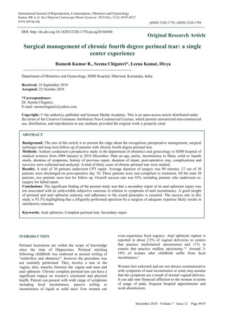
Figure 1: Grossly deficient perineum with absence of
anal puckering around the anterior aspect of anus at 3
and 9’0 clock position.
Surgery
Anesthesia-All the patients underwent CPT repair under
regional anesthesia.
Foley catheter-A Foley catheter is placed before the
procedure. The Foley catheter is typically left in
overnight and removed the morning after surgery because
pain from the anal surgery often inhibits spontaneous
voiding.
Positioning-Patients were positioned in dorsal lithotomy.
Surgical goals-The goal of sphincter repair was to
reconstruct a cylindrical anal canal utilizing both external
sphincter muscle and perineal body scar, to allow
circumferential contraction of the anal musculature when
the patient attempts to defer defecation.
Procedure
On either side of the lower edge of labia minora, two
lateral ends of rectovaginal septum are held with Allis
tissue forceps (Figure 2). Postero-laterally over the
dimple, two more Allis are applied representing the torn
edges of external anal sphincter. Along the junction of
posterior vaginal wall and rectal mucosa an elliptical
incision is made extended postero-laterally over the
dimple.
Figure 2: The vaginal and rectal walls are separated
from each other by dissection to the levator ani
complex.
Sharp dissection is done to separate anterior wall of rectal
mucosa from posterior vaginal wall creating a good
rectovaginal space. Incision is deepened over the dimple
to expose the torn ends of EAS (Figure 3) and are held
with Allis tissue holding forceps to feel the tightness of
the sphincter. Two main aspects of sphincter repair are
the removal of scar tissue, identifying the torn edges of
EAS and the mobilization of rectum. Anorectal mucosa is
repaired with 3-0 continuous polyglactin 910 suture to
reach the mucocutaneous junction of the anal opening.
Internal anal sphincter: The IAS is identified, repaired
separately from the EAS. The IAS lies between the EAS
and the anal epithelium. It is thinner and paler than the
striated EAS. The appearance of the IAS can be described
as being analogous to the flesh of raw fish, as opposed to
the red meat appearance of the EAS. Ends of the IAS are
3. Kumar RR et al. Int J Reprod Contracept Obstet Gynecol. 2018 Dec;7(12):4919-4923
International Journal of Reproduction, Contraception, Obstetrics and Gynecology Volume 7 · Issue 12 Page 4921
identified and sutured with 3-0 polygalactin 910 (Figure
4). The torn ends of the EAS is freshened then be
overlapped in a “double-breasted” fashion using 3-0
Polyglactin 910. A proper overlap is possible only when
the full length of the torn ends of the EAS is identified.
Figure 3: The scar and the external sphincter muscles
are carefully freed from the internal sphincter to
allow the left and right external sphincter and scar
complexes to overlap anteriorly.
Perineal body reconstruction: The two levator ani
muscles are drawn together in between the posterior
vaginal wall and anterior wall of the rectum and anal
canal is sutured with interrupted 3-0 vicryl.
Figure 4: The anterior defect of the IAS extended
through the anal mucosa and was plicated with 3-0
polyglactin suture.
Repair of posterior vaginal wall, subcutaneous tissue and
skin are done using 2-0 delayed absorbable suture. A
rectovaginal examination is performed to confirm
complete repair and ensure that all tampons or swabs
have been removed. Detailed note is made of the findings
and repair. Completion of a pre-designed proforma and a
pictorial representation of the tear is done.
Postoperative care
In the immediate post-operative period,
1: Syrup Lactulose 15 ml thrice daily was given for the
first 3 weeks to avoid constipation and to ensure that the
patient is having soft stools.
2: Five days of intravenous antibiotics ceftriaxone 1gm
and ornidazole 500mg, both twice a day, were given to all
the patient to cover the enteric flora.
3: Sitz bath was given for all the patients twice daily and
the patients were allowed to take clear liquids for the first
48 hours and for the next 5 days were put on low residual
diet.
4: For pain relief injection diclofenac was used for the
first 48 hours for all patients and later on it was used as
needed.
5: Patients were mobilized after 24 hours.
On the 10th
postoperative day, wound site was examined
and resting anal sphincter tone was assessed by digital
examination.
Figure 5: Post-operative image the distance between
the posterior fourchette and the anal canal was seen to
be lengthened.
RESULTS
A total of thirty patients underwent CPT repair during the
study period. Data analysis was done using SPSS
software. Cleveland clinic scoring system (Table1) for
fecal incontinence was assessed and surgical repair was
recommended to a patient with a score of 12 or greater.
73.3% of the patients were of age group between 20 and
30 years. All of them were multiparous .80% of the
patients had deliveries at home, unsupervised and
conducted by dais. Nearly 75% of the patients were
completely incontinent, CCIS score of more than 20
(Table 2). 30.3 %were completely ignorant about the
disease for more than 10 years and assumed that the
symptoms are a result of normal vaginal delivery (Table
3). Only about 26.3% of the patients could recognize
symptoms within 5 years of their delivery.
4. Kumar RR et al. Int J Reprod Contracept Obstet Gynecol. 2018 Dec;7(12):4919-4923
International Journal of Reproduction, Contraception, Obstetrics and Gynecology Volume 7 · Issue 12 Page 4922
Table 1: Cleveland clinic incontinence score for the assessment of faecal incontinence.
Never Rarely Sometimes Usually Always
Solid 0 1 2 3 4
Liquid 0 1 2 3 4
Flatus 0 1 2 3 4
Use of pads 0 1 2 3 4
Lifestyle alteration 0 1 2 3 4
Complete chronic fourth degree perineal tear is shown in
Figure 1. Average duration of surgery was 90 minutes.
Figure 5 shows final post-operative image showing
reconstruction of the perineal body. 27 out of 30 patients
were discharged on post-operative day 10. Three patients
went against medical advice on day 3 and were non-
compliant to treatment.
Table 2: Degree of incontinence.
Incontinence n Percentage
Complete incontinence 22 73.3
Severe incontinence 8 26.6
Six months later they came with complaints of fecal
incontinence for which re-surgery was done using the
same protocol and they had a successful repair. Of the
total 30 patients, two patients were lost for follow up.
Follow up was done once a month for the first 6 months
then once in 3 months for the next one year and then at
six monthly interval for a period of 24 months. Overall
success rate was 93% including patients who underwent
re surgery for failed repair. All patients were successfully
treated and were asymptomatic till the last follow-up.
Table 3: Duration of symptoms.
Duration of symptoms n Percentage
<5 years 8 26.3
5-10 years 13 43.3
>10 years 9 30.3
Rarely: less than once a month; sometimes: more than
once per month or less than once a week; usually: more
than once a week but less than once a day.
Figure 6: Age distribution of the patients.
Always: more than once a day. CCIS 0, perfect
continence; CCIS 1-7, good continence; CCIS 8-14,
moderate incontinence; CCIS 15-20, severe incontinence;
CCIS .20, completely incontinent.
DISCUSSION
Perineal injuries are common in obstetric practice.
However, CPT is very infrequent. A third or fourth-
degree tear (involving the anal sphincter complex) occurs
in 0.5-2%. Eighty-five per cent will have a persistent
defect of the sphincter despite the immediate (primary)
repair by the obstetrician.4
However, due to lack of awareness and due to deliveries
at home, it can go unnoticed. Though there are very
sparse case reports of delayed presentation, large case
studies are not reported. This single center study reports
thirty cases of chronic fourth degree perineal tear.
The significant finding of the present study was that a
secondary repair of an anal sphincter injury was not
associated with an unfavourable subjective outcome in
relation symptoms of anal incontinence. The incidence of
perineal wound dehiscence after repair of third or a
fourth-degree perineal tear has been reported to occur in
10% of the cases.5
Several studies gives an estimated
complication rate of 15% for fourth degree perineal
laceration repair. Most common complications among
them are wound dehiscence, rectovaginal fistula,
hematoma, perineal abscess, dysperunia.6,7
In the present
study there were no such complications noted. All
patients underwent repair under optimal conditions in the
operating theatre and received pre-operative antibiotics.
A good insight of perineal and anal sphincter anatomy,
perioperative use of antibiotics and appropriate bowel
care all are essential to ensure a successful outcome.
There are no randomized trials reporting the role of
prophylactic antibiotics in the management of perineal
tears. Even in the ACOG bulletin, this recommendation
has been omitted. In present study, 73.3% of the patients
were completely incontinent and 43.3% had symptoms
for the duration between 5 and 10 years. In present study,
authors used overlap technique of repair in all patients.
This study was not conducted to compare the techniques
of repair. There have been several clinical trials that
compared the end to end technique with the overlapping
technique, but it remains unresolved if one technique is
0
10
20
30
40
50
60
0 10 20 30 40
5. Kumar RR et al. Int J Reprod Contracept Obstet Gynecol. 2018 Dec;7(12):4919-4923
International Journal of Reproduction, Contraception, Obstetrics and Gynecology Volume 7 · Issue 12 Page 4923
superior to the other.4,8,9
The success rate in this study is
93.3% highlighting that a diligently performed operation
by a surgeon of adequate expertise likely results in
satisfactory results.
In present study 80% of the patients had home deliveries.
There has to be an emphasis that nursing personnel and
midwives should be effectively trained so as to manage
the second stage to prevent disasters that can significantly
affect the quality of life.
Primary repair is exemplary as it is cost effective and
significantly improves the overall quality of life. For the
patients who are presenting very late and previous failed
repairs, Cleveland clinic scoring system for fecal
incontinence was assessed and surgical repair was
recommended to a patient with a score of 12 or greater an
underlying amenable abnormality and gross fecal
continence.10-12
Improvement in the functional length of
the sphincter correlated to a successful outcome .In
resourced settings usually an endoanal ultrasound will be
performed to know the extent and the type of anal
sphincter injury and the pudendal nerve damage. Several
previous studies conducted over a long period of time
suggest that symptoms of anal incontinence deteriorate
over time after repair. However, in the present study
symptomatic improvement is seen from 3-6 months of
time. Details regarding improvement in the symptoms
were evaluated using questionnaire method though many
studies suggest that evaluating the symptoms of fecal
incontinence should be delayed for at least 12 months.
Long term follow-up is thus essential.13,14
CONCLUSION
Authors have reported a large series of CPT repair with a
successful outcome. Structured training model for
beginners in recognizing and repair of primary tear is
beneficial. Secondary repair is usually offered to patients
with gross faecal incontinence. The outcome depends on
the extent of the anal sphincter damage and associated
neurological injuries.
Funding: No funding sources
Conflict of interest: None declared
Ethical approval: Not required
REFERENCES
1. Sultan AH. Anal incontinence after childbirth. Curr
Opin Obstet Gynecol. 1997;9:320-4.
2. Sultan AH, Kim MA, Hudson CN, Thomas JM, Bartam
CI. Anal-sphincter disruption during vaginal delivery. N
Engl J Med. 1993;329:1905-11.
3. Fernando RJ, Sultan AH, Kettle C, Radley S, Jones P,
O’Brien PMS. Repair techniques for obstetric anal
sphincter injuries: a randomized controlled trial. Obstet
Gynecol. 2006;107:1261-8.
4. Fitzpatrick M, Behan M, O’Connell PR, O’Herlihy C.
A randomized clinical trial comparing primary overlap
with approximation repair of third-degree obstetric
tears. Am J Obstet Gynecol. 2000;183:1220-4.
5. Goldaber KG, Wendel PJ, McIntire DD, Wendel GD.
Postpartum perineal morbidity after fourth-degree
perineal repair. American Journal of Obstetrics &
Gynecology. 1993 Feb 1;168(2):489-93.
6. Venkatesh KS, Ramanujam PS, Larson DM, Haywood
MA. Anorectal complications of vaginal delivery. Dis
Colon Rectum. 1989;32:1039-41.
7. Pezim ME, Spencer RJ, Stanhope CR, Beart RW Jr,
Ready RL, Ilstrup DM. Sphincter repair for fecal
incontinence after obstetrical or iatrogenic injury. Dis
Colon Rectum 1987;30:521-5.
8. Corman ML. Anal sphincter reconstruction. Surg Clin
North Am. 1980;60:457-63.
9. Garcia V, Rogers RG, Kim SS, Hall RJ, Kammerer-
Doak DN. Primary repair of obstetric anal sphincter
laceration: a randomized trial of two surgical
techniques. Am J Obstet Gynecol. 2005;192:1697-701.
10. Williams A, Adams EJ, Tincello DG, Alfirevic Z,
Walkinshaw SA, Richmond DH. How to repair an anal
sphincter injury after vaginal delivery: results of a
randomised controlled trial. BJOG. 2006;113:201-7.
11. National institute of Clinical Excellence (NICE). Faecal
incontinence: the management of faecal incontinence in
adults. Clinical guidelines CG :49 National Institute for
Health and care Excellence (NICE), 2013.
12. Fernando R, Sultan AH, Kettle C, Thakar R, Radley S.
Methods of repair for obstetric anal sphincter injury.
Cochrane Database Syst Rev. 2006;3:CD002866.
13. Jorge JM, Wexner SD. Etiology and management of
fecal incontinence. Dis Colon Rectum. 1993;36:77-97.
14. Tan EK, Jacovides M, Khullar V, Teoh TG, Fernando
RJ, Tekkis PP. A cost-effectiveness analysis of delayed
sphincteroplasty for anal sphincter injury. Colorectal
Dis. 2008;10:653-62.
Cite this article as: Kumar RR, Chigateri S, Kamat
L, Divya. Surgical management of chronic fourth
degree perineal tear: a single center experience. Int J
Reprod Contracept Obstet Gynecol 2018;7:4919-23.