Development of mandible - Dr. Shweta Yadav - Oral and Maxillofacial Surgery
•Download as PPTX, PDF•
3 likes•149 views

Development of mandible

Recommended
More Related Content
What's hot
What's hot (20)
Similar to Development of mandible - Dr. Shweta Yadav - Oral and Maxillofacial Surgery
Similar to Development of mandible - Dr. Shweta Yadav - Oral and Maxillofacial Surgery (20)
Recently uploaded
Recently uploaded (20)
Development of mandible - Dr. Shweta Yadav - Oral and Maxillofacial Surgery
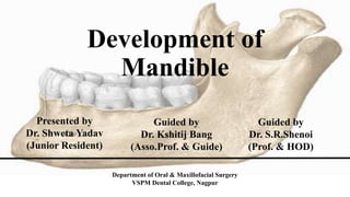
- 1. Development of Mandible Presented by Dr. Shweta Yadav (Junior Resident) Guided by Dr. Kshitij Bang (Asso.Prof. & Guide) Guided by Dr. S.R.Shenoi (Prof. & HOD) Department of Oral & Maxillofacial Surgery VSPM Dental College, Nagpur
- 2. INTRODUCTION • Mandible – Largest and Strongest bone of the face • Greek word ‘mandere’- to masticate • Latin word ‘mandibula’-lower jaw
- 3. History and Background • JOHN HUNTER (1771) compared a series of dried mandibles and concluded that to attain space for permanent molar teeth the mandible must grow by posterior apposition of ramus accompanied by anterior ramus resorption. • HUMPHRY (1866) studied growth of mandible by inserting metal wires in the mandible of young pigs. • BELCHIE (1936) fed pigs the madder plant root which labeled appositional growth
- 4. • BJORK (1955): conducted implant studies on jaws to determine the growth pattern & rotation ,when subjected to serial cephalometric methods. • DONALD ENLOW : proposed the V principle of growth and counterpart principle.
- 5. Prenatal Growth and Development
- 6. In sixth week ,a single ossification Centre for each half arises in the bifurcation of inferior alveolar nerve into mental and incisive 7th week-bone begin to develop lateral to Meckels cartilage and continues until the posterior aspect is covered with bone Between 8th & 12th week ,mandibular growth accelerate ,as a result mandibular length increases
- 7. Ossification stops at a point , which later become lingula , the remaining part of meckels cartilage continues to form sphenomandibular ligament & spinous process of sphenoid Secondary accessory cartilage appears between 10th & 14th week to form head of condyle , part of coronoid process & mental protuberance
- 8. SECONDARY CARTILAGES IN MANDIBULAR DEVELOPMENT CONDYLAR PROCESS • About 5th week of I.U.L. area of mesenchymal condensation above the ventral part of developing mandible • About 10th week develops into cone shaped cartilage • By 14th week starts ossification • By 4 months migrates inferiorly and fuses with ramus • 4th month onwards replaced by bone but proximal end persists into adulthood acting as Growth cartilage & Articular cartilage
- 9. CORONOID PROCESS • By 10th to 14th week of I.U.L. secondary cartilages seen in region of coronoid • Cartilage becomes incorporated into expanding intramembranous bone of ramus and disappears before birth MENTAL REGION • Secondary cartilages seen on both sides -- ossify by 7th week I.U.L. • They ossify to form mental ossicles in fibrous tissue of symphysis.
- 10. Postnatal Growth and Development MANDIBLE AT BIRTH • Ascending ramus- low and wide • Coronoid process- relatively large • Body – containing buds and partial crowns of deciduous teeth • Mandibular canal- runs low in the body
- 11. MANDIBULAR GROWTH DURING FIRST YEAR Appositional growth especially active at • Alveolar border • Distal and superior surface of ramus • Condyle • Lower border of mandible • Lateral surface of mandible By the end of first year mandible appears as a single bone.
- 12. GROWTH PROGRESSION AFTER FIRST YEAR- MECHANISM & SITE Ramal remodeling is important 1. Positions the lower arch in co-ordination with the growth of upper arch 2. Adaptive to changing cranio facial conditions Hunterian Concept • The principal vectors of mandibular growth are posterior and superior • Mandible as a whole becomes displaced antero-inferiorly.
- 13. MANDIBULAR FORAMEN • Relocates backward and upward by deposition on the anterior and resorption from the posterior part of the rim • From childhood throughout the old age maintains a constant position about midway between the anterior and posterior borders of the ramus.
- 14. RAMUS TO BODY REMODELING CONVERSION In general the arch length is increased and body has been lengthened by 1. Deposits on the posterior surface of lingual tuberosity and the contiguous lingual side of ramus 2. A resultant lingual shift of anterior part of the ramus to become added to the body
- 15. Condyle and mandibular growth
- 16. Theories of mandibular growth GENETIC THEORY: states that all growth is compelled by genetic influence ie: genetic encoding of mandible determines its growth. CARTILAGENOUS THEORY: states that the cartilage is the primary determinant of skeletal growth while bone responds secondarily & passively. ENLOW’S EXPANDING ‘V’ PRINCIPLE: states that many facial bones or a part of the bone follows a ‘v’ pattern of enlargement. FUNCTIONAL MATRIX THEORY: mandible as a group of microskeletal units and a basal core part related to the growth of macroskeletal unit.
- 17. THANK YOU