
Recommended
More Related Content
Similar to MIDDLE EAR MSN
Similar to MIDDLE EAR MSN (20)
Recently uploaded
Recently uploaded (20)
MIDDLE EAR MSN
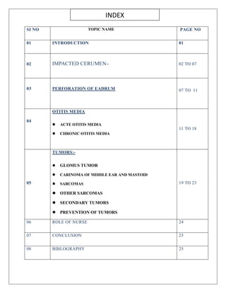
- 1. SI NO TOPIC NAME PAGE NO 01 INTRODUCTION 01 02 IMPACTED CERUMEN:- 02 TO 07 03 PERFORATION OF EADRUM 07 TO 11 04 OTITIS MEDIA ACTE OTITIS MEDIA CHRONIC OTITIS MEDIA 11 TO 18 05 TUMORS:- GLOMUS TUMOR CARINOMA OF MIDDLE EAR AND MASTOID SARCOMAS OTHER SARCOMAS SECONDARY TUMORS PREVENTION OF TUMORS 19 TO 23 06 ROLE OF NURSE 24 07 CONCLUSION 25 08 BIBLOGRAPHY 25 INDEX
- 2. MIDDLE EAR– IMPACTED WAX, TYMPANIC MEMBRANE PERFORATION, OTITIS MEDIA , TUMORS. INTRODUCTION: The middle ear is a 2cm space between the tympanic membrane and the cochlea .it consists of the cochlea .it consists of three bone (the malleus ,inches and stapes)surrounded by air .together these bones are known as the ossicles .the function of the middle ear is to correct for an impedance mismatch between the air in the outer ear relatively small oval window that sit sat the base of the stapes . IMPACTED CERUMEN INTRODUCTION: Ear wax (cerumen) is a natural protective oily substance which is produced in the outer third of the ear canal. Its function is to remove small foreign particles such as dust from the canal . DEFINITION: Impacted cerumen is accumulated ear wax that obstructs the external acoustic meatus.
- 3. CAUSES: It is most common people who have excessive thick or dry cerumen. 1. Swimmer’s ear 2. Dermatological conditions 3. Autoimmune conditions 4. Blockage with in ear canal 5. Unnecessary ear cleaning PATHOPHYSIOLOGY: Due to etiology ↓ Formation excessive thick cerumen ↓ It interferes with the transmission of sound carried on airwaves. ↓ Diminished hearing CLINICAL MANIFESTATION: Sense of fullness or pain in the ear, refers to as otalgia
- 4. Diminished hearing Patients ask that words be repeated misinterpreted questions or raises the volume on the television or radio. DIAGNOSTIC EVALUATION: History collection Physical examination Otoscope examination MEDICAL MANAGEMENT: 1The directed cerumen is hydrated by installing 1 to 2 drops of half strength peroxide, warm glycerin or mineral oil or it is softened with commercial agents, such as carbamide peroxide and triethanolamine. 2. Cerumen is removed mechanically by irrigating the ear if the eardrum is intact or using instrument called a cerumen spoon. NURSING MANAGEMENT: 1. Examination of ear for identify the disease condition. 2 .Irrigation of the ear or instillation of liquids is ordered, the nurse warm the liquids to body temperature in order to removing the cerumen. ROLE OF NURSE: Examine the outer ear and he ear canal. Assess the level of cerumen impactation Educate the patient not to insert anything in to ear canal, including cotton tipped applicators, hairpins ,matchsticks, safety pins, toothpicks, paperclips, or fingers. Irrigate ears to remove ear wax or remove foreign objects from their patients’ ear canal. SELF CARE TIPS TO EAR WAX BUILD UP: 1. Don’t spray liquids inside the ear with force.
- 5. 2. Never put cold water in your ear 3. Take a well diet. 4. Check the temperature of cleaning solution. 5. Follow up with an ENT. PERFORATION OF EAR DRUM INTRODUCTION A perforated eardrum is a hole or tear in the eardrum. It may be uncomfortable but usually heals on its own without treatment within two months. The eardrum, also known as the tympanic membrane, is a thin layer of tissue that separates the outer ear from the middle ear. DEFINITION: Perforation eardrum (tympanic membrane ) is a rupture or perforation (hole)of the eardrum .the hole exposes the middle and inner ear to damage or infection .perforation of ear drum lead conductive hearing loss. CAUSES: Perforation of the eardrum or tympanic membrane can be caused by : Foreign bodies usually pointed objects. Improper curetting or syringing. Infection of the middle ear. Trauma can also cause perforation. Rapid changes in atmospheric pressure
- 6. PATHOPHYSIOLOGY: Infection or trauma ↓ Increase pressure in the middle ear ↓ Pressure builds up and pushes against the eardrum. ↓ When the pressure further increases ↓ Eardrum perforates. ↓ Injury may extend to the ossicles and to the inner ear. CLINICAL MANIFESTATION: Pain Vertigo(spinning sensation) Hearing change or loss Disorientation Fullness of ear Blood-tinged discharge from the ear.
- 7. MANAGEMENT: If the patient is seen shortly after injury , the external ear or canal is packed with a sterile cotton plug Systemic antibiotics and decongestant drops in the nose are used to prevent infection of the middle ear through the Eustachian tube. Advice the patient to keep the ear clean and dry while healing Because most perforated eardrum injuries heal on their own within 2 months, treatment may include analgesics to alleviate pain and antibiotics to prevent infection. If the patient perforated eardrum is due to a foreign object in the ear, do not try to remove it yourself. Only a medical professional should attempt to remove any foreign bodies in the ear. SURGICAL MANAGEMENT: Some large holes/non-healing small holes require surgery. Surgical procedures are performed with general anesthetic. Most people go home from the hospital or clinic on the same day. 1. Tympanoplasty: it is surgical correction of the perforated eardrum. 2. Myringoplasty: closure of perforation is called myringoplasty.
- 8. 3. Ossiculoplasty: surgical procedure of ossicular reconstruction. PREVENTION: Some causes of ruptured eardrums cannot be prevented or avoided. A little caution can lower the risk. 1. Treat ear infection early. 2. Avoid flying or scuba diving if you have sinus infection or upper respiratory tract infection. 3. If you must fly or scuba dive, pinch your nose and swallow air frequently to help equalize the pressure. 4. Never put anything in your ear, even to clean it. 5. Wear proper ear protection such as ear plugs or protection designed for sports activities. OTITIS MEDIA Otitis means inflammation of the ear, and media means middle. This inflammation often begins with infections that causes sore throats, colds or other respiratory problems, and spreads to the middle ear. These can be because by viruses or bacteria, and can be acute or chronic. DEFINITION: It is an inflammation of middle ear.
- 9. CLASSIFICATION: Otitis media has many degrees of severity, various names are used to describe each the terminology is sometimes confusing because of multiple terms because of multiple terms being used to describe the same condition. TYPES: Acute otitis media Chronic otitis media ACUTE OTITIS MEDIA: It is also called acute suppurative otitis media or purulent otitis media. AOM is an acute inflammation and infection of the middle ear mucosa. Acute otitis media is usually of rapid onset and short duration. CAUSES OF ACUTE OTITIS MEDIA: More common Common cold Influenza Whooping cough Acute tonsillitis Less common Sinusitis
- 10. Temporal bone fracture Trauma to the tympanic membrane RISK FACTORS: Risk factors for acute otitis media have been indentified and can generally be divided into those associated with the host and those associated with the environment. Host risk factors Low birth weight Family history Altered immunity History of seasonal allergies Perforation of tympanic membrane. Environmental risk factors Frequent upper airway infection. Low socioeconomic status Tobacco and pollutant exposure Pacifier use increases risk for AOM. Crowded living conditions. PATHOPHYSIOLOGY OF OTITIS MEDIA: Etiology ↓ Infection and inflammation in ↓ Otitis media ↓
- 11. Recurrent infection → Cholesteatoma CLINICAL MANIFESTATIONS: Severe earache (otalgia) Hearing loss Tugging and rubbing the ear Mastoid tenderness DIAGNOSTIC EVALUATINS: 1. History collection 2. Physical examination 3. Otoscopy 4. Sinus x-ray 5. MRI 6. CT scan of the temporal lobe MEDICAL MANAGEMENT: 1. Antibiotics: the first line antibiotic treatment, if warranted is amoxicillin. If the bacteria is resistant, then amoxicillin –clavulanate or another penicillin derivative plus beta-lactamase inhibitors is second line.5 days of treatment has been found to be as effective as ten days in otherwise healthy children. 2. Management of pain and fever: the management of AOM should always include assessment of pain and fever. Antipyretics and analgesics may be necessary and should be prescribed liberally. To treat the pain caused by otitis media oral as well as topical analgesics are effective. Oral agents may include ibuprofen, acetaminophen and or narcotics. Topical agents shows to be effective include antipyrine and benzocaine ear drops. Steroids, decongestants, and antihistamines are not effective in the treatment of AOM, and they may causes complication
- 12. CHRONIC OTITIS MEDIA: It is the result of repeated episodes of acute otitis media causing irreversible tissue pathology and persistent perforation of the tympanic membrane. it is also leads to mastoid bone infection. CAUSES : Late treatment of acute otitis media Inadequate or inappropriate antibiotic therapy. Upper airway sepsis Lowered resistance Particularly virulent infection CLINICAL MANIFESTATION: Hearing loss Chronic foul smelling ear damage Facial weakness Fever Confusion Persistent deep ear pain or headache COMPLICATION: Cholesteatoma- is an growth of the skin of the external layer of the eardrum in to the middle ear.
- 13. MEDICAL MANAGEMENT: Antibiotic- fluoroquinolone otic preparation, with or without a corticosteroids are excellent options for topical treatment. Aminoglycoside otics may also be used. Systemic therapy should be continued fir 3-4 weeks. Aural Toilet: aural toilet is a critical process in the treatment of chronic otitis media .a solution of 50% peroxide and 50% sterile water is generally painless and effective .30-40 ml of this solution can be irrigated through the external auditory canal, using a small syringe or bulb type aspirator. Control of granulation tissue: the use of topical antimicrobial drops is the first steps in controlling granulation. These drops help reduce granulation tissue by eliminating infection and by removing the inciting irritating inflammation SURGICAL MANAGEMENT: 1. Myringoplasty: it is the operation specifically designed to close tympanic membrane defect with use of an operating microscope 2. Myringotomy: a small incision is made in the eardrum to allow fluid to drain and keep the eardrum from rupturing. 3. Tympanoplasty:the purpose of tympanoplasty is to repair the perforated eardrum, and sometimes the middle ear bones that consist of the incus, malleus, and stapes. 4. Typanocentesis: insert a needle through the anterior portion of the tympanic membrane, and aspirate the contents of the middle ear in to sterile trap for identification of microbes and their properties 5. Ossiculoplasty: it is a surgical reconstruction of the middle ear bone to restore hearing. 6. Masitoidectomy:mastoiditis involves incision ,drainage and surgical repair of the mastoid process.
- 14. POST OPERATIVE CARE OF CHRONIC OTITIS MEDIA: After the surgery place the patient in flat and side,lyingposition. It is normal to have impaired hearing during the postoperative period if there is packing the ear. Apply cotton ball dressing is used for an endrual incision. The patient should be educated to change the cotton packing and dressing daily. If a post auricular incision and a drain placed a mastoid dressing is used Monitor the tightness of the dressing to prevent tissue necrosis. Monitor the amount of drainage. PREVENTION OF OTITIS MEDIA : Sleep for at least 8 hours Healthy diet Avoid cleaning the ear too deep it can cause dirt to accumulate in the ear canal. Avoid soaking too long in warm water in humid climate. Avoid swimming I contaminated water. Drink enough water. Do not listen to loud music with headphones. TUMORS DEFINITION: Abnormal growth of cell in ear. TUMORS ARE DIVIDED IN TO: 1. Primary tumors-
- 15. Benign: glomus tumor Malignant : carcinoma , sarcoma. 2.Secondary tumors- a. From adjacent areas E.g., Nasopharynx, external meatus, and parotid. b. Metastatic E.g. From carcinoma of bronchus, breast, kidney, thyroid, prostate, and gastrointestinal tract. GLOMUS TUMOR It is the most common benign neoplasm of middle ear and is so named because of its origin from the glomus bodies. The tumor consists of paraganglionic cells derived from the neural crest. CAUSES: Glomus tumors can affect anyone, but they are more common in olde adults and have no known risk factors .while the exact causes are unknown ,genetic factors may play a role in their development , including the SDHD gene(succinate dehydrogenase gene). For purpose of diagnosis and treatment ,two types differentiated:- Glomus jugulare:they arise from the dome of jugular bulb invade the hypotympanum and jugular foramen causing neurological signs of IXth to XIIth cranial nerve involvement. They may compress jugular vein or invade its lumen. Glomus tympanicum: they arisefrom the promontory of the middle ear and cause aural symptoms sometimes with facial paralysis.
- 16. CLINICAL MANIFESTATIONS: a. When tumor is intratympanic:- symptoms are deafness and tinnitus. b. When tumors present as a polyp:- deafness and tinnitus ,there is a history of profuse bleeding from the ear , dizziness facial paralysis. c. Cranial nerve palsies: dysphagia and hoarseness with unilateral paralysis of the soft palate DIAGNOSTIC EVALUATION: History collection and physical examination CT scan- head MRI Four vessel angiography TREATMENT: It consists of: Surgical removal Radiation Embolisation Combination of the above technique Depending on the extent of tumors, surgical removal may be done through trans meatal ,transmasoid,or skull base approach. Radiation treatment does not cure the tumor but may reduce its vascularity and arrest its growth. Embolisation is used to treat the vascularity of tumor before surgery. CARCINOMA OF MIDDLE EAR AND MASTOID: It is a rare condition. There is one caste in 20,000 new patients examine but it is the commonest primary middle ear malignancy.
- 17. ETIOLOGY: Long standing ear discharge Chronic irrigation CLINICAL MANIFESTATIONS: Chronic foul-smelling discharge especially when blood stained Pain which is usually severe and come at night. Facial palsy Hemorrhagic granulation or polyp Appearance of or increase in deafness or vertigo. DIAGNOSTIC EVALUATION: CT scan Biopsy Radiological examination Angiography TREATMENT: A combination of surgery and radiotherapy gives better results. Surgery consists of: Radical mastoidectomy Subtotal or total petrosectomy on the external ear Radiotherapy alone is given as a palliative measure when tumors involve cranial nerves or spreads in to cranial cavity or the nasopharynx.
- 18. SARCOMAS Rhabdomyosarcoma: -it is a rare tumor, mostly affecting children. It arises from the embryonic muscles tissue or pluripotentialmesenchyme. In early stages it mimics chronic suppurative otitis media with ear discharge, polyp or granulation. Facial palsy occurs early. Diagnosis is made only on biopsy. Prognosis is poor. a combination of radiation and chemotherapy is the treatment of choice. Surgery is done in selected localized lesions. OTHER SARCOMAS: Osteosarcoma, lymphoma, fibrosarcoma and chondrosarcoma are rare distant metastases are seen in the lungs and bones. prognosis is poor. SECONDARY TUMORS Tumors of external auditory metus, parotid gland or nasopharynx may invade middle ear cleft either through the preformed pathways or bone erosion. Sometimes temporal bone is the site of distant metastases in advanced cases of carcinoma of the breast, bronchus, prostate, kidney or gastrointestinal tract. PREVENTION OF TUMORS: While ear cancer can’t always be prevented, Pacticing safe sun Staying in the shade Avoiding tanning beds Lower your risk of skin cancer that could turn in to ear cancer. ROLE OF NURSE: Assisting physicians in assessing, diagnosing, and treating patients with injuries and disorders of the ear. Collecting samples and maintaining records of patients’ medical histories and symptoms.
- 19. Examining and assessing the health of a patient ear using instruments like an otoscope etc. Educating patients on how to properly manage their condition at home. Monitor vital signs and administer themedications. Assisting physician with surgical procedures Preparing patients for surgery CONCLUSION: Among the ear disease are most common followed. Most of these are of acute onset with less than a year of disease duration. They exist mainly in low socioeconomic class and most of the patients avail the medical treatment. BIBLIOGRAPHY: PL Dhingra, shruti Dhingra textbook of disease of ear, nose and throat,5 edition published by ELSEVIER page no 12 to 123 Javed Ansari and Davinder Kaur textbook medical surgical nursing published by medical publishers’ page no 327 to 332. B Venkatesan textbook of medical surgical nursing 1 edition published by EMMESS Page no 788 to 793 http://www.verywellhealth.com