2. process results in cell shrinkage by nuclear and cytoplasmic
condensation and fragmentation, membrane blebbing and the
formation of apoptotic bodies. Necrosis is the term used when
cells lose membrane integrity no matter how the cell reached
that point, so there are two types of necrotic cells those
derived from apoptosis or oncosis (9). Interestingly oncosis is
an ATP independent process whilst apoptosis is an ATP de-
pendent process (10).
Until recently the flow cytometry community has
searched for a means to distinguish between cells under-
going apoptosis or oncosis by standard flow cytometry
(6,11–14). There are a plethora of assays for the study of
apoptosis several hours after induction, by measurement of
mitochondrial membrane potential, caspases, annexin V
binding through to cell death or necrosis as measured by
lack of viability and DNA fragmentation. These assays
include the use of Hoechst 33342 (Ho33342) and propi-
dium iodide (PI; 15–17) and annexin V binding to externa-
lized phosphatidylserine (PS) in the presence of a viability
dye such as PI to show the presence of apoptotic and
necrotic cells derived from apoptosis (4). Classically cas-
pases become activated during apoptosis; this activation can
be detected by fluorogenic caspase peptide substrates (Phi-
PhiLux) and Fluorochrome-Labeled Inhibitors of Caspases
or FLICA, which become fluorogenic when acted on by
active caspases (18,19). These fluorescence signals resulting
from activation of caspases can be detected flow cytometri-
cally and multiplexed with annexin V, mitochondrial poten-
tial and reactive oxygen dyes further subdivisions of apo-
ptosis and necrosis can be studied (5,6,20–22). Another
approach has been the use of SYTO dyes, originally thought
of as nucleic acid binding dyes that could be used to detect
apoptosis (23–26). SYTO dyes have been used as a cheaper
replacement for annexin V (7,26). Interestingly, SYTO16
was shown not to significantly reduce during sodium azide
or heat shock induction of oncosis, the cells rapidly lost
membrane integrity, a classic profile of oncosis (7). A few
earlier studies have tried to find a means to show a differ-
ence between cells undergoing apoptosis and oncosis with-
out much success by standard flow cytometry (12,13,22).
One study by Waring et al. (11) has shown that thymocytes
undergoing oncosis have a higher level of annexin V bind-
ing than cells undergoing apoptosis, however this is not the
case for all cell lines (12). Interestingly changes in mito-
chondrial function at hourly time points have been
reported in cells undergoing apoptosis and necrosis by the
use of Etoposide and sodium azide (30 mM or 0.2%; 22).
However it was concluded that there were no differences in
changes in mitochondrial function in cells undergoing apo-
ptosis or oncosis. Measurement of mitochondrial function
was reported more recently to show differences in cells
undergoing apoptosis and oncosis in a drug dose response
study after 24 h exposure (13). Although most earlier stu-
dies concluded that it is not possible to distinguish between
cells generated from an apoptotic or oncotic process the
recent use of SYTO16 does afford a means to show differ-
ences in cells undergoing apoptosis and oncosis by standard
flow cytometry in a time dependent manner (7,12,22).
More recently a major advance in the real-time study of
apoptosis developed by Wlodkowic and colleagues has shown
that a range of viability dyes, including PI and SYTO16 are
not toxic to cells and can be used in Lab-on-a-Chip-based
real-time imaging systems in the study of the induction of
apoptosis by real-time fluorescence and morphological analy-
sis (2,27–29). In a similar vein a polarity-sensitive annexin-
based biosensor (pSIVA) with switchable fluorescence states
allows the real-time detection of apoptotic cells by time-lapse
imaging from 5–42 h (30). Here we describe a standard flow
cytometric assay that distinguishes between apoptosis and
oncosis based on real-time kinetic measurement of potential
differences of the plasma and mitochondrial membranes
directly after the addition of agonists by use of the fluorescent
dyes bis-oxonol and carbocyanine dye, DiIC1(5) (31).
Necrotic cells derived from apoptosis and oncosis were
also further investigated by use of bis-oxonol in conjunction
with DNA quantification and confocal microscopy to deter-
mine if it was possible to distinguish between early and late
necrosis.
MATERIALS AND METHODS
Cell Lines
Jurkat T-cell line were grown in RMPI-1640 with L-Glu-
tamine (Cat No 21875-034, Invitrogen, Paisley, UK) supple-
mented with 10% Foetal Bovine Serum (FBS, Cat No 16000-
044, Invitrogen, Paisley, UK) and penicillin and streptomycin
(Cat No 15140-122, Invitrogen, Paisley, UK) in the presence of
5% CO2 at 378C.
Induction of Cell Death
Jurkat cells were treated with 1 lM Staurosporine (STS;
Cat No S6942, Sigma Chemicals, Poole, UK) for up to 30 h to
induce apoptosis. Oncosis was induced by various approaches,
first by heat treatment at 428C for 1.5 h and then incubated at
378C for up to 30 h; or with 1% Sodium Azide (Cat No S8032
Sigma Chemicals, Poole, UK) for up to 30 h; or with 0.01%
Triton X-100 (Cat No X-100, Sigma Chemicals, Poole, UK) up
to 30 h. Time points analyzed were 0,1, 2, 3, 4, 5, 20, 24, and
30 h, (n 5 3) see cell labeling section below.
Cell Labeling
Before or after induction of apoptosis or necrosis cells
were loaded with DiBAC4(5) bis-oxonol (100 nM, Cat No
B436, Invitrogen, Paisley, UK) and carbocyanine dye
DiIC1(5) (40 nM, Cat No H14700, Invitrogen, Paisley, UK)
by incubating cells with dyes for 15 min at 378C. Cells
were then washed in PBS buffer (Cat No D8537, Invitrogen,
Paisley, UK) and resuspended in 100 ll calcium-rich buffer
with Annexin-FITC (2.5 ll) (Cat No 556547 Becton Dickin-
son, San Jose, CA). Cells were then incubated at Room
Temperature (RT) for 15 min. DNA viability dyes, DAPI
(200 ng/ml) (Cat No D9542, Sigma Chemicals, Poole, UK)
or 7-Amino-actiomycin D (7-AAD) 25 lg/ml (Cat No
559925 Becton Dickinson, San Jose, CA) was added just
before flow cytometric analysis.
ORIGINAL ARTICLE
182 Real-Time Flow Cytometry for the Kinetic Analysis of Oncosis
3. Flow Cytometry
Single color controls for annexin V-FITC, bis-oxonol,
DilC1(5) and DAPI or 7-AAD were used to set compensations.
Annexin V-FITC was detected in the 530/25 nm channel on
the argon laser octagon (BD LSRII 500 Volts; BD FACSCanto
II 357 V; BD FACSAria I 676 V). Bis-oxonol was detected in
the 575/26 nm (BD FACSCanto II 495 V; BD FACSAria I
901 V) or 610/10 nm channel (BD LSRII 610 V) on the argon
laser octagon. 7-AAD was detected in the 670LP detector on the
argon laser octagon (voltage 516 linear). DilC1(5) was detected
in the 660/20 nm channel on Red HeNe trigon (BD LSRII 350
V; BD FACSCanto II 253 V; BD FACSAria I 291 V). DAPI was
detected in the 440/40 nm channel on the violet diode trigon
(BD LSRII 400 V; BD FACSCanto II 319 V; BD FACSAria I 577
V). The compensation matrix used on the BD FACSCanto II
without 7-AAD was PE-FITC 59.5%; DAPI-FITC 0.12%; FITC-
PE 3.12%; DAPI-PE 0.44%; APC-PE 1.5%; FITC-DAPI 0.08%,
and APC-DAPI 0.06%. The compensation matrix used on the
BD FACSCanto II with 7-AAD was PE-FITC 66.7%; DAPI-
FITC 0.12%; FITC-PE 4.12%; DAPI-PE 0.44%; APC-PE 0.2%;
PE-7-AAD 25%; FITC-DAPI 0.08%, and APC-DAPI 0.06%.
The compensation matrix used on the BD LSR II was bis-oxo-
nol-FITC 34.29%; APC-FITC 0.01%; FITC-bis-oxonol 0.74%;
APC-bis-oxonol 6.59%; bis-oxonol-APC 0.33%; DAPI-APC
1.41%; FITC-DAPI 0.32%; bis-oxonol-DAPI 0.54% and APC-
DAPI 0.02%. The compensation matrix used on BD FACSAria I
was bis-oxonol-FITC 25%; APC-FITC 0.01%; FITC-bis-oxonol
1.01%; APC-bis-oxonol 1.25%; bis-oxonol-APC 5.5%; FITC-
DAPI 0.125%; APC-DAPI 0.5%. The bis-oxonol dye measures
pmp depolarization by showing an increase in fluorescence whilst
cells undergoing hyperpolarization show a decrease in fluores-
cence. In contrast the carbocyanine dye, DiIC1(5) acts in a reverse
manner to bis-oxonol, depolarization of mmp giving a decrease
in fluorescence whilst hyperpolarization of mmp shows an
increase in fluorescence.
Cells (100,000) were analyzed or sorted on a Becton
Dickinson FACSCanto II or FACSAria I cell sorter fitted with a
488 nm Ti-Sapphire Argon laser, Red HeNe 633 nm diode and
violet diode 405 nm with FACSDiva Software ver 6.1.2. Kinetic
experiments lasting 25 min were performed on a BD LSRII
fitted with a 488 nm Ti-Sapphire Argon laser, Red HeNe 633
nm diode, UV laser (350–360 nm) and violet diode 405 nm
with FACSDiva Software ver 6.1.2. All data was analyzed on
FlowJo (Treestar Inc, CA) in the form of list-mode data files
version FCS 3.00 using the default bio-exponential transfor-
mation. Optical filters and mirrors in The BDFACS Aria I and
LSRII were installed in 2005 and in 2008 for the BD FACS-
Canto II, respectively.
DNA Fragmentation Determinations
Cells undergoing the various apoptotic and oncotic treat-
ments including STS, heat shock 428C, 1% sodium azide and
0.01% Triton X-100 were analyzed for DNA fragmentation af-
ter 20 h incubation (n 5 2). To characterize dead cells under-
going necrosis further the annexinV1ve
/DAPI1ve
population
were analyzed for bis-oxonol intensity, with low and high
populations being analyzed for DNA content by the addition
of 7-AAD at 25 lg/ml. Sub G1 analysis was performed on a
Becton Dickinson FACSCanto II using the 670LP channel set
to linear and the width parameter used to allow doublet dis-
crimination.
Sub G1 Analysis
Cells undergoing apoptosis or oncosis treatments as
described above were sampled at 24 h for DNA fragmentation
(n 5 3). Cell preparations were fixed in 70% ice-cold ethanol
and left on ice at least for 30 min. Cells were washed twice (at
2,000 rpm) in PBS buffer. Cells were then incubated with 100
lg/ml RNAse (Cat No R5125, Sigma Chemicals, Poole, UK) at
378C for 15 min. Cells were resuspended in 50 lg/ml propi-
dium iodide (Cat No P4170, Sigma Chemicals, Poole, UK).
Samples were analyzed (20,000 events collected) on a Becton
Dickinson FACS Canto II cytometer using the 576/25 nm
channel (288 V) from the argon laser to detect PI in a linear
manner with the width parameter used to exclude doublets of
cells. Histogram analysis of the propidium iodide signal
allowed the determination of the percentage of cells in Sub G1,
G1, S phase, and G2m phases. Data was analyzed by FACSDiva
software ver 6.1.2 and FlowJo software (Treestar Inc. CA).
Paired Student t tests were performed in Microsoft
Office Excel with P 5 [0.05 not significant (NS), P 5
0.05*, and P 5 0.01**.
Cell Sorting
Cells were treated with STS, heat shock 428C, 1% sodium
azide, or 0.01% Triton X-100 for 20 h. Necrotic cells from
such cultures were sorted into two populations by gating on
DAPI1ve
/Annexin V-FITC1ve
events, which were either bis-
oxonol low or high intensity. Single color controls were used
to set compensations as described above using a BD FACSAria
I instrument.
Fluorescent Imaging
Treated cells (30 min) or sorted bis-oxonol low and high
intensity necrotic cells (20 h treatments) were pelleted and
10 ll placed on glass microscope slide, mounted with a cover-
slip, and sealed with nail varnish. Cells were imaged using a
Zeiss 510 confocal microscope (Jena, Germany) fitted with a
meta-head detection system and argon laser (488 nm), violet
diode (405 nm), green HeNe (543 nm), and red HeNe (633
nm). Annexin V-FITC was excited with the 488 nm laser and
imaged in the 530/30 nm channel; bis-oxonol was excited with
the 543 nm laser and imaged in the 560LP channel; DAPI was
excited with the 405 nm laser and imaged in the 440/40 nm
channel; DilC1(5) was excited by the red HeNe (633 nm) laser
and imaged in the 660LP channel.
RESULTS
Real-Time Kinetic Flow Cytometric Analysis
of Oncosis and Apoptosis
Induction of apoptosis or oncosis was measured flow
cytometrically by a live cell rapid real-time kinetic analysis of
changes in fluorescent signals of plasma and mitochondrial
ORIGINAL ARTICLE
Cytometry Part A 79A: 181À191, 2011 183
4. membrane potential dyes, bis-oxonol and DiIC1(5) respec-
tively. Live cells (annexin V2ve
/DAPI2ve
) were gated according
to the gating strategy shown in Supporting Information Figure 1.
Live cells treated with STS showed a relatively slow mmp hyper-
polarization (increase in fluorescence) and pmp hyperpolariza-
tion (reduction in fluorescence) respectively over a 25 min period
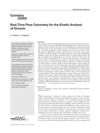
Figure 1. Real-time flow cytometric measurement of mmp and pmp with confocal microscopy. Jurkat cells were labeled with bis-oxonol,
DiIC1(5), annexin V-FITC, and DAPI. A live cell gate was applied and a 30 s baseline recorded for bis-oxonol and DiIC1(5) on the 610/10 nm
and 660/20 nm channels on a BD LSR II shown as overlaid line graphs (see Supporting Information Fig. 1 for gating strategy). Cells were
then treated with 1 lM STS (A), 1% sodium azide (C), heat shocked at 428C (E), and 0.01% Triton X-100 (G). The time course was run from 0
to 25 min, agonists were added after 30 sec as indicated by the arrow. Time course plots are representative experiments, n 5 2. Confocal
microscopy of treated samples after 25 min show bis-oxonol (red) and DiIC1(5) (purple) staining in cells treated with STS (B), 1% sodium
azide (D), 428C heat shock (F) and 0.01% Triton X-100 (H). Live cells were imaged on a Zeiss 510 confocal Meta microscope using 543 and
633 nm lasers to excite bis-oxonol and DiIC1(5) and 600LP and 650LP emissions collected respectively. Scale bars equal 10 lm. (n 5 2).
ORIGINAL ARTICLE
184 Real-Time Flow Cytometry for the Kinetic Analysis of Oncosis
5. (Fig. 1A). In contrast the oncotic agents sodium azide and Triton
X-100, induced in live cells a depolarization of the pmp with an
increase in bis-oxonol fluorescence and depolarization of mmp,
resulting in reduced fluorescence of DiIC1(5) (Figs. 1C and 1G).
Interestingly sodium azide initially induced a hyperpolarization
of the mitochondrial membrane before abrogation of mitochon-
drial function (Fig. 1C). Heat shock treatment (428C) of cells
showed little change in pmp and a gradual reduction in DiIC1(5)
fluorescence (Fig. 1E). This was in contrast to that found in cells
undergoing heat shock at 568C were there was an abrogation of
mitochondrial function (data not shown). Thus heat shock at
428C seems to be a relatively mild treatment of cells compared to
the other treatments employed in this study.
The relative rapid reduction in mitochondrial function
(reduction of fluorescence) observed with oncosis treatments
was expected as cells undergoing oncosis have been reported
to show a rapid depletion of ATP (10). In contrast apoptosis is
known to be an ATP dependent process and thus mitochon-
drial function does not rapidly reduce, these differences being
detected by real-time flow cytometric measurement of the
mmp dye, DiIC1(5).
Confocal Microscopy
The effects on membrane potentials within live cells were
also observed by confocal microscopy after the various treat-
ments for 30 min (Fig. 1). Cells undergoing apoptosis with
STS treatment for 0.5 h showed no obvious observable change
compared to controls (data not shown) in DiIC1(5) fluores-
cence or bis-oxonol fluorescence as detected by kinetic flow
cytometric analysis (Figs. 1A and 1B). Induction of oncosis by
1% sodium azide showed a complete abrogation of mmp indi-
cated by the lack of DiIC1(5) fluorescence (Fig. 1D) and
matches that observed by kinetic flow cytometry (Fig. 1C).
The induction of oncosis by heat shock temperature of 428C
showed no such change in fluorescent staining patterns reveal-
ing only a slight visual change in bis-oxonol and DiIC1(5)
fluorescence’s mirroring that shown by kinetic flow cytometric
analysis (Fig. 1E and 1F). Induction of oncosis by Triton X-
100 showed an increase in bis-oxonol fluorescence and a
decrease in DiIC1(5) fluorescence by confocal microscopy,
again mirroring that shown by kinetic flow cytometry (Figs.
1G and 1H). The image and kinetic flow cytometric analysis
not only showed the clear differences between the induction
processes in apoptosis and oncosis but the different mechan-
isms of action of the oncotic agents used in this study, that is,
physical and chemical treatment.
Mitochondrial Function
Live cell gating (see Supporting Information Fig. 1) of the
time dependent data points (0–30 h) and analysis of DiIC1(5)
fluorescence allows the determination of mitochondrial func-
tion at each time point. The live resting cell population
(CTRL) showed a steady [90% of cells with mitochondrial
function (Fig. 2). Live cells undergoing apoptosis (STS)
showed a gradual fall in mitochondrial function over the 30 h
time course, from 0–5 h there was a steady fall mitochondrial
function from 85% at 1 h down to 60% after 5 h, with only
15% functional mitochondria present after 20 h (Fig. 2). In
contrast all methods of induction of oncosis show a rapid fall
in mitochondrial function in a remarkably similar degree over
the 30 h time course (Fig. 2). After 1 h of treatment the mito-
chondrial function of live cells had fallen to 15–30%, rapidly
falling to 5% after 5 h (Fig. 2). Interestingly the level of mito-
chondrial function of live cells which had undergone heat
shock treatment showed a rapid increase in mitochondrial
function, rising from 10 to 70% from 20–30 h. This apparent
increase in mitochondrial function maybe due to the fact
that the cells that have survived the heat shock maintain their
mitochondrial function and proliferate with other cells going
on to bind annexin V and undergo necrosis.
The time dependent profiles of mitochondrial dysfunc-
tion indicates that the real-time kinetic analysis of mito-
chondrial function in live cells undergoing oncosis has
measured an actual fall in mitochondrial function within
the first minutes of treatment in a reproducible manner
given the relatively small SEMs of the data points, with the
exception of those observed in cells undergoing heat treat-
ment (Fig. 2).
Time Course Study of Oncosis and Apoptosis
Standard flow cytometric analysis of untreated cells, apo-
ptotic and oncotic cells by the annexin V assay showed that
annexin V was detectable during oncosis as previously
reported in the literature with sodium azide, heat shock and
Triton X-100 after 24 h of treatment, see Supporting Informa-
tion Figures 2A–2D (7,11–14,22).
However, time course studies revealed that although
annexin V rapidly binds to cells undergoing apoptosis in a
significant manner after 3 h of treatment the same cannot
Figure 2. Time course study of mitochondrial function. Jurkat
cells were loaded with DiIC1(5) and bis-oxonol and labeled with
annexin V-FITC and DAPI for each time point from 0—30 h. One
hundred thousand events were collected. Live cells (annexin V-
FITCneg
/DAPIneg
events) were analyzed for DiIC1(5) fluorescence
and the level of mitochondrial function determined for each time
point. Jurkat cells were untreated for controls (CTRL), treated with
STS, 428C heat shock (HS 42 C), 1% sodium azide (1% SA), and
0.01% Triton X-100 (T-X 0.01%), n 5 3, error bars indicate SEM.
ORIGINAL ARTICLE
Cytometry Part A 79A: 181À191, 2011 185
6. be said of cells undergoing oncosis (Figs. 3A–3D). Chemical
induction of oncosis by sodium azide showed a similar
binding of annexin V to that observed in cells undergoing
428C heat shock but was approximately 50% lower than
that observed in cells undergoing apoptosis (Figs. 3B and
3C). Detergent induction of oncosis showed few cells with
annexin V (10%) binding capacity and a rapid increase in
the dead cell population, this was very different from that
observed by treatment with sodium azide and 428C heat
shock (Fig. 3D). In contrast, apoptosis showed a significant
amount of death only at the latter stages of the time course
(Fig. 3A). The various oncotic reagents induced a marked
difference in the appearance of a rising dead cell popula-
tion. Triton X-100 (0.01%) showed significant cell death at
2 h, whereas sodium azide and 428C heat shock showed
significant cell death at the latter stages of the time course
respectively (Figs. 3B–3D).
Sub G1 Analysis of Oncotic and Apoptotic
Treated Cells
Cells undergoing apoptosis showed a significantly higher
level of DNA fragmentation than controls, whereas heat
treated cells showed no significant increase in subG1 levels
(Supporting Information Fig. 3, and Table 1). However, cells
undergoing oncosis with sodium azide or Triton X-100 showed
a higher level of DNA fragmentation than that observed with
apoptosis (Supporting Information Fig. 3, and Table 1).
Time Course Study of Cell Plasma Membrane
Potential Depolarization During Oncosis
and Apoptosis
The time course study of pmp depolarization, (Fig. 4)
showed a steady incidence of cells with pmp depolarization
whether living (annexin V2ve
/DAPI2ve
), apoptotic (annexin
V1ve
/DAPI2ve
) or dead (annexin V1ve
/DAPI1ve
, DP and
Figure 3. Time course study of annexin V binding. Jurkat T cells treated with 1 lM staurosporine (STS) to induce apoptosis (A), 1% sodium
azide (B), heat shock 428C (C), 0.01% Triton X-100 (D) for 30 h. At time points 0, 1, 2, 3, 4, 5, 20, 24, and 30 h cells were loaded with DiIC1(5)
and bis-oxonol and labeled with annexin V-FITC and DAPI. 100,000 events were collected for each time point. Annexin V versus DAPI dot-
plots were analyzed for each time point to determine percent live cells (annexin V2ve
/DAPI2ve
), apoptotic or annexin V1ve
/DAPI2ve
) and
dead cells (annexin V1ve
/DAPI1ve
and annexin V2ve
/DAPI2ve
), n 5 3, error bars indicate SEM.
ORIGINAL ARTICLE
186 Real-Time Flow Cytometry for the Kinetic Analysis of Oncosis
7. annexin V2ve
/DAPI1ve
, SP) until the 20 h time point for all
treatments compared to controls (Figs. 4A–4E). Plasma mem-
brane depolarization increased after this time during apopto-
sis, sodium azide and Triton X-100 treatments (Figs. 4B, 4D,
and 4E, Supporting Information Figs. 4D and 4E). In contrast
heat shock treatment showed no change in levels of pmp
depolarization even after 20 h and was similar to that found in
controls, (Figs. 4A and 4C, Supporting Information Figs. 4A
and 4C). Live cells undergoing apoptosis, showed a slight rise
in pmp depolarization compared to control cells at 24 h (Figs.
4A and 4B and Supporting Information Figs. 4A and 4B).
Annexin V binding populations in all treatments at 24 h
showed a higher incidence of pmp depolarization compared
to that displayed by untreated live cells (Figs. 4A–4E). The two
necrotic cell populations i.e. annexin V1ve
/DAPI1ve
(DP) and
annexin V2ve
/DAPI1ve
(SP), showed different levels of pmp
depolarization with a higher incidence in the double positive
population compared to the single positive necrotic cell and
annexin V1ve populations respectively (Figs. 4A–4D). This
was with the exception of Triton X-100 treatment were the
depolarization levels in all these populations was of a similar
order (Fig. 4E).
The ‘live cells’ still present with all treatments at the latter
time points had little mitochondrial function, see Figure 2
(with the exception of heat shock) and a significant number
had depolarized plasma membranes (above control levels).
This partially fulfills the criteria for cell death although these
cells maintained membrane integrity. The mitochondrial func-
tion time course study showed clear differences in the mecha-
nism of action of apoptotic and oncotic agents.
Phenotyping of Late and Early Necrosis
To determine the significance of high and low levels of
bis-oxonol in necrotic cells, DNA content, sorting and image
analysis of such cells in apoptotic and oncotic cultures was
performed. The side scatter of such cells also varied signifi-
cantly in the different oncotic cell cultures but not in cells
undergoing apoptosis (Figs. 5A, 5E, 5I, and 5M). DNA content
analysis of necrotic cells which were bis-oxonolhi1ve
and bis-
oxonollow1ve
clearly showed that bis-oxonollow1ve
had a higher
percentage of cells located in the Sub G1 zone with little DNA,
compared to the bis-oxonolhi1ve
which had a higher DNA
content (Figs. 5B, 5F, 5J, and 5N).
Confocal microscopy of these two types of necrotic cells
showed cells of approximately 2–20 lm in size depending on
the treatment. Oncotic treatments generated necrotic cells of
varying size which correlated generally to the level of bis-oxo-
nol fluorescence, except in the case of Triton X-100 treatment
were the reverse was true (Figs. 5O and 5P). Cells were other-
wise generally larger if they were bis-oxonolhi1ve
and smaller if
they were bis-oxonollow1ve
(Figs. 5G, 5H, 5K, and 5L);
whereas, cells undergoing apoptosis were of a similar size, in
both low and high intensity bis-oxonol events (Figs. 5C
and 5D). Thus dead cells derived from oncosis, with bis-
oxonolhi1ve
and bis-oxonollow1ve
staining appears to be indi-
cative of early and late necrosis respectively as supported by
the differences in side scatter, DNA content and the variation
in size of bis-oxonollow1ve
and bis-oxonolhi1ve
stained cells,
and generally verified by image analysis of bis-oxonolhi1ve
and
bis-oxonollow1ve
cells.
DISCUSSION
Previous studies employing flow cytometry to character-
ize cells undergoing oncosis has focused on the use of annexin
V binding to externalized PS to such cells (5,11,12,14). DNA
content has also been used to show a difference between apo-
ptotic and heat shock treatment at 568C (11). Both of these
approaches have proved to be nonspecific for characterizing
differences between cells undergoing apoptosis and oncosis
(22,13). More recently the use of SYTO16 and viability dyes
has allowed the discrimination of apoptosis and oncosis in
that no reduction in SYTO16 was observed in cells undergoing
oncosis compared to that observed in cells undergoing apo-
ptosis (7). In another advance, propidium iodide has been
used in a real-time image analysis study of apoptosis from the
start of the induction process and continuously over a 24 h
period (27–29). In this approach adherent cells were preloaded
with Hoechst and grown in culture with a low concentration
of PI (0.25 lg/ml) and then exposed to STS for 24 h with
images taken every 15 min in a real-time manner (27). The
use of annexin V in real-time image analysis has also allowed
the study of apoptosis by real-time live cell microscopy but
only covering the 5–42 h period after the induction of apopto-
sis (30). The method described here, of a rapid real-time anal-
ysis of membrane potentials by standard flow cytometry has
shown to be able to not only differentiate between apoptosis
and oncosis but also show differences in the mechanism of
action of different types of oncotic inducing reagents, includ-
ing drug, chemical, heat, and detergent treatments. Longer
time course studies has allowed further elucidation of differ-
ences occurring during apoptosis and oncosis, using annexin
V, cell viability dyes, mitochondrial inner membrane, and
plasma membrane potentials.
The changes in membrane potentials during the first 25
min of induction showed clearly that mitochondrial function
was disrupted very early in oncosis over the range of types of
reagents tested. It has been previously reported that oncosis is
characterized by the rapid depletion of ATP (10). The kinetic
Table 1. Sub G1 analysis was performed after 24 h of treatment
for controls, apoptosis induced by STS, heat shock 428C, 1%
sodium azide, and 0.01% Triton X-100 respectively
TREATMENT MEAN PERCENTAGE SUBG1
Control 12.61/2 3
Apoptosis 37.21/2 6.8a
Heat Shock 428C 25.41/2 7NS
1% Sodium Azide 49.51/2 4a
0.01% Triton X-100 55.51/2 7b
n 5 3, Mean 1/2 SEM.
Paired Student t-test were performed,
a
P 50.05.
b
P 50.01.
NS 5 not significant.
ORIGINAL ARTICLE
Cytometry Part A 79A: 181À191, 2011 187
8. changes in DiIC1(5) fluorescence indicates a rapid reduction
in mitochondrial function and thus a depletion of ATP in cells
undergoing oncosis. These changes in mitochondrial function
were not observed in cells undergoing apoptosis as measured
by real-time kinetic measurements of mmp. The time course
study of mitochondrial function during apoptosis showed a
40% fall after 5 h whilst all oncotic agents showed a rapid
reduction to 10% functionality over the same time period.
The apoptotic reagent induced no significant changes in pmp,
which was contrary to that observed with most cases of onco-
sis in which cells showed plasma membrane depolarization.
Thus the measurement of mitochondrial membrane and
plasma membrane potentials in real-time affords a rapid easy
method to distinguish oncosis and apoptosis at the induction
Figure 4. Time course study of plasma membrane depolarization. Jurkat T cells were untreated [control (A)] or treated with 1 lM stauro-
sporine, apoptosis (B), heat shock at 428C (C), 1% sodium azide (D), and 0.01% Triton X-100 (E) for 30 h. At time points 0, 1, 2, 3, 4, 5, 20, 24,
and 30 h cells were loaded with DiIC1(5) and bis-oxonol and labeled with annexin V-FITC and DAPI. One hundred thousand events were
collected for each time point and data analyzed to show changes in bis-oxonol fluorescence. Live (annexinV2ve
/DAPI2ve
), annexin V1ve
,
and both dead cell populations annexinV1ve
/DAPI1ve
(DP) and annexinV2ve
/ DAPI1ve
(SP) were analyzed for bis-oxonol depolarization, n 5
3, errors bars indicate SEM.
ORIGINAL ARTICLE
188 Real-Time Flow Cytometry for the Kinetic Analysis of Oncosis
9. stage of these two different necrobiological processes. This
approach to study oncosis could used in conjunction with that
employed to study apoptosis in real-time and SYTO16 and
thus enable differentiation between the induction of oncosis
and apoptosis in vitro and potentially ex vivo (27–29).
Time course studies of annexin V binding, mitochondrial
function, DNA content and plasma membrane depolarization
indicate that PS externalization is part of the process of apo-
ptosis, but is not an integral part of oncosis because of the
observed low incidence of cells binding annexin V. Oncotic
Figure 5. Characterization of early and late necrotic cells. Jurkat T cells were treated with 1 lM staurosporine (apoptosis), 1% sodium az-
ide, heat shock at 428C and 0.01% Triton X-100 for 20 h, respectively. Cells were labeled with bis-oxonol, DiIC1(5), annexin V-FITC, and
DAPI. Apoptosis and necrosis was determined by annexin V versus DAPI analysis. Necrotic cells with bis-oxonol low and high intensities
were then analyzed for side scatter and DNA content (7-AAD 670LP) from apoptotic cell cultures (A), (B), 1% sodium azide (E), (F), heat
shock 428C (I), (J), and 0.01% Triton X-100 (M), (N). The low and high intensity bis-oxonol cells from the dead populations for each treat-
ment were then sorted and imaged by confocal microscopy. DAPI staining indicated by blue, annexin V labeling indicated by green, bis-
oxonol (pm) staining indicated by red, DiIC1(5) staining of mitochondria (mito) indicated by purple, these were then merged into a final
image. Sorted dead cells undergoing apoptosis with bis-oxonol high (C) and low (D) were imaged followed by dead cells after treatments
with 1% sodium azide with bis-oxonol high (G) and low intensities (H), dead cells from heat shock 428C bis-oxonol high (K) and low (L),
and 0.01% Triton X-100 bis-oxonol high (O) and low (P), respectively. Scale bars as indicated in lm. Histograms and confocal imaging rep-
resentative experiments, n 5 2.
ORIGINAL ARTICLE
Cytometry Part A 79A: 181À191, 2011 189
10. cells thus appear to rapidly lose membrane integrity and
undergo necrosis, this was also observed when combining
SYTO16 and PI and there was no decrease in SYTO16 signal
to indicate an apoptotic response to sodium azide and heat
shock (7). The different modes of action of the reagents
employed in this study were also reflected in the varying
degrees of DNA fragmentation. The degree of DNA fragmen-
tation increased from heat shock treatments (although not sig-
nificant), to significance with STS, then sodium azide and Tri-
ton X-100 treatments. The use of DNA fragmentation estima-
tions can thus be useful in revealing the mechanism of action
of the reagent employed in terms of the latter stages of the cell
death process under investigation.
The varying degrees of plasma membrane depolariza-
tion revealed within the four population’s studied, that is
live cells, annexin V binding cells, as well dead cells that
bind or do not bind annexin V is an interesting observation
in that it further subdivides these cell populations. Further
studies into the significance of these observations are cur-
rently underway. Untreated cells showed few live cells with
depolarized membranes. There was a general increase in the
proportion of cells showing depolarization in the order of,
live cells, dead cells not binding annexin V, then annexin V
binding cells, then lastly dead cells that bind annexin V
showed a very high degree of plasma membrane depolariza-
tion. The proportion of cells displaying depolarization
remained constant and only increased at the latter stages of
treatments. It is interesting that a subpopulation of live cells
undergoing oncosis had no mitochondrial function and had
a depolarized plasma membrane, parameters indicative of
dead cells and yet maintained plasma membrane integrity
as measured by vital dye exclusion.
Necrotic cells were shown via bis-oxonol intensity to
be defined as in an early or late phase of necrosis as
defined by side scatter and DNA content. The use of bis-
oxonol in this manner allows easy discrimination of necro-
sis into early and late phases and at the same time as meas-
uring levels of annexin V binding and mitochondrial activ-
ity. This type of approach has also been employed by using
SYTO16 to discriminate live, apoptotic, and necrotic and
showed reductions in forward scatter, increases in side scat-
ter and an increase in Sub G1, as the cells move through
the apoptotic process (7). However here we show that the
necrotic cell population can be further subdivided into early
and late necrosis by use of the bis-oxonol signal intensity.
This potentially adds another level of complexity to the
necrobiological process.
The measurement of plasma membrane and mitochon-
drial inner membrane potentials can thus be used to investi-
gate the mechanism of action of different oncotic inducing
agents in real-time and show, which cells are in an early or late
stage of necrosis. Previously SYTO16 has been used to differ-
entiate oncosis and apoptosis in a time dependent manner
(7). However, the real-time imaging of cells loaded with
Hoechst in the presence of PI allowed the detection of apopto-
sis an hour after induction (27). This major advance in the
study of apoptosis could be used to study the oncotic process
too by the employment fluorescent mitochondrial dyes as
tested in this study (27). This new approach of rapid real-time
flow cytometric analysis of changes in mitochondrial function
and plasma membrane depolarization allows the investigation
of oncosis in an immediate manner by standard flow cytome-
try. This new assay will hopefully prove useful in the study of
oncosis and drug treatment of tumors.
ACKNOWLEDGMENTS
I would like to thank Dr Paul Allen and Prof Marion
Macey for their assistance with the preparation of the
manuscript.
LITERATURE CITED
1. Darzynkiewicz Z, Juan G, Li X, Gorcyzya W, Murakami T, Traganos F. Cytometry in
cell necrobiology: Analysis of apoptosis and accidental cell death (necrosis). Cytome-
try 1997;27:1–20.
2. Wlodkowic D, Skommer J, Darzynkiewicz Z. Cytometry in cell necrobiology revis-
ited. Recent advances and new vistas. Cytometry A 2010;77A:591–606.
3. Fink SL, Cookson BT. Apoptosis, pyrotosis and necrosis: mechanistic description of
dead and dying eukaryotic cells. Infect Immun 2005;73:1907–1916.
4. Vermes I, Haanen C, Steffens-Nakken H, Reutelingsperger C. A novel assay for
apoptosis: Flow cytometric detection of phosphatidylserine expression on early
apoptotic cells using fluorescein labeled annexin V. J Immunol Methods
1995;184:39–51.
5. Rasola A, Geuna M. A flow cytometric assay simultaneously detects independent apo-
ptotic parameters. Cytometry A 2001;45A:151–157.
6. Lugli E, Troaino L, Ferraresi R, Roat E, Prada N, Nasi M, Pinti M, Cooper EL,
Cossaizza A. Characterization of cells with different mitochondrial membrane poten-
tial during apoptosis. Cytometry A 2005;68A:28–35.
7. Wlodkowic D, Skommer J, Pelkonen J. Towards an understanding of apoptosis detec-
tion by SYTO dyes. Cytometry A 2007;71A:61–72.
8. Schwartz SM, Bennett MR. Death by another name. Am J Pathol 1995;147:229–234.
9. Majno GJI. Apoptosis, oncosis and necrosis: an overview of cell death. Am J Pathol
1995;146:3–15.
10. Eguchi Y, Shimizu S, Tsujimoto Y. Intracellular ATP levels determine cell death fate
by apoptosis or necrosis. Cancer Res 1997;57:1835–1840.
11. Waring P, Lambert D, Sjaarda A, Hurne A, Beaver J. Increased cell surface exposure of
phosphatidylserine on propidium iodide negative thymocytes undergoing death by
necrosis. Cell Death Diff 1999;6:624–637.
12. Lecoeur H, Prevost MC, Gougeon ML. Oncosis is associated with exposure of phospha-
tidylserine residues on the outside layer of the plasma membrane: A reconsideration of
the specificity of the annexin V/propidium iodide assay. Cytometry 2001;44:44–65.
13. Chang MC, Chan CP, Wang YJ, Lee PH, Chen LI, Tsai YL, Lin BR, Wang YL, Jeng JH.
Induction of necrosis and apoptosis to KB cancer cells by sanguinarine is associated
with reactive oxygen species production and mitochondrial membrane depolariza-
tion. Toxicol Appl Pharmacol 2007;128:143–151.
14. Matteucci C, Grelli S, De Smaele E, Fontana C, Mastino A. Identification of nuclei
from apoptotic, necrotic and viable lymphoid cells by using multiparameter flow
cytometry. Cytometry 1999;35:145–153.
15. Pollack A, Ciancio G. Cell cycle phase-specific analysis of cell viability using Hoechst
33342 and propidium iodide after ethanol preservation. Methods Cell Biol
1990;33:19–24.
16. Ormerod MG, Sun XM, Snowden RT, Davies R, Fearnhead H, Cohen GM. Increased
membrane permeability of apoptotic thymocytes: a flow cytometric study. Cytometry
1993;14:595–602.
17. Belloc F, Dumain P, Boisseau MR, Jalloustre C, Reiffers J, Bernard P, Lacombe F. A
flow cytometric method using Hoechst 33342 and propidium iodide for simultane-
ous cell cycle analysis and apoptosis determination in unfixed cells. Cytometry
1994;17:59–65.
18. Telford WG, Komoriya A, Packard BZ. Detection of localized caspase activity in early
apoptotic cells by laser scanning cytometry. Cytometry A 2002;47A:81–88.
19. Bedner E, Smolewski P, Amstad P, Darzynkiewicz Z. Activation of caspases measured
in situ by binding of fluorochromes-labeled inhibitors of caspases (FLICA): Correla-
tion with DNA fragmentation. Exp Cell Res 2000;259:308–313.
20. Darzynkiewicz Z, Bruno S, Del Bino G, Gorczyca W, Hotz MA, Lassota P, Traga-
nos F. Features of apoptotic cells measured by flow cytometry. Cytometry
1992;13:795–808.
S21. Poot M, Pierce RH. Detection of changes in mitochondrial function during apopto-
sis by simultaneous staining with multiple fluorescent dyes and correlated multi-
parameter flow cytometry. Cytometry 1999;35:311–317.
22. Lizard G, Fournel S, Genestier L, Dhedin N, Chaput C, Flacher M, Mutin M, Panaye
G, Revillard JP. Kinetics of plasma membrane and mitochondrial alterations in cells
undergoing apoptosis. Cytometry 1995;21:275–283.
23. Frey T. Nucleic acid dyes for detection of apoptosis in live cells. Cytometry
1995;21:265–274.
24. Poot M, Gibson LL, Singer VL. Detection of apoptosis in live cells by mitotracker red
CMXRos and SYTO dye flow cytometry. Cytometry 1997;27:358–364.
ORIGINAL ARTICLE
190 Real-Time Flow Cytometry for the Kinetic Analysis of Oncosis
11. 25. Van Zandvoort MAMJ, de Grauw CJ, Gerritsen HC, Broers JLV, Oude Egbrink MGA,
Ramaekers FCS, Slaaf DW. Discrimination of DNA and RNA in cells by a vital fluo-
rescent probe: Lifetime imaging of SYTO 13 in healthy and apoptotic cells. Cytometry
2002;47:226–235.
26. Sparrow RL, Tippet E. Discrimination of live and early apoptotic mononuclear cells
by the fluorescent SYTO 16 vital dye. J Immunol Methods 2005;305:173–187.
27. Wlodkowic D, Skommer J, McGuinness D, Faley S, Kolch W, Darzynkiewicz Z,
Cooper JM. Chip-Based dynamic real-time quantification of drug-induced cytotoxic-
ity in human tumor cells. Anal Chem 2009;81:6952–6959.
28. Wlodkowic D, Skommer J, Skommer J, Faley S, Darzynkiewicz Z, Cooper JM.
Dynamic analysis of apoptosis using cyanine SYTO probes: From classical to micro-
fluidic cytometry. Exp Cell Res 2009;315:1706–1714.
29. Zhao H, Oczos J, Janowski P, Trembecka D, Dobrucki J, Darzynkiewicz Z, Wlodkowic
D. Rationale for the real-time and dynamic cell death assays using propidium iodide.
Cytometry A 2010;77A:399–405.
30. Kim YE, Chen J, Chan JR. Langen R. Engineering a polarity-sensitive biosensor for
time-lapse imaging of apoptotic processes and degeneration. Nat Methods
2010;7:67–73.
31. Klapperstuck T, Glanz D, Klapperstuck M, Wohlrab J. Methodological aspects of
measuring absolute values of membrane potential in human cells by flow cytometry.
Cytometry A 2009;75A:593–608.
32. Dawson TL, Gores GJ, Nieminem AL, Herman B, Lemasters JJ. Mitochondria as a
source of reactive oxygen species during reductive stress in rat hepatocytes. Am J Phy-
siol 1993;261:C961–C967.
ORIGINAL ARTICLE
Cytometry Part A 79A: 181À191, 2011 191