
Recommended
More Related Content
Similar to LARYNX.pptx
Similar to LARYNX.pptx (20)
Recently uploaded
Recently uploaded (20)
LARYNX.pptx
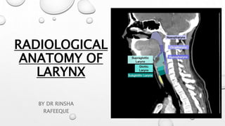
- 1. RADIOLOGICAL ANATOMY OF LARYNX BY DR RINSHA RAFEEQUE
- 2. LARYNX • The larynx is an inferior continuation of the oropharynx. • 5-7 cm structure • Upper boundary starts at tip of epiglottis opposite to 3rd to 4th cervical vertebra • Lower end at cricoid cartilage opposite to 6th cervical vertebra.
- 3. RELATIONS Anterior: strap muscles Posterior: esophagus, laryngopharynx Superior: hyoid bone, laryngopharynx Inferior: trachea
- 5. LARYNGEAL CARTILAGES UNPAIRED • Thyroid cartilage • Cricoid cartilage • Epiglottic cartilage PAIRED • Arytenoid cartilage • Accessory cartilages 1. Cuneiform cartilage 2. Corniculate cartilage
- 9. 1) EPIGLOTTIS Superior most, midline leaf-shaped cartilage. Free margin and a fixed portion (stem). The free margin of the epiglottis is closely related to the base of tongue – the median glosso-epiglottic fold and projects above the level of the hyoid
- 11. 2) THYROID CARTILAGE • Largest • 2 anterior lamina meet anteriorly at acute angle • Superior thyroid notch present at anterior superior aspect • Superior cornua are elongated and narrow attaching to thyrohyoid ligament • Inferior cornua are short and thick and articulating medially with cricoid cartilage.
- 12. 3) CRICOID CARTILAGE • Only complete ring in endolarynx • Has 2 portions : anterior arch and posterior lamina • Lower border of cricoid cartilage is junction between larynx above and trachea below
- 13. ARYTENOID CARTILAGE Paired pyramidal cartilages that sit at top of cricoid cartilage lamina posteriorly VOCAL PROCESS: attaches to the vocal fold MUSCULAR PROCESS: lateral projection attaches to the posterior and lateral cricoarytenoid muscles SUPERIOR PROCESS: superior projection articulates corniculate cartilage CORNICULATE CARTILAGE : rests on top of superior process of arytenoid within aryepiglottic folds CUNEIFORM CARTILAGE: lateral and superior to corniculate within ary epiglottic folds.
- 17. SUPRA GLOTTIS THE VENTRICLE itself is seen as a small air-filled outpouching between the false and true cords FALSE VOCAL CORDS : ARY EPIGLOTTC FOLD : projects from tip of arytenoid cartilage to inferolateral part of epiglottis Represents superolateral part of supraglottis dividing into pyriform sinus.
- 18. PRE-EPIGLOTTIC SPACE Fat-filled space, rich in lymphatics. Superiorly by the hyoepiglottic ligament Anteriorly by the thyrohyoid membrane, Inferiorly by the thyroepiglottic ligament and Posteriorly by the epiglottis. The PES and PGS communicate with each other superiorly.
- 19. THE PARAGLOTTIC SPACE located deep to the true and false cords and bound laterally by the thyroid and cricoid cartilages It extends upto the undersurface of the true vocal cords continuous with the extralaryngeal soft tissues between the thyroid and cricoid cartilages; an important pathway for extralaryngeal tumor spread
- 20. GLOTTIS ANTERIOR COMMISSURE : midline anterior meeting point of the true vocal cords. It comprises of the 1. Anterior cord, 2. The anterior junction of the two vocal cords, 3. The thyroid cartilage and 4. Broyle’s ligament, a fibrous structure connecting the vocal ligaments to the cartilage POSTERIOR COMMISSURE is the mucosal surface on the anterior surface of the cricoid cartilage between the arytenoid cartilages. Composed of THYROARYTENOID muscle
- 23. SUB GLOTTIS Extends from under surface of true vocal cord to inferior cricoid cartilage CONUS ELASTICUS : fibro elastic membrane extending from medial margin of true vocal cord to cricoid below.
- 24. INTRINSIC MUSCLES OF THE LARYNX 1) Muscles of the inlet Aryepiglottic muscle Oblique arytenoid muscle Thyroepiglottic muscle
- 25. 2) Muscles of the vocal folds Cricothyroid muscle Thyroarytenoid muscle Posterior cricoarytenoid muscle Lateral cricoarytenoid muscle Interarytenoid muscle Vocalis muscle
- 26. VASCULAR SUPPLY • Above the vocal cords: superior laryngeal vessels • Below the vocal cords: inferior laryngeal vessels
- 27. LYMPHATIC SUPPLY • Supraglottic: superior deep cervical nodes and the pre-epiglottic nodes • Subglottic: inferior deep cervical nodes and the prelaryngeal (delphian) nodes NERVE SUPPLY : MOTOR: the recurrent laryngeal nerve supplies all the intrinsic muscles of the larynx apart from the cricothyroid muscle SENSORY: • SUPRAGLOTTIC: internal laryngeal nerve (branch of the superior laryngeal nerve) • INFRAGLOTTIC: recurrent laryngeal nerve
- 42. CT FINDINGS IN LARYNX Post contrast- no enhancement of mucosal surface of larynx but hypopharyngeal mucosa enhances False vocal cords, aryepiglottic folds , pre and para epiglottic spaces are fat containing hence they appear hypo attenuating Ossified cartilages appear hyperdense in outer and inner cortex with a hypodense central area (medullary fat) Cortical bone, fatty marrow and non ossified hyaline cartilage do not show post contrast enhancement Collapsed pyriform fossa may mimic a tumor on CT ( do Valsalva to exclude)
- 43. THANK YOU