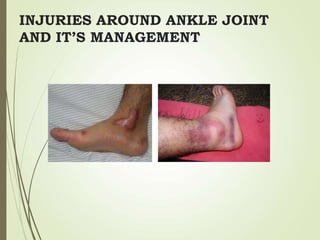
2. INTRODUCTION
Ankle injury refers to disruption of any
component or components of the ankle
joint following trauma.
Ankle injuries occur frequently, and have
high propensity for complications.
4. Bony mortise- quadrilateral
shape
Posterolateral position of
fibula
Ligaments
3 groups
-Lateral
-Medial
-Syndesmotic
5. ANKLE JOINT IS SUPPORTED BY
Fibrous capsule
Deltoid ligament
A. Superficial
a. Anterior-
Tibionavicular
b. Middle-
Tibiocalcanean
c. Posterior- Posterior
tibiotalar
B. Deep : Anterior-
Tibiotalar
11. LAUGE HANSEN
1. Position of foot at
injury-
Pronation/Supination
2. Deforming force-
Abduction/ adduction/
external rotation
Most Common
mechanism of injury- SER
Most Common unstable
ankle fracture variant- SER
20. What else to see in x-rays
LAT MALLEOLUS
Level of fracture
Orientation of fracture
Fracture comminution
MED/POST MALLEOLUS
Size
Assoc plafond #
Assoc syndesmotic injury
25. Pott’s Fracture
Fracture involving the ankle joint
loosely referred to as Pott’s Fracture
1. First degree single malleolus fractured.
2. In second degree two malleoli are
fractured.
3. In third degree there is bimalleolar
fracture with a fracture of posterior part
of inferior articular surface of the tibia
referred to as third malleolus. (Tri
Malleolar fracture)
26. MANAGEMENT
RICE
Definitive
Aim- restoration of complete normal anatomical alignment of
ankle.
Patients if needs operation should be operated within 24hrs of
injury or after one week once the swelling subsides.
Undisplaced fracture medial malleolus :
Below knee POP cast for 6 weeks.
Reduction fails (may be due to soft tissue (periosteal) inter
position)
27. Displaced:
Open reduction and internal fixation by
Cancellous screws group
Tension band wiring
Fracture lateral malleolus:
Lateral Malleolus helps in length maintenance &
maintenance of ankle mortice.
Hence, lateral malleolus has to be fixed internally.
28. TIBIAL PILON FRACTURES
Intraarticular fracture of distal tibia.
Fibula is fractured in 85% of these patients.
29.
30. TIBIAL PILON FRACTURE
1. Plaster immobilization
2. Traction
3. Lag screw fixation
4. OR & IF with plates
5. External fixation with or without limited
internal fixation
If articular incongruity <2 mm
and reserved for low energy
injuries
31. COMPLICATIONS
Malunion- may result in posttraumatic arthritis and
painful movements.
Nonunion of medial malleolus- commonly due to
interposition of fractured periosteum between two
fragments.
Repeated edema
Sudeck’s Osteodystrophy
34. Blood supply
Extraosseous supply
Posterior tibial a. tarsal canal a.
Anterior tibial a. sinus tarsi a
Peroneal a. sinus tarsi a.
Intraosseous supply
Talar head
Talar body
-anastomosis between tarsal canal a. and
tarsal sinus a.
36. Talar neck fracture
Aviator’s astragalus
High energy injury, hyperdorsiflexion
15~20% open fracture
Associated with malleloar fracture(25% of cases), medial
malleolus is more common
High risk of soft tissue injury and compartment syndrome
38. Treatment
Hawkins type I
4~6 weeks of no weightbearing in a short leg cast
walking cast for 1~2 months
Percutaneous screw fixation
39. Treatment
Hawkins type II
Orthopaedic emergency: traction and plantar flexion by
manipulation anatomic reduction(50%) treated as type
I
Open reduction: screw placed across the neck fracture
40. Treatment
Hawkins type III
ORIF and Skeletal traction
through the calcaenus
Open fracture (> type III)
:talar body excision followed
By primary tibiocalcaneal or
Blair-type arthrodesis
Hawkins type IV
Rare injury
As type II
41. Complication
Skin necrosis and infection
Delayed union or nonunion
Malunion
Posttraumatic arthritis
Osteonecrosis
43. Anatomy
Largest, most irregularly shaped bone in foot
Large calcellous bone and multiple processes
Achilles tendon posteriorly and plantar fascia inferiorly : tuberosity
Posterior facet: talar lateral process and body
Middle facet: Sustentacular fragment (flexor hallucis longus pass)
Anterior process: cuboid
48. Intraarticular fracture
Joint-depression type, in which the
primary fracture line exited the bone
close to the subtalar joint
tongue-type, in which the primary
fracture line exited the bone posteriorly
49. Intraarticular fracture
--Treatment
Nondisplaced articular fractures
Bulky (Robert-jones) dressing: active subtalar ROM, prohibit
weightbearing walking 8~12 wks later
Displaced intraarticular fracture with large fragment
ORIF
50. Intraarticular fracture
--Treatment
Displaced intraarticular fracture with severe comminution
Increasing intraarticualr comminution leads to less satisfactory
results
ORIF primary arthrodesis
Restoring the heel width and height
52. ANKLE AND FOOT INJURIES
Q1) The stability of the ankle joint is maintained by all of
the following except
a. Spring ligament
b. Deltoid ligament
c. Lateral ligament
d. Shape of the superior talar articular surface
53. Q2) The most commonly affected component of lateral
collateral ligament complex in an ankle sprain
a. Anterior talo fibular ligament
b. Posterior talo fibular ligament
c. Calcaneofibular Ligament
d. None
54. Q3) Ankle sprain is due to
a. Rupture of anterior talo-fibular ligament
b. Rupture of posterior talo-fibular ligament
c. Rupture of deltoid ligament
d. Rupture of calcaneo-fibular ligament
55. Q4) Mechanism of injury of transverse fracture of medial
malleolus is
a. Abduction injury
b. Adduction injury
c. Rotation injury
d. Direct injury
56. Q5) Cottons fracture is
a. Avulsion fracture of C7
b. Bimalleolar fracture
c. Trimalleolar fracture
d. Burst fracture of the Atlas
e. None of the above