SlideShare a Scribd company logo
1 of 6
Download to read offline
Circadian clock protein cryptochrome regulates the
expression of proinflammatory cytokines
Rajesh Narasimamurthya
, Megumi Hatorib
, Surendra K. Nayakb
, Fei Liua
, Satchidananda Pandab,1
, and Inder M. Vermaa,1
a
Laboratory of Genetics, Salk Institute for Biological Studies, La Jolla, CA 92037; and b
Regulatory Biology Laboratory, Salk Institute for Biological Studies, La
Jolla, CA 92037
Contributed by Inder M. Verma, June 13, 2012 (sent for review May 18, 2012)
Chronic sleep deprivation perturbs the circadian clock and increases
susceptibility to diseases such as diabetes, obesity, and cancer.
Increased inflammation is one of the common underlying mech-
anisms of these diseases, thus raising a hypothesis that circadian-
oscillator components may regulate immune response. Here we
show that absence of the core clock component protein crypto-
chrome (CRY) leads to constitutive elevation of proinflammatory
cytokines in a cell-autonomous manner. We observed a constitutive
NF–κB and protein kinase A (PKA) signaling activation in Cry1−/−
;
Cry2−/−
cells. We further demonstrate that increased phosphoryla-
tion of p65 at S276 residue in Cry1−/−
;Cry2−/−
cells is due to in-
creased PKA signaling activity, likely induced by a significantly
high basal level of cAMP, which we detected in these cells. In
addition, we report that CRY1 binds to adenylyl cyclase and limits
cAMP production. Based on these data, we propose that absence
of CRY protein(s) might release its (their) inhibition on cAMP pro-
duction, resulting in elevated cAMP and increased PKA activation,
subsequently leading to NF–κB activation through phosphoryla-
tion of p65 at S276. These results offer a mechanistic framework
for understanding the link between circadian rhythm disruption
and increased susceptibility to chronic inflammatory diseases.
biological clock | immune system
In response to the rotation of the earth, many of the mamma-
lian physiological processes such as sleep–wake cycle and
feeding pattern undergo an ∼24-h oscillation referred as the
circadian clock. Circadian oscillation helps organisms antici-
pate and adapt to predictable daily changes in the environment.
The hypothalamic suprachiasmatic nucleus (SCN) functions
as a master circadian oscillator, which controls behaviors. At
molecular level, the circadian oscillator is based on a cell-au-
tonomous transcription–translation feedback loop, in which
transcriptional activators circadian locomotor output cycles
kaput (CLOCK)/brain and muscle aryl hydrocarbon receptor
nuclear translocator (ARNT)-like (BMAL)1 activate the ex-
pression of Cryptochrome (Cry) and Period (Per), in turn their
protein products repress BMAL1 and CLOCK activity, thus
producing Cry and Per expression rhythms (1, 2). Reverse
orientation c-erb (REV-ERB)s and RAR-related orphan re-
ceptor (ROR)s also participate in the rhythmic transcriptional
activity of the molecular oscillator (3). Molecular components
of the circadian clock network are conserved in the SCN and
also in peripheral cells. The oscillator uses direct and indirect
mechanisms to impose rhythms and regulate several physiologi-
cal processes, such as rest–activity cycle, endocrine system, and
metabolism (4).
Sleep loss or chronic sleep deprivation disrupts the circadian
rhythm. It has been reported that people exposed to constant
circadian disruption, such as shift-workers, show increased in-
cidence of chronic diseases such as diabetes, obesity, and also
cancer (5–7). Chronic inflammation is one of the important
pathogenic features of these diseases, which prompted us to
hypothesize that circadian clock components may play a role in
the regulation of inflammation. Moreover, recent evidence
demonstrated that major players in immune function, such as
spleen, lymph node, and macrophage and natural killer (NK)
cells, contain a cell-autonomous circadian clock (8–10). More-
over, secretion of cytokines, TNF-α and IL-6 has been reported
to display circadian oscillation in macrophages, where ∼8%
of transcriptome is under circadian regulation (10). Clinical
evidence and sleep-loss studies have identified physiological
connections between the circadian clock and immune system
(11, 12). A molecular understanding of the mechanism of clock
modulation of immune function, however, remains unclear.
In this study, we found that lack of a functional clock system
due to absence of the core clock component cryptochrome
(CRY) leads to elevated proinflammatory cytokine activation
mediated through the NF–κB signaling pathway. We also found
that absence of CRY proteins leads to constitutive activation of
PKA, which results in phosphorylation of p65 at S276, which
ultimately lead to increased NF–κB activation and expression of
proinflammatory cytokines. Our results provide a possible mo-
lecular link between the arrhythmic clock system and increased
inflammatory response.
Results
Absence of Cryptochrome Proteins Constitutively Activates the Pro-
inflammatory Cytokine Expression. To gain insight into the role of
circadian clock components on the immune function, we mea-
sured expression of inflammatory mediators in the hypothalamus
of Cry1 and Cry2 double knockout (Cry1−/−
;Cry2−/−
) mice, which
show arrhythmic behavior due to lack of a functional clock sys-
tem (13, 14). Significant increase in expression of IL-6, TNF-α,
and iNOS was observed in Cry1−/−
;Cry2−/−
mice relative to WT
mice (Fig. 1A). We next tested whether the increased expression
is cell-autonomous or systemic result of oscillator dysfunction.
Fibroblasts possess a cell-autonomous and self-sustained circa-
dian clock system (15, 16) that recapitulates key features of
whole organism’s circadian function. Cry1−/−
;Cry2−/−
fibroblasts,
which lack circadian oscillation (17), exhibited significantly in-
creased expression of IL-6, TNF-α, and iNOS (Fig. 1B) and other
genes involved in inflammation (Fig. S1A), thus exhibiting cell-
autonomous roles of CRY proteins in regulation of key cyto-
kines. Compared with Cry1−/−
or Cry2−/−
single knockout fibro-
blasts, cytokine expression showed a synergistic increase in
double knockout Cry1−/−
;Cry2−/−
fibroblasts (Fig. S1B). IL-6
protein was detectable only in Cry-deficient fibroblasts, but not in
other clock mutants that lack either Per1 or Per2 or Bmal1 (Fig.
1C). These results demonstrate that lack of CRY constitutively
activates the proinflammatory cytokines, such as IL-6, TNF-α,
and other inflammatory molecules, indicating a potential role for
CRY proteins in regulation of inflammatory cytokine expression.
Author contributions: R.N., S.P., and I.M.V. designed research; R.N., M.H., S.K.N., and F.L.
performed research; R.N., S.K.N., F.L., S.P., and I.M.V. analyzed data; and R.N., M.H., S.P.,
and I.M.V. wrote the paper.
The authors declare no conflict of interest.
1
To whom correspondence may be addressed. E-mail: verma@salk.edu or panda@salk.
edu.
This article contains supporting information online at www.pnas.org/lookup/suppl/doi:10.
1073/pnas.1209965109/-/DCSupplemental.
12662–12667 | PNAS | July 31, 2012 | vol. 109 | no. 31 www.pnas.org/cgi/doi/10.1073/pnas.1209965109
Because IL-6 is inducible with TNF-α, we next asked if the high
expression of IL-6 observed in Cry1−/−
;Cry2−/−
cells can be fur-
ther induced by TNF-α. Both WT and Cry1−/−
;Cry2−/−
cells
showed ∼8- to 10-fold increase of IL-6 expression following
TNF-α induction (Fig. S1C), suggesting that both cells are re-
sponsive for an inflammatory stimuli.
Innate Immune System Gets Hypersensitive in the Absence of CRY.
Macrophages play a critical role in secretion of most in-
flammatory cytokines upon immune response. It has recently
been shown that the expression of cytokines in macrophages is
circadian clock–gated (10). Therefore, we next analyzed bone
marrow–derived macrophages (BMDM) from WT and Cry1−/−
;
Cry2−/−
mice for expression of immune mediators. BMDM from
Cry1−/−
;Cry2−/−
mice showed a marked increase in expression of
inflammatory cytokines such as IL-6, Cxcl1, and iNOS, compared
with WT cells (Fig. 1D). Analysis of proinflammatory cytokines
TNF-α and IL-6 by ELISA in the cell supernatants did not show
any significant difference between WT and Cry1−/−
;Cry2−/−
BMDM cells (Fig. 1E), which prompted us to check whether
Cry1−/−
;Cry2−/−
BMDM cells are hypersensitive to LPS stimu-
lation. WT and Cry1−/−
;Cry2−/−
BMDM cells were treated with
LPS and supernatants were collected after 12 h. ELISA analysis
showed a marked increase in TNFα and IL-6 protein secretions
in Cry1−/−
;Cry2−/−
BMDM cells compared with WT (Fig. 1E),
which suggests that Cry-deficient macrophages are hypersensitive
to immune response.
To determine the contribution of the hematopoietic cells from
WT and Cry1−/−
;Cry2−/−
mice in vivo in systemic cytokine pro-
duction during inflammatory response, we performed bone
marrow transplantation (BMT) experiments. Bone marrow from
either WT or Cry1−/−
;Cry2−/−
mice were transplanted and
reconstituted into lethally irradiated recipient mice (B6.129S4-
Il2rgtm1Wjl/J; Jackson Lab), which lack T, B, and NK cells and
the macrophages in blood and spleen. Transplanted mice were
allowed to repopulate for 8 wk and then the blood lymphocytes
of these mice were analyzed by FACS for repopulation
efficiency. FACS analysis showed that nearly 95% of lympho-
cytes in the transplanted mice were derived from donor mice,
and in particular almost 100% of macrophages were derived
from donor cells in transplanted mice (Fig. 2A). Furthermore,
donor-derived T cells, B cells, monocytes, and macrophages
IL-6pg/ml
IL-6
pg/ml
Fibroblasts
0
100
200
300
400
500
600
pg/ml
C
E
TNF
4,000
3,000
2,000
0
1,000
-/-
Per1WT
-/-
Per2 -/-
Bmal1 -/-
Cry1 -/-
Cry2 -/-
Cry1 ;
-/-
Cry2
BMDM
0
0.1
0.2
0.3
0.4
0.5
0.6
WT
-/-
Cry1 ;
-/-
Cry2
IL-6
RelativemRNAexpression
0
0.2
0.4
0.6
0.8
1.0
iNOS
0
0.04
0.08
0.12
0.16
Cxcl1
Hypothalamus
IL-6
0
0.4
0.8
1.2
1.6
2.0
RelativemRNAexpression
*
0
1
2
3
4
5
iNOS
0
0.01
0.02
0.03
0.04
0
40
80
120
160
RelativemRNAexpression
IL-6
0
4
8
12
16
0
40
80
120
160
iNOS
A
B
D
TNF
WT
-/-
Cry1 ;
-/-
Cry2
WT
-/-
Cry1 ;
-/-
Cry2
WT
-/-
Cry1 ;
-/-
Cry2
WT
-/-
Cry1 ;
-/-
Cry2
WT
-/-
Cry1 ;
-/-
Cry2
TNF
WT
-/-
Cry1 ;
-/-
Cry2
WT
-/-
Cry1 ;
-/-
Cry2
WT
-/-
Cry1 ;
-/-
Cry2
* *
150
100
50
20
10
0
Fibroblasts
BMDM
LPS
WT
-/-
Cry1 ;
-/-
Cry2
Fig. 1. Lack of a functional circadian clock system by
absence of the circadian oscillator component CRY con-
stitutively upregulates expression of inflammatory cyto-
kines. (A) Estimation of mRNA levels of indicated genes by
quantitative real-time PCR (qRT–PCR) in the hypothalamus
region of brain of WT and Cry1−/−
;Cry2−/−
mice are shown.
Data are mean ± SEM (n = 4 mice per group); *P < 0.05.
(B) Quantification of mRNA levels of indicated genes by
qRT–PCR in WT and Cry1−/−
;Cry2−/−
fibroblasts are shown.
Data are mean ± SD (n = 3). (C) WT, Per1−/−
, Per2−/−
,
Bmal1−/−
, Cry1−/−
, Cry2−/−
, and Cry1−/−
;Cry2−/−
fibroblasts
were left untreated for 12 h and the supernatants were
analyzed for IL-6 protein levels by ELISA. Data are mean ±
SD (n = 3). (D) BMDM from WT and Cry1−/−
;Cry2−/−
mice
were analyzed for mRNA levels of indicated genes by
qRT–PCR. Data are mean ± SD (n = 3). (E) WT and Cry1−/−
;
Cry2−/−
BMDM cells were either untreated or treated with
LPS (1 μg/mL for TNF-α and 10 ng/mL for IL-6 analysis) for
12 h and the supernatants were estimated for TNF-α or
IL-6 protein levels by ELISA are shown. Data are mean ± SD
(n = 4). Relative mRNA expression levels after normaliza-
tion to actin are shown in A, B, and D.
CD 45.1
ng/ml
ng/mlCount
Count
B
A
IL-6
600
500
400
300
30
20
10
0
20,000
16,000
12,000
8,000
4,000
0
5
10
4
10
3
10
2
100
TNF
BMT-mice
LPS
WT
-/-
Cry1 ;
-/-
Cry2
BMT-mice
LPS
WT
-/-
Cry1 ;
-/-
Cry2
1,500 200
500
1,000
0
CD 45.1
5
10
4
10
3
10
2
100
10095.2
150
100
50
0
Fig. 2. Absence of cryptochrome potentiate immune system of the mice to
secrete increased levels of inflammatory cytokines upon LPS challenge. (A)
Reconstitution analysis by FACS of blood lymphocytes derived from recipient
mice, B6.129S4-Il2rgtm1Wjl/J (CD45.2 positive), 8 wk after injection with
bone marrow of donor mice (CD45.1 positive) of one representative animal.
Left shows typical donor contribution to total lymphocytes. Right shows the
percentage of CD45.1 positive cells (donor) gated on CD11b positive cells. (B)
WT or Cry1−/−
;Cry2−/−
BMT were injected with either PBS or LPS (5 mg/kg)
and the 2-h serum samples collected from these mice were analyzed for TNF-
α and IL-6 protein levels by ELISA. Data are mean ± SEM (n = 3 mice per
group for LPS injection and n = 1 for PBS injection).
Narasimamurthy et al. PNAS | July 31, 2012 | vol. 109 | no. 31 | 12663
IMMUNOLOGY
were efficiently repopulated in both WT and Cry1−/−
;Cry2−/−
bone marrow–transplanted mice (Fig. S2 A–D). Eight weeks
after transplantation, mice transplanted with either WT (BMT-
WT) or Cry1−/−
;Cry2−/−
(BMT-KO) bone marrow were injected
with PBS or LPS and serum samples of TNF-α and IL-6 protein
were measured 2 h later. LPS-injected BMT-KO mice showed
a significantly high level of both TNF-α and IL-6 (Fig. 2B) in their
serum relative to LPS-injected BMT-WT mice. These re-
sults clearly indicate that absence of CRY proteins potentiates
the immune system and significantly elevates secretion of
proinflammatory cytokines in a physiological system upon
LPS challenge.
NF–κB Signaling Pathway Is Constantly Activated in Cry1−/−
;Cry2−/−
Cells. Next, we sought to identify the signaling pathway that is
responsible for constitutive cytokine activation in Cry1−/−
;Cry2−/−
cells. Although CRY proteins are known to inhibit the tran-
scription from CLOCK/BMAL1 target genes, IL-6 promoter is
not known to be a CLOCK/BMAL1 target (18). Because NF–κB
signaling is one of the major regulators and integrators of the
inflammatory response in mammals (19, 20), we tested whether
blocking of NF–κB signaling can suppress the constitutive IL-6
activation in Cry1−/−
;Cry2−/−
cells. In canonical NF–κB signaling,
inducers such as TNF-α or LPS trigger inhibitor of nuclear fac-
tor-κB (IκB) kinase 2 (IKK2) activation, leading to phosphory-
lation, ubiquitination, and proteasomal degradation of IκBα,
allowing the transport of NF–κB into the nucleus to activate its
target genes (21). Cry1−/−
;Cry2−/−
cells were infected with either
lentiviral GFP or lentiviral-GFP-tagged IκBα superrepressor
(IκBαM), which is a nondegradable form of IκBα. Expression of
a dominant negative form of IκBα, IκBαM led to significant re-
duction of IL-6 expression in Cry1−/−
;Cry2−/−
cells (Fig. 3A),
which suggests that constitutive activation of the NF–κB sig-
naling pathway likely underlies constitutive IL-6 activation in
Cry1−/−
;Cry2−/−
cells. We next wondered whether IKK2 is also
involved in constitutive NF–κB activation in Cry1−/−
;Cry2−/−
cells, because in most NF–κB signaling, IKK2 is necessary and
sufficient for phosphorylation of IκBα and its degradation (21).
Inhibition of IKK2 function by a chemical inhibitor MLN120B
(10 μM) (22) showed a marked reduction of constitutive IL-6
expression in Cry1−/−
;Cry2−/−
(Fig. 3B), further suggesting that
the constitutive activation of NF–κB pathway in Cry1−/−
;Cry2−/−
cells most likely signals, at least partly, through the canonical
IKK2 kinase complex.
The rate-limiting step in NF–κB signaling is phosphorylation
of IκBα and its subsequent degradation (21). We assessed the
phosphorylation status of IκBα in Cry1−/−
;Cry2−/−
cells. Com-
pared with WT cells, we observed a markedly increased phos-
pho-IκBα in Cry1−/−
;Cry2−/−
cells, which is substantially reduced
by inhibition of IKK2 with MLN120B (Fig. 3C). This result is
further supported by the high level of phosphorylated and active
IKK2 in Cry1−/−
;Cry2−/−
cells compared with WT cells (Fig. 3D).
Because increased IκBα degradation will lead to increased entry
of p65 into the nucleus (21), we compared the nuclear p65 levels
in WT and Cry1−/−
;Cry2−/−
cells. Western blot analysis after
nucleocytoplasmic subcellular fractionation of WT and Cry1−/−
;
Cry2−/−
cells showed a substantially elevated level of nuclear p65
in Cry1−/−
;Cry2−/−
cells, which is significantly reduced by in-
hibition of IKK2 with MLN120B (Fig. 3E).
Upon entering into the nucleus, transcription factor p65 binds
to NF–κB binding sites to activate its target genes (21). We
therefore analyzed the p65 binding activity in the nuclear lysates
assessed by affinity to oligonucleotides containing a consensus
NF–κB binding sequence. This assay showed a good functional
A B C
D E
F H
IL-6 IL-6
WT
-/-
Cry1 ;
-/-
Cry2
TNF
G
GFP GFP-I B M
RelativemRNAexpression
WT
-/-
Cry1 ;
-/-
Cry2 WT
-/-
Cry1 ; -/-
Cry2
DMSO
p-IB/IB
DMSO
MLN
120B
TNF
WT
-/-
Cry1 ;
-/-
Cry2 WT
p-IKK2
IKK2
-Tub
p-IKK2/IKK2
6
4
2
0
160
120
80
40
0
160
120
80
40
0
8
6
4
2
0
p-I B
I B
-Tub
WT
-/-
Cry1 ;
-/-
Cry2 WT
-/-
Cry1 ;
-/-
Cry2
-/-
Cry1 ;
-/-
Cry2
DMSO
MLN
120B
Nuclear fraction
p65/TBP
(Nuclear)
p65
TBP
-Tub
WT
-/-
Cry1 ;
-/-
Cry2 WT
-/-
Cry1 ;
-/-
Cry2
TNF
RelativemRNAexpression
GFP
GFP-
p65
GFP-
p65 S276A
WT
-/-
Cry1 ;
-/-
Cry2
p-p65 S276
p65
-Tub
WT
-/-
Cry1 ;
-/-
Cry2
p-p65S276/p65
12
8
4
0
IL-6100
80
60
40
20
0
2.0
1.5
1.0
0.5
0
Competitor
p65bindingactivity
(RelativetoWT)
WT
-/-
Cry1 ;
-/-
Cry2 WT
-/-
Cry1 ;
-/-
Cry2
-/-
Cry1 ;
-/-
Cry2
DMSO
MLN
120B
Cytoplasmic fraction
4
3
2
1
0
MLN
120B
GFP-
I B M
WT
Fig. 3. Blocking the canonical NF–κB pathway sup-
presses constitutive activation of IL-6 in Cry1−/−
;Cry2−/−
fibroblasts. (A) Estimation of IL-6 mRNA by qRT–PCR
of Cry1−/−
;Cry2−/−
fibroblasts infected with lentivirus
expressing GFP- or GFP-tagged superrepressor mutant of
IκBα (GFP-IκBαM) with increasing dose are shown. (B) IL-6
mRNA expression was quantified by qRT–PCR of Cry1−/−
;
Cry2−/−
fibroblasts treated with either DMSO or
MLN120B (10 μM) for 2 h. (C) Total cell lysates of WT and
Cry1−/−
;Cry2−/−
fibroblasts either untreated or treated
with DMSO or with the IKK2 inhibitor, MLN 120B (10 μM,
1 h) were analyzed by Western blot for phospho-IκBα,
IκBα, and tubulin. (D) Total cell lysates of WT and Cry1−/−
;
Cry2−/−
fibroblasts analyzed by Western blot for phos-
pho-IKK2, IKK2, and tubulin are shown. (E) Nuclear and
cytoplasmic fractions of untreated or DMSO-treated WT
and untreated or DMSO- or MLN120B-treated Cry1−/−
;
Cry2−/−
fibroblasts analyzed by Western blot for p65,
TATA-binding protein (TBP) and tubulin are shown. De-
tection of TBP and tubulin are used as markers of nuclear
and cytoplasmic fractions, respectively. (F) Nuclear
extracts (5 μg) from WT and Cry1−/−
;Cry2−/−
fibroblasts
analyzed for p65 binding activity by ELISA are shown.
Competition assay was performed with wild-type NF–κB
consensus oligonucleotides. Values shown are relative to
WT, value of WT p65 binding activity is set to 1, which is
a representative of three independent experiments. (G)
Western blot show the level phospho-p65 S276 in total
cell lysates of WT and Cry1−/−
;Cry2−/−
fibroblasts either
untreated or treated with TNF-α (10 ng/mL, 30 min). (H)
Estimation of IL-6 mRNA levels by qRT–PCR of Cry1−/−
;
Cry2−/−
fibroblasts expressing either GFP or super-
repressor form of IκBα (GFP-IκBαM) or GFP-p65 or mutant
form of p65 (GFP-p65 S276A) are shown. Relative mRNA
expression levels after normalization to actin are shown
in A, B, and H, and data are mean ± SD (n = 3).
12664 | www.pnas.org/cgi/doi/10.1073/pnas.1209965109 Narasimamurthy et al.
efficiency and specificity for p65 binding in fibroblasts (Fig. S3).
We observed a marked increase in p65-binding activity in Cry1−/−
;
Cry2−/−
fibroblasts compared with WT cells, and this increased
activity is dramatically reduced upon addition of competitor
oligonucleotides (Fig. 3F). It has been reported that p65 is
phosphorylated at S276 residue for its subsequent acetylation
and increase of its transcriptional activity (23). We found a sig-
nificant elevation of phospho-p65 at S276 in Cry1−/−
;Cry2−/−
cells
compared with WT cells (Fig. 3G). Moreover, expression of
a mutant form of p65 (p65 S276A) significantly suppressed IL-6
activation in Cry1−/−
;Cry2−/−
cells (Fig. 3H). Together, these
results suggest that the increased phosphorylation of both IκBα
and p65 at S276 may play an important role in the constitutive
activation of IL-6 transcription in Cry1−/−
;Cry2−/−
cells.
PKA Signaling Activity Is Constitutive in Cry1−/−
;Cry2−/−
Cells. Pro-
tein kinase A (PKA) is involved in the phosphorylation of p65 at
S276 (24). Owing to the significantly elevated phosphorylation
level of p65 at S276 (Fig. 3G), we speculated that PKA signaling
activity may also be high in Cry1−/−
;Cry2−/−
cells. We assessed
the phosphorylation status of a known substrate of PKA, vaso-
dialator-stimulated phosphoprotein (VASP) (25) and detected
its increased phosphorylation in Cry1−/−
;Cry2−/−
cells (Fig. 4A).
We also observed a near complete suppression of VASP phos-
phorylation in Cry1−/−
;Cry2−/−
cells by a PKA inhibitor H89 (20
μM), but not with the IKK2 inhibitor MLN120B (Fig. 4A), which
suggests that PKA signaling is constitutively activated in Cry1−/−
;
Cry2−/−
cells.
We next investigated whether inhibition of PKA signaling can
suppress IL-6 activation detected in Cry1−/−
;Cry2−/−
cells. IL-6
mRNA activation was remarkably reduced when Cry1−/−
;Cry2−/−
cells were treated with increasing concentrations of the PKA
inhibitor H89 (Fig. 4B). Consistent with this result with in-
hibitor, a marked reduction of IL-6 activation in Cry1−/−
;Cry2−/−
cells was also observed by expression of a mutant form of PKA
(GFP-DN PKA) (Fig. 4C), which acts as a dominant negative
protein by blocking the dissociation of regulatory subunit of
PKA from its catalytic subunit (26). Inhibition of PKA signaling
also resulted in a marked reduction in phospho-p65 at S276 in
Cry1−/−
;Cry2−/−
cells (Fig. 4D).
A nuclear protein, A-kinase-interacting protein 1 (AKIP1),
binds both to PKA (27) and p65 (28). This interaction has been
suggested to enhance PKA-mediated transcriptional activity of
p65 by promoting its nuclear retention and phosphorylation at
S276, which potentially increases recruitment of transcriptional
coactivators such as p300 to p65 (28). Given the constitutive
activation of NF–κB and PKA signaling in Cry1−/−
;Cry2−/−
cells
(Figs. 3 and 4 A–D), we tested whether knockdown of AKIP1
can suppress the constitutive activation of IL-6 in Cry1−/−
;Cry2−/−
cells. RNAi-mediated depletion of AKIP1 reduced IL-6 acti-
vation in Cry1−/−
;Cry2−/−
cells (Fig. 4E and Fig. S4). Thus, our
data supports the notion that NF–κB activation through PKA
contributes to the constitutive activation of IL-6 in Cry1−/−
;
Cry2−/−
cells.
Rise in intracellular levels of cAMP leads to PKA activation
(29). We compared cAMP concentration in WT and Cry1−/−
;
Cry2−/−
cells before and following induction with forskolin,
which triggers cAMP production by activating adenylyl cyclase
(30). WT and Cry1−/−
;Cry2−/−
cells were first incubated with
3-Isobutyl-1-methylxanthine (IBMX) (500 μM), an inhibitor of
phosphodiesterase, for 30 min, followed by addition of forskolin
(10 μM) for 30 min, and cAMP levels were quantified. As an-
ticipated, cAMP levels are elevated in WT and Cry1−/−
;Cry2−/−
cells upon forskolin treatment (Fig. 4F). However, surprisingly,
we observed substantially high levels of cAMP even in uninduced
state (control and DMSO treated) of Cry1−/−
;Cry2−/−
cells
compared with WT cells, suggesting that the increase in PKA
signaling activation in Cry1−/−
;Cry2−/−
cells is most likely due to
high cAMP concentration at the basal level.
H89
p-p65 S276
p65
Tub
pmol/ml
BA
RelativemRNAexpression
C
ED F
RelativemRNAexpression
IL-6 IL-6
IL-6 cAMP
WT
-/-
Cry1 ;
-/-
Cry2 WT
WT
-/-
Cry1 ; -/-
Cry2
160
120
80
40
0
100
80
60
40
20
0
GFP
80
40
0
60
20
1,600
1,200
800
400
0
sh-E6 sh-AKIP1 DMSO Fsk DMSO Fsk
WT
-/-
Cry1 ;
-/-
Cry2
H89
p-VASP
VASP
Actin
8Br-cAMP
MLN120B
DMSO
H89
p-VASP/VASP
12
8
4
0
p-p65S276/p65
0
1
2
3
4
5
WT
-/-
Cry1 ; -/-
Cry2
WT
-/-
Cry1 ;
-/-
Cry2 WT
-/-
Cry1 ;
-/-
Cry2
GFP-
DN-PKA
Fig. 4. Increased cAMP and PKA signaling activity and also the PKA-mediated phosphorylation of p65 at S276 contributes to elevated NF–κB target gene
activation in Cry1−/−
;Cry2−/−
. (A) WT fibroblasts were either untreated or treated with 8Br–cAMP (100 μM) for 30 min and Cry1−/−
;Cry2−/−
fibroblasts were
either untreated or treated with H89 (20 μM), DMSO, or MLN120B (10 μM) for 1 h, and total cell lysates were analyzed by Western blot for phospho-VASP,
VASP and actin. (B) Quantification of IL-6 mRNA expression by qRT–PCR of Cry1−/−
;Cry2−/−
fibroblasts treated with H89 (10 and 20 μM for 1 h). (C) Estimation
of IL-6 mRNA expression by qRT–PCR of Cry1−/−
;Cry2−/−
fibroblasts expressed with a dominant–negative (GFP–DN–PKA) form of PKA. (D) Total cell lysates of
untreated WT and untreated or H89 (20 and 30 μM, 1 h) treated Cry1−/−
;Cry2−/−
fibroblasts were analyzed by Western blot for phospho-p65 S276, p65 and
tubulin. (E) RNA extracted from Cry1−/−
;Cry2−/−
fibroblasts infected with lentivirus expressing sh-RNA against a nonspecific target gene E6 as a control or
against AKIP1 were analyzed for IL-6 mRNA expression by qRT–PCR. (F) Cellular concentration of cAMP measured by ELISA in the lysates of WT and Cry1−/−
;
Cry2−/−
fibroblasts either untreated or treated with DMSO or forskolin (10 μM, 30 min) are shown. Data are mean ± SD (n = 4). Relative mRNA expression
levels after normalization to actin are shown in B, C, and E, and data are mean ± SD (n = 3).
Narasimamurthy et al. PNAS | July 31, 2012 | vol. 109 | no. 31 | 12665
IMMUNOLOGY
CRY1 Expression Limits cAMP Production Likely by Binding to and
Inhibiting the Function of Adenylyl Cyclase. On the basis of above
results, we hypothesized that CRY regulates the intracellular
cAMP levels to regulate NF–κB pathway. We addressed whether
overexpression of CRY1 protein can suppress the cAMP
production induced by either forskolin, prostaglandin E2 (PGE2),
or isoproterenol (each 10 μM). Stimulation with forskolin directly
activates adenylyl cyclase (30); whereas, induction with PGE2 and
isoproterenol activates their cognate Gαs–coupled receptors,
which ultimately activate adenylyl cyclase leading to transient
increase in intracellular cAMP (31, 32). Dynamic changes in the
intracellular cAMP levels were measured by a cAMP-sensitive
luciferase reporter system (GloSensor cAMP assay; Promega)
expressed in 293T cells. Overexpression of CRY1 resulted in
a significant reduction in cAMP production induced by forskolin,
PGE2, and also by isoproterenol (Fig. 5 A–C). Because we ob-
served a substantial suppression in cAMP production induced by
all three stimuli tested, we hypothesize that CRY1 may acts on
adenylyl cyclase to block its function, thereby suppressing cAMP
production. To test this proposal, lysates of 293T cells over-
expressing Flag-tagged CRY1 or GFP were analyzed by immu-
noprecipitation (IP). In 293T cell lysates, expressing Flag-CRY1
when adenylyl cyclase was immunoprecipitated with an antibody
against adenylyl cyclase III (AC III), we detected Flag-CRY1
by immune blot (Fig. 5D). These results indicate that CRY1 bind
to adenylyl cyclase to block its function and suppress the
cAMP production.
Discussion
In summary, results presented here indicate that both PKA and
IKK2–IκBα-dependent phosphorylation of NF–κB contributes to
increased activation of inflammatory cytokines, for example IL-6,
in Cry1−/−
;Cry2−/−
fibroblasts. Here, we propose that circadian
clock modulates NF–κB activity via CRY-mediated control of
PKA activity. Circadian clock–controlled expression of CRY
proteins regulate the production of cAMP, which in turn con-
tributes to the observed circadian rhythms in cytosolic cAMP,
which is a necessary component to core clock function in SCN
neurons and in peripheral cell types (33). We presume that
temporally gated oscillations of cAMP levels (33) and resultant
PKA activity temporally modulates the basal activation state of
the NF–κB pathway and may explain the well-described circa-
dian rhythms in immune responses in mammals (34). Our data
support the hypothesis that when CRY proteins are present they
bind to adenylyl cyclase (Fig. 5D) and downregulate cAMP
production (Fig. 5 A–C). In the absence of CRY, inhibition on
adenylyl cyclase is relieved, which subsequently leads to in-
creased cAMP production (Fig. 4F) and PKA activation
(Fig. 4A) that cause increased phosphorylation of p65 at S276
(Fig. 4D), ultimately resulting in activation of its target genes
such as IL-6 (Fig. 5E).
We observed increased activation of TNF-α in Cry1−/−
;Cry2−/−
fibroblasts and also in the hypothalamus of Cry1−/−
;Cry2−/−
mice.
Hashiramoto et al. has also previously shown high expression of
TNF-α in Cry1−/−
;Cry2−/−
fibroblasts (35). We also observed in-
creased phospho-IKK2 in Cry1−/−
;Cry2−/−
fibroblasts (Fig. 3D).
We put forward that the most likely initiator of phosphorylation
and activation of IKK2 signaling complex in Cry1−/−
;Cry2−/−
cells
is the increased activation of TNF-α (a well-known activator of
IKK2 complex), which has been observed in these cells. This
activated IKK2 complex subsequently leads to phosphorylation
(Fig. 3C) and degradation of IκBα, which in turn cause increased
p65 accumulation into the nucleus (Fig. 3E) and high level of
DNA binding activity (Fig. 3F), ultimately resulting in increased
expression of NF–κB target genes such as IL-6. Phosphorylation
and degradation of IκBα is a critical step required to release p65
and PKA from IκBα, which allows PKA to phosphorylate p65
(36) and enter into nucleus, which is why overexpression of
superrepressor mutant form of IκBα (IκBαM) dramatically
suppressed up to 80% of IL-6 expression in Cry1−/−
;Cry2−/−
fibroblasts (Fig. 3A).
Recent reports have demonstrated the role of CRY in regu-
lation of metabolism and immune function. CRY proteins are
known to regulate glucose homeostasis by at least two different
mechanisms. They bind to and repress glucocorticoid receptors
(37), which regulate transcription of gluconeogenic genes. CRY
proteins also bind to and inhibit Gαs function, thereby attenu-
ating GPCR-activation-dependent increase in intracellular
cAMP (38). However, we detected CRY1 in complex with ade-
nylyl cyclase (Fig. 5D), and overexpression of CRY1 reduced
AC III
IgG
GFP
Flag-CRY1
AC III
Tub
Flag
Lysate
BA E
DC
IB
IP
Prostaglandin E2
Time (min)
0 6 12 18 24 30
Forskolin
Foldstimulation
200
160
120
80
40
0
Vector/ Fsk
CRY1/ Fsk
Vector/ Vehicle
Foldstimulation
200
160
120
Isoproterenol
Vector/ ISP
CRY1/ ISP
Vector/ Vehicle
Foldstimulation
240
200
160
80
40
0
120
Time (min)
0 6 12 18 24 30
Input
cAMP ATP
Forskolin
GPCR
IL-6
Ubiquitination
& proteasomal
degradation
AC III
IgG
GFP
Flag-CRY1RR
AC III
Tub
Flag
E
IB
IP
Prostaglandin E2
200
160
120
Input
cAMP ATPAA
Forskolin
IL-6
Ubiquitination
& proteasomal
degradation
YCR
G
NEMO
IKK1
P P
IKK2
p65p50
p50 p65
CBP/p300
p50 p65
Ac P
AKIP1
p65p50
P
ADCY
Sα
P
PKA
P
BακI
Vector/ PGE2
CRY1/ PGE2
Vector/ Vehicle
80
40
0
Time (min)
0 6 12 18 24 30
Fig. 5. CRY1 inhibits the forskolin-, PGE2-, and isoproterenol-induced generation of intracellular cAMP, likely by binding to and inhibiting the function of
adenylyl cyclase. (A–C) Luciferase assay performed to measure the kinetics of cAMP production after stimulation in the presence of 500 μM of IBMX in
293T cells are shown. Fold stimulation of cAMP production are shown after stimulation with either forskolin, PGE2, or isoproterenol (each 10 μM) in the
absence (control vector) or presence of CRY1 expression. Data are mean ± SD (n = 3). (D) CRY1 interacts with adenylyl cyclase. All of the indicated im-
munoprecipitate and lysate samples analyzed by Western blot for Flag tag (CRY1), adenylyl cyclase III and tubulin are shown. (E) The proposed model
showing the likely function of CRY in binding to and suppressing the action of adenylyl cyclase in cAMP production and the pathways activated in the
absence of CRY proteins.
12666 | www.pnas.org/cgi/doi/10.1073/pnas.1209965109 Narasimamurthy et al.
cAMP production in response to PGE2, isoproterenol, and even
the direct adenylyl cyclase activator, forskolin (30) (Fig. 5A).
These results clearly suggest an additional cell-autonomous role
of CRY in modulating cellular response to different stimuli,
which contributes to our understanding of altered immune re-
sponse under circadian disruption. It has previously been shown
that p53 mutant mice lacking functional CRY proteins are more
susceptible to TNF-α-initiated apoptosis through NF–κB acti-
vation (39). Monje et al. had reported that depression like be-
havior in mice induced by constant darkness is associated with
increased levels of IL-6 in plasma and hippocampus mediated
through NF–κB signaling pathway (40). Interestingly, NF–κB has
been shown to play a role in metabolic adaptation both in normal
and also in cancer cells by regulation of mitochondrial oxidative
phosphorylation (41), and the current finding will suggest an
additional CRY–NF–κB-mediated pathway in metabolic adap-
tation of cancer cells.
In this study, we analyzed the inflammatory response of mam-
malian cells only lacking a functional circadian clock system,
without any additional manipulations. Our results strongly
indicate that an arrhythmic clock system, induced by the absence
of CRY proteins, alone is sufficient to increase the stress levels
of cells leading to constant expression of inflammatory cytokines
and causing a low-grade chronic inflammatory status called
metaflammation (42) and Cry-deficient cells could be a good
model system to study this phenomenon. Compelling evidence
has shown that low-grade constant inflammation could be the
underlying cause for chronic diseases such as diabetes, obesity,
and also cancer (43–45). Moreover, people exposed to frequent
circadian clock disturbances, such as night-shift workers, are
increasingly susceptible to these diseases. Therefore, further
understanding connection between the circadian clock, me-
tabolism, and inflammation will help to identify therapeu-
tic drug targets to cure diseases like metabolic syndrome
and cancer.
Materials and Methods
All animal experiments were carried out in accordance with the guidelines of
the Institutional Animal Care and Use Committee of the Salk Institute. Male
C57BL/6J and Cry1−/−
;Cry2−/−
mice were used (12 wk old). TRIzol (Invitrogen)
reagent was used to extract RNA from animal tissues by following the
manufacturer’s protocol. Immortalized fibroblasts and 293T cells were cul-
tured in Dulbecco-modified Eagle medium (Invitrogen) supplemented with
10% (vol/vol) FBS (Atlanta Biologicals) and 1% (vol/vol) of antibiotic–anti-
mycotic (Invitrogen). The production of lentiviruses was performed accord-
ing to the protocol described elsewhere (46). Additional details are in SI
Materials and Methods.
ACKNOWLEDGMENTS. This work was supported in part by grants from the
National Institutes of Health (NIH), Ipsen/Biomeasure, Sanofi-aventis, and
the H. N. and Frances C. Berger Foundation. The project described was
supported by Grant R37AI048034 from the National Institute of Allergy and
Infectious Diseases. R.N. was supported in part by a grant from the Swiss
National Science Foundation (Bern, Switzerland). S.P. was supported by NIH
Grant DK091618; M.H. was supported by Japan Society for Promotion of
Science Fellowship. S.K.N. was supported by NIH Grant P30 CA014195-38.
I.M.V. is an American Cancer Society Professor of Molecular Biology, and
holds the Irwin and Joan Jacobs Chair in Exemplary Life Science.
1. Bass J, Takahashi JS (2010) Circadian integration of metabolism and energetics. Sci-
ence 330:1349–1354.
2. Liu AC, Lewis WG, Kay SA (2007) Mammalian circadian signaling networks and
therapeutic targets. Nat Chem Biol 3:630–639.
3. Cho H, et al. (2012) Regulation of circadian behaviour and metabolism by REV-ERB-α
and REV-ERB-β. Nature 485:123–127.
4. Saini C, Suter DM, Liani A, Gos P, Schibler U (2011) The mammalian circadian timing
system: Synchronization of peripheral clocks. Cold Spring Harb Symp Quant Biol 76:39–47.
5. Imeri L, Opp MR (2009) How (and why) the immune system makes us sleep. Nat Rev
Neurosci 10:199–210.
6. Rajaratnam SM, Arendt J (2001) Health in a 24-h society. Lancet 358:999–1005.
7. Willyard C (2008) Hungry for sleep. Nat Med 14:477–480.
8. Arjona A, Sarkar DK (2005) Circadian oscillations of clock genes, cytolytic factors, and
cytokines in rat NK cells. J Immunol 174:7618–7624.
9. Hayashi M, Shimba S, Tezuka M (2007) Characterization of the molecular clock in
mouse peritoneal macrophages. Biol Pharm Bull 30:621–626.
10. Keller M, et al. (2009) A circadian clock in macrophages controls inflammatory im-
mune responses. Proc Natl Acad Sci USA 106:21407–21412.
11. Bass J, Turek FW (2005) Sleepless in America: A pathway to obesity and the metabolic
syndrome? Arch Intern Med 165:15–16.
12. Bollinger T, Bollinger A, Oster H, Solbach W (2010) Sleep, immunity, and circadian
clocks: A mechanistic model. Gerontology 56:574–580.
13. van der Horst GT, et al. (1999) Mammalian Cry1 and Cry2 are essential for mainte-
nance of circadian rhythms. Nature 398:627–630.
14. Vitaterna MH, et al. (1999) Differential regulation of mammalian period genes and
circadian rhythmicity by cryptochromes 1 and 2. Proc Natl Acad Sci USA 96:12114–12119.
15. Nagoshi E, et al. (2004) Circadian gene expression in individual fibroblasts: Cell-au-
tonomous and self-sustained oscillators pass time to daughter cells. Cell 119:693–705.
16. Welsh DK, Yoo SH, Liu AC, Takahashi JS, Kay SA (2004) Bioluminescence imaging of
individual fibroblasts reveals persistent, independently phased circadian rhythms of
clock gene expression. Curr Biol 14:2289–2295.
17. Yagita K, Tamanini F, van Der Horst GT, Okamura H (2001) Molecular mechanisms of
the biological clock in cultured fibroblasts. Science 292:278–281.
18. Rey G, et al. (2011) Genome-wide and phase-specific DNA-binding rhythms of BMAL1
control circadian output functions in mouse liver. PLoS Biol 9:e1000595.
19. Li Q, Verma IM (2002) NF-kappaB regulation in the immune system. Nat Rev Immunol
2:725–734.
20. Vallabhapurapu S, Karin M (2009) Regulation and function of NF-kappaB transcrip-
tion factors in the immune system. Annu Rev Immunol 27:693–733.
21. Hayden MS, Ghosh S (2008) Shared principles in NF-kappaB signaling. Cell 132:
344–362.
22. Hideshima T, et al. (2006) MLN120B, a novel IkappaB kinase beta inhibitor, blocks
multiple myeloma cell growth in vitro and in vivo. Clin Cancer Res 12:5887–5894.
23. Chen LF, et al. (2005) NF-kappaB RelA phosphorylation regulates RelA acetylation.
Mol Cell Biol 25:7966–7975.
24. Zhong H, Voll RE, Ghosh S (1998) Phosphorylation of NF-kappa B p65 by PKA stim-
ulates transcriptional activity by promoting a novel bivalent interaction with the
coactivator CBP/p300. Mol Cell 1:661–671.
25. Butt E, et al. (1994) cAMP- and cGMP-dependent protein kinase phosphorylation sites
of the focal adhesion vasodilator-stimulated phosphoprotein (VASP) in vitro and in
intact human platelets. J Biol Chem 269:14509–14517.
26. Clegg CH, Correll LA, Cadd GG, McKnight GS (1987) Inhibition of intracellular cAMP-
dependent protein kinase using mutant genes of the regulatory type I subunit. J Biol
Chem 262:13111–13119.
27. Sastri M, Barraclough DM, Carmichael PT, Taylor SS (2005) A-kinase-interacting pro-
tein localizes protein kinase A in the nucleus. Proc Natl Acad Sci USA 102:349–354.
28. Gao N, Asamitsu K, Hibi Y, Ueno T, Okamoto T (2008) AKIP1 enhances NF-kappaB-
dependent gene expression by promoting the nuclear retention and phosphorylation
of p65. J Biol Chem 283:7834–7843.
29. Taylor SS, et al. (2005) Dynamics of signaling by PKA. Biochim Biophys Acta 1754:
25–37.
30. Seamon KB, Padgett W, Daly JW (1981) Forskolin: Unique diterpene activator of
adenylate cyclase in membranes and in intact cells. Proc Natl Acad Sci USA 78:
3363–3367.
31. Furuyashiki T, Narumiya S (2011) Stress responses: The contribution of prostaglandin
E(2) and its receptors. Nat Rev Endocrinol 7:163–175.
32. Kobilka BK (2007) G protein coupled receptor structure and activation. Biochim Bio-
phys Acta 1768:794–807.
33. O’Neill JS, Maywood ES, Chesham JE, Takahashi JS, Hastings MH (2008) cAMP-de-
pendent signaling as a core component of the mammalian circadian pacemaker.
Science 320:949–953.
34. Hrushesky WJ, Langevin T, Kim YJ, Wood PA (1994) Circadian dynamics of tumor
necrosis factor alpha (cachectin) lethality. J Exp Med 180:1059–1065.
35. Hashiramoto A, et al. (2010) Mammalian clock gene Cryptochrome regulates arthritis
via proinflammatory cytokine TNF-alpha. J Immunol 184:1560–1565.
36. Zhong H, SuYang H, Erdjument-Bromage H, Tempst P, Ghosh S (1997) The tran-
scriptional activity of NF-kappaB is regulated by the IkappaB-associated PKAc subunit
through a cyclic AMP-independent mechanism. Cell 89:413–424.
37. Lamia KA, et al. (2011) Cryptochromes mediate rhythmic repression of the gluco-
corticoid receptor. Nature 480:552–556.
38. Zhang EE, et al. (2010) Cryptochrome mediates circadian regulation of cAMP signal-
ing and hepatic gluconeogenesis. Nat Med 16:1152–1156.
39. Lee JH, Sancar A (2011) Regulation of apoptosis by the circadian clock through NF-
kappaB signaling. Proc Natl Acad Sci USA 108:12036–12041.
40. Monje FJ, et al. (2011) Constant darkness induces IL-6-dependent depression-like
behavior through the NF-kappaB signaling pathway. J Neurosci 31:9075–9083.
41. Mauro C, et al. (2011) NF-κB controls energy homeostasis and metabolic adaptation
by upregulating mitochondrial respiration. Nat Cell Biol 13:1272–1279.
42. Hotamisligil GS (2006) Inflammation and metabolic disorders. Nature 444:860–867.
43. Ben-Neriah Y, Karin M (2011) Inflammation meets cancer, with NF-κB as the match-
maker. Nat Immunol 12:715–723.
44. Gregor MF, Hotamisligil GS (2011) Inflammatory mechanisms in obesity. Annu Rev
Immunol 29:415–445.
45. Sun B, Karin M (2012) Obesity, inflammation, and liver cancer. J Hepatol 56:704–713.
46. Tiscornia G, Singer O, Verma IM (2006) Production and purification of lentiviral
vectors. Nat Protoc 1:241–245.
Narasimamurthy et al. PNAS | July 31, 2012 | vol. 109 | no. 31 | 12667
IMMUNOLOGY

More Related Content

What's hot

Regulation of immune response
Regulation of immune responseRegulation of immune response
Regulation of immune responsegirish sawala
 
Annotated bibliography paola caballero
Annotated bibliography  paola caballeroAnnotated bibliography  paola caballero
Annotated bibliography paola caballeropcaballero21
 
Islet society meeting 2015 sydney
Islet society meeting 2015 sydneyIslet society meeting 2015 sydney
Islet society meeting 2015 sydneyMd. Shahidul Islam
 
Research Day Poster FINAL VERSION
Research Day Poster FINAL VERSIONResearch Day Poster FINAL VERSION
Research Day Poster FINAL VERSIONAnastasia Pimenova
 
PERTURBAÇÕES INTUITIVAS DE 1901 CONFISCO (Baseados em conclusões do - b b...
PERTURBAÇÕES INTUITIVAS  DE  1901   CONFISCO (Baseados em conclusões do - b b...PERTURBAÇÕES INTUITIVAS  DE  1901   CONFISCO (Baseados em conclusões do - b b...
PERTURBAÇÕES INTUITIVAS DE 1901 CONFISCO (Baseados em conclusões do - b b...1901 Atos das Policias Internacionais
 
Cobb ross cell signaling chapter
Cobb ross cell signaling chapterCobb ross cell signaling chapter
Cobb ross cell signaling chapterNusrat Gulbarga
 
Bioinformatic data analysis – comparison from three human studies using diffe...
Bioinformatic data analysis – comparison from three human studies using diffe...Bioinformatic data analysis – comparison from three human studies using diffe...
Bioinformatic data analysis – comparison from three human studies using diffe...Agnieszka Caruso
 
Analysis of Human Embryonic Stem Cells with Regulatable Expression of the Cel...
Analysis of Human Embryonic Stem Cells with Regulatable Expression of the Cel...Analysis of Human Embryonic Stem Cells with Regulatable Expression of the Cel...
Analysis of Human Embryonic Stem Cells with Regulatable Expression of the Cel...Christopher S Park
 
Kumar-Ricker-Poster-mesa_2013_V2
Kumar-Ricker-Poster-mesa_2013_V2Kumar-Ricker-Poster-mesa_2013_V2
Kumar-Ricker-Poster-mesa_2013_V2shantanu kumar
 
1111-O-GlcNAc Signalling in Porcine Skeletal Muscle ver2.0
1111-O-GlcNAc Signalling in Porcine Skeletal Muscle ver2.01111-O-GlcNAc Signalling in Porcine Skeletal Muscle ver2.0
1111-O-GlcNAc Signalling in Porcine Skeletal Muscle ver2.0Alexa Turner
 
Research Paper - Ayla Kanber
Research Paper - Ayla KanberResearch Paper - Ayla Kanber
Research Paper - Ayla KanberAyla Kanber
 
Dis. Model. Mech.-2013-Bersell-CRE_TOX paper
Dis. Model. Mech.-2013-Bersell-CRE_TOX paperDis. Model. Mech.-2013-Bersell-CRE_TOX paper
Dis. Model. Mech.-2013-Bersell-CRE_TOX paperBalakrishnan Ganapathy S
 
J. Biol. Chem.-2008-Lin-15003-14
J. Biol. Chem.-2008-Lin-15003-14J. Biol. Chem.-2008-Lin-15003-14
J. Biol. Chem.-2008-Lin-15003-14Da-Wei Lin
 
Achilles is a circadian clock controlled gene that regulates immune function ...
Achilles is a circadian clock controlled gene that regulates immune function ...Achilles is a circadian clock controlled gene that regulates immune function ...
Achilles is a circadian clock controlled gene that regulates immune function ...Edith Csiki-Fejér
 

What's hot (20)

Regulation of immune response
Regulation of immune responseRegulation of immune response
Regulation of immune response
 
Annotated bibliography paola caballero
Annotated bibliography  paola caballeroAnnotated bibliography  paola caballero
Annotated bibliography paola caballero
 
Islet society meeting 2015 sydney
Islet society meeting 2015 sydneyIslet society meeting 2015 sydney
Islet society meeting 2015 sydney
 
Research Day Poster FINAL VERSION
Research Day Poster FINAL VERSIONResearch Day Poster FINAL VERSION
Research Day Poster FINAL VERSION
 
Am J Physiol 284 2003
Am J Physiol 284 2003Am J Physiol 284 2003
Am J Physiol 284 2003
 
PERTURBAÇÕES INTUITIVAS DE 1901 CONFISCO (Baseados em conclusões do - b b...
PERTURBAÇÕES INTUITIVAS  DE  1901   CONFISCO (Baseados em conclusões do - b b...PERTURBAÇÕES INTUITIVAS  DE  1901   CONFISCO (Baseados em conclusões do - b b...
PERTURBAÇÕES INTUITIVAS DE 1901 CONFISCO (Baseados em conclusões do - b b...
 
Th9
Th9Th9
Th9
 
Insulin and Neurogenesis 2
Insulin and Neurogenesis 2Insulin and Neurogenesis 2
Insulin and Neurogenesis 2
 
Cobb ross cell signaling chapter
Cobb ross cell signaling chapterCobb ross cell signaling chapter
Cobb ross cell signaling chapter
 
Bioinformatic data analysis – comparison from three human studies using diffe...
Bioinformatic data analysis – comparison from three human studies using diffe...Bioinformatic data analysis – comparison from three human studies using diffe...
Bioinformatic data analysis – comparison from three human studies using diffe...
 
Analysis of Human Embryonic Stem Cells with Regulatable Expression of the Cel...
Analysis of Human Embryonic Stem Cells with Regulatable Expression of the Cel...Analysis of Human Embryonic Stem Cells with Regulatable Expression of the Cel...
Analysis of Human Embryonic Stem Cells with Regulatable Expression of the Cel...
 
Kumar-Ricker-Poster-mesa_2013_V2
Kumar-Ricker-Poster-mesa_2013_V2Kumar-Ricker-Poster-mesa_2013_V2
Kumar-Ricker-Poster-mesa_2013_V2
 
Herrmann STAT3 2008
Herrmann STAT3 2008Herrmann STAT3 2008
Herrmann STAT3 2008
 
1111-O-GlcNAc Signalling in Porcine Skeletal Muscle ver2.0
1111-O-GlcNAc Signalling in Porcine Skeletal Muscle ver2.01111-O-GlcNAc Signalling in Porcine Skeletal Muscle ver2.0
1111-O-GlcNAc Signalling in Porcine Skeletal Muscle ver2.0
 
Research Paper - Ayla Kanber
Research Paper - Ayla KanberResearch Paper - Ayla Kanber
Research Paper - Ayla Kanber
 
Epistasis
EpistasisEpistasis
Epistasis
 
Dis. Model. Mech.-2013-Bersell-CRE_TOX paper
Dis. Model. Mech.-2013-Bersell-CRE_TOX paperDis. Model. Mech.-2013-Bersell-CRE_TOX paper
Dis. Model. Mech.-2013-Bersell-CRE_TOX paper
 
J. Biol. Chem.-2008-Lin-15003-14
J. Biol. Chem.-2008-Lin-15003-14J. Biol. Chem.-2008-Lin-15003-14
J. Biol. Chem.-2008-Lin-15003-14
 
Achilles is a circadian clock controlled gene that regulates immune function ...
Achilles is a circadian clock controlled gene that regulates immune function ...Achilles is a circadian clock controlled gene that regulates immune function ...
Achilles is a circadian clock controlled gene that regulates immune function ...
 
HHMI Poster Final
HHMI Poster FinalHHMI Poster Final
HHMI Poster Final
 

Viewers also liked

Ingreso moodle capa1_le_eng_links
Ingreso moodle capa1_le_eng_linksIngreso moodle capa1_le_eng_links
Ingreso moodle capa1_le_eng_linksletisoles
 
Research: Thriller
Research: Thriller Research: Thriller
Research: Thriller Jodie Turini
 
Constantin Munteanu_Some Detailed Activities
Constantin Munteanu_Some Detailed ActivitiesConstantin Munteanu_Some Detailed Activities
Constantin Munteanu_Some Detailed ActivitiesConstantin Munteanu
 
Parasitological case study
Parasitological case studyParasitological case study
Parasitological case studyengyvictor
 
Research: Taken Research
Research: Taken ResearchResearch: Taken Research
Research: Taken ResearchJodie Turini
 
Chem9 chapt 4 chemical_reactions_stoichiometry
Chem9 chapt 4 chemical_reactions_stoichiometryChem9 chapt 4 chemical_reactions_stoichiometry
Chem9 chapt 4 chemical_reactions_stoichiometryletisoles
 
Identifying Reasons in Arguments
Identifying Reasons in Arguments Identifying Reasons in Arguments
Identifying Reasons in Arguments letisoles
 
How to use tone in writing
How to use tone in writingHow to use tone in writing
How to use tone in writingletisoles
 
Letter format le
Letter format leLetter format le
Letter format leletisoles
 
Borussia Dortmund's defensive organization/transition
Borussia Dortmund's defensive organization/transitionBorussia Dortmund's defensive organization/transition
Borussia Dortmund's defensive organization/transitionNikola Janjić
 

Viewers also liked (15)

Evaluation 7
Evaluation 7Evaluation 7
Evaluation 7
 
Madonna
MadonnaMadonna
Madonna
 
Ingreso moodle capa1_le_eng_links
Ingreso moodle capa1_le_eng_linksIngreso moodle capa1_le_eng_links
Ingreso moodle capa1_le_eng_links
 
Eval 2
Eval 2Eval 2
Eval 2
 
Research maga
Research magaResearch maga
Research maga
 
Research: Thriller
Research: Thriller Research: Thriller
Research: Thriller
 
Constantin Munteanu_Some Detailed Activities
Constantin Munteanu_Some Detailed ActivitiesConstantin Munteanu_Some Detailed Activities
Constantin Munteanu_Some Detailed Activities
 
Parasitological case study
Parasitological case studyParasitological case study
Parasitological case study
 
evaluation 1
evaluation 1 evaluation 1
evaluation 1
 
Research: Taken Research
Research: Taken ResearchResearch: Taken Research
Research: Taken Research
 
Chem9 chapt 4 chemical_reactions_stoichiometry
Chem9 chapt 4 chemical_reactions_stoichiometryChem9 chapt 4 chemical_reactions_stoichiometry
Chem9 chapt 4 chemical_reactions_stoichiometry
 
Identifying Reasons in Arguments
Identifying Reasons in Arguments Identifying Reasons in Arguments
Identifying Reasons in Arguments
 
How to use tone in writing
How to use tone in writingHow to use tone in writing
How to use tone in writing
 
Letter format le
Letter format leLetter format le
Letter format le
 
Borussia Dortmund's defensive organization/transition
Borussia Dortmund's defensive organization/transitionBorussia Dortmund's defensive organization/transition
Borussia Dortmund's defensive organization/transition
 

Similar to Narasimamurthy et al.-PNAS

2014 IP3K J Cell Biol-2014-Heller-1219-36
2014 IP3K    J Cell Biol-2014-Heller-1219-362014 IP3K    J Cell Biol-2014-Heller-1219-36
2014 IP3K J Cell Biol-2014-Heller-1219-36Monica Ghidinelli
 
Sejal Mistry IAB poster
Sejal Mistry IAB posterSejal Mistry IAB poster
Sejal Mistry IAB posterSejal Mistry
 
ANZBMS Poster osteoclasts cpdm 2014 v4
ANZBMS Poster osteoclasts cpdm 2014 v4ANZBMS Poster osteoclasts cpdm 2014 v4
ANZBMS Poster osteoclasts cpdm 2014 v4Jade Lozankoski
 
Research Symposium Poster (Spring - April 2014)
Research Symposium Poster (Spring - April 2014)Research Symposium Poster (Spring - April 2014)
Research Symposium Poster (Spring - April 2014)William Parker
 
hemichannel makes it a major contributor toionic dysregula
hemichannel makes it a major contributor toionic dysregulahemichannel makes it a major contributor toionic dysregula
hemichannel makes it a major contributor toionic dysregulaSusanaFurman449
 
Paper 2_updated manuscript for thesis
Paper 2_updated manuscript for thesisPaper 2_updated manuscript for thesis
Paper 2_updated manuscript for thesisMelissa Benson
 
Immunology - 2014 - Malavez - Distinct contribution of protein kinase C and ...
Immunology - 2014 - Malavez - Distinct contribution of protein kinase C  and ...Immunology - 2014 - Malavez - Distinct contribution of protein kinase C  and ...
Immunology - 2014 - Malavez - Distinct contribution of protein kinase C and ...YadiraMalavez
 
Westrich & Sprouse, 2010 - COID - Circadian rhythm dysregulation in bipolar d...
Westrich & Sprouse, 2010 - COID - Circadian rhythm dysregulation in bipolar d...Westrich & Sprouse, 2010 - COID - Circadian rhythm dysregulation in bipolar d...
Westrich & Sprouse, 2010 - COID - Circadian rhythm dysregulation in bipolar d...Ligia Westrich
 
1-s2.0-S0085253815474071-main.pdf
1-s2.0-S0085253815474071-main.pdf1-s2.0-S0085253815474071-main.pdf
1-s2.0-S0085253815474071-main.pdfBinti22
 
Direct conversion of neurons to fibroblasts
Direct conversion of neurons to fibroblastsDirect conversion of neurons to fibroblasts
Direct conversion of neurons to fibroblastssyed shafiq
 
SEMINARIO DE BIOLOIGA MOLECULAR. MARIANA
SEMINARIO DE BIOLOIGA MOLECULAR. MARIANASEMINARIO DE BIOLOIGA MOLECULAR. MARIANA
SEMINARIO DE BIOLOIGA MOLECULAR. MARIANAMarianaChicacorrea
 
Rho Kinase Promotes Alloimmune Responses by Regulating the Proliferation and ...
Rho Kinase Promotes Alloimmune Responses by Regulating the Proliferation and ...Rho Kinase Promotes Alloimmune Responses by Regulating the Proliferation and ...
Rho Kinase Promotes Alloimmune Responses by Regulating the Proliferation and ...Federal University of Bahia
 

Similar to Narasimamurthy et al.-PNAS (20)

2014 IP3K J Cell Biol-2014-Heller-1219-36
2014 IP3K    J Cell Biol-2014-Heller-1219-362014 IP3K    J Cell Biol-2014-Heller-1219-36
2014 IP3K J Cell Biol-2014-Heller-1219-36
 
Sejal Mistry IAB poster
Sejal Mistry IAB posterSejal Mistry IAB poster
Sejal Mistry IAB poster
 
ANZBMS Poster osteoclasts cpdm 2014 v4
ANZBMS Poster osteoclasts cpdm 2014 v4ANZBMS Poster osteoclasts cpdm 2014 v4
ANZBMS Poster osteoclasts cpdm 2014 v4
 
473410
473410473410
473410
 
Research Symposium Poster (Spring - April 2014)
Research Symposium Poster (Spring - April 2014)Research Symposium Poster (Spring - April 2014)
Research Symposium Poster (Spring - April 2014)
 
hemichannel makes it a major contributor toionic dysregula
hemichannel makes it a major contributor toionic dysregulahemichannel makes it a major contributor toionic dysregula
hemichannel makes it a major contributor toionic dysregula
 
Paper 2_updated manuscript for thesis
Paper 2_updated manuscript for thesisPaper 2_updated manuscript for thesis
Paper 2_updated manuscript for thesis
 
Immunology - 2014 - Malavez - Distinct contribution of protein kinase C and ...
Immunology - 2014 - Malavez - Distinct contribution of protein kinase C  and ...Immunology - 2014 - Malavez - Distinct contribution of protein kinase C  and ...
Immunology - 2014 - Malavez - Distinct contribution of protein kinase C and ...
 
meinkoth1993.pdf
meinkoth1993.pdfmeinkoth1993.pdf
meinkoth1993.pdf
 
AN_A&D2016
AN_A&D2016AN_A&D2016
AN_A&D2016
 
Westrich & Sprouse, 2010 - COID - Circadian rhythm dysregulation in bipolar d...
Westrich & Sprouse, 2010 - COID - Circadian rhythm dysregulation in bipolar d...Westrich & Sprouse, 2010 - COID - Circadian rhythm dysregulation in bipolar d...
Westrich & Sprouse, 2010 - COID - Circadian rhythm dysregulation in bipolar d...
 
JCI0936541
JCI0936541JCI0936541
JCI0936541
 
1-s2.0-S0085253815474071-main.pdf
1-s2.0-S0085253815474071-main.pdf1-s2.0-S0085253815474071-main.pdf
1-s2.0-S0085253815474071-main.pdf
 
Curcuma
CurcumaCurcuma
Curcuma
 
Direct conversion of neurons to fibroblasts
Direct conversion of neurons to fibroblastsDirect conversion of neurons to fibroblasts
Direct conversion of neurons to fibroblasts
 
SEMINARIO DE BIOLOIGA MOLECULAR. MARIANA
SEMINARIO DE BIOLOIGA MOLECULAR. MARIANASEMINARIO DE BIOLOIGA MOLECULAR. MARIANA
SEMINARIO DE BIOLOIGA MOLECULAR. MARIANA
 
Reboll-2012-Compl
Reboll-2012-ComplReboll-2012-Compl
Reboll-2012-Compl
 
Knsx13
Knsx13Knsx13
Knsx13
 
Rho Kinase Promotes Alloimmune Responses by Regulating the Proliferation and ...
Rho Kinase Promotes Alloimmune Responses by Regulating the Proliferation and ...Rho Kinase Promotes Alloimmune Responses by Regulating the Proliferation and ...
Rho Kinase Promotes Alloimmune Responses by Regulating the Proliferation and ...
 
Inflammation and epilepsy
Inflammation and epilepsyInflammation and epilepsy
Inflammation and epilepsy
 

Narasimamurthy et al.-PNAS

  • 1. Circadian clock protein cryptochrome regulates the expression of proinflammatory cytokines Rajesh Narasimamurthya , Megumi Hatorib , Surendra K. Nayakb , Fei Liua , Satchidananda Pandab,1 , and Inder M. Vermaa,1 a Laboratory of Genetics, Salk Institute for Biological Studies, La Jolla, CA 92037; and b Regulatory Biology Laboratory, Salk Institute for Biological Studies, La Jolla, CA 92037 Contributed by Inder M. Verma, June 13, 2012 (sent for review May 18, 2012) Chronic sleep deprivation perturbs the circadian clock and increases susceptibility to diseases such as diabetes, obesity, and cancer. Increased inflammation is one of the common underlying mech- anisms of these diseases, thus raising a hypothesis that circadian- oscillator components may regulate immune response. Here we show that absence of the core clock component protein crypto- chrome (CRY) leads to constitutive elevation of proinflammatory cytokines in a cell-autonomous manner. We observed a constitutive NF–κB and protein kinase A (PKA) signaling activation in Cry1−/− ; Cry2−/− cells. We further demonstrate that increased phosphoryla- tion of p65 at S276 residue in Cry1−/− ;Cry2−/− cells is due to in- creased PKA signaling activity, likely induced by a significantly high basal level of cAMP, which we detected in these cells. In addition, we report that CRY1 binds to adenylyl cyclase and limits cAMP production. Based on these data, we propose that absence of CRY protein(s) might release its (their) inhibition on cAMP pro- duction, resulting in elevated cAMP and increased PKA activation, subsequently leading to NF–κB activation through phosphoryla- tion of p65 at S276. These results offer a mechanistic framework for understanding the link between circadian rhythm disruption and increased susceptibility to chronic inflammatory diseases. biological clock | immune system In response to the rotation of the earth, many of the mamma- lian physiological processes such as sleep–wake cycle and feeding pattern undergo an ∼24-h oscillation referred as the circadian clock. Circadian oscillation helps organisms antici- pate and adapt to predictable daily changes in the environment. The hypothalamic suprachiasmatic nucleus (SCN) functions as a master circadian oscillator, which controls behaviors. At molecular level, the circadian oscillator is based on a cell-au- tonomous transcription–translation feedback loop, in which transcriptional activators circadian locomotor output cycles kaput (CLOCK)/brain and muscle aryl hydrocarbon receptor nuclear translocator (ARNT)-like (BMAL)1 activate the ex- pression of Cryptochrome (Cry) and Period (Per), in turn their protein products repress BMAL1 and CLOCK activity, thus producing Cry and Per expression rhythms (1, 2). Reverse orientation c-erb (REV-ERB)s and RAR-related orphan re- ceptor (ROR)s also participate in the rhythmic transcriptional activity of the molecular oscillator (3). Molecular components of the circadian clock network are conserved in the SCN and also in peripheral cells. The oscillator uses direct and indirect mechanisms to impose rhythms and regulate several physiologi- cal processes, such as rest–activity cycle, endocrine system, and metabolism (4). Sleep loss or chronic sleep deprivation disrupts the circadian rhythm. It has been reported that people exposed to constant circadian disruption, such as shift-workers, show increased in- cidence of chronic diseases such as diabetes, obesity, and also cancer (5–7). Chronic inflammation is one of the important pathogenic features of these diseases, which prompted us to hypothesize that circadian clock components may play a role in the regulation of inflammation. Moreover, recent evidence demonstrated that major players in immune function, such as spleen, lymph node, and macrophage and natural killer (NK) cells, contain a cell-autonomous circadian clock (8–10). More- over, secretion of cytokines, TNF-α and IL-6 has been reported to display circadian oscillation in macrophages, where ∼8% of transcriptome is under circadian regulation (10). Clinical evidence and sleep-loss studies have identified physiological connections between the circadian clock and immune system (11, 12). A molecular understanding of the mechanism of clock modulation of immune function, however, remains unclear. In this study, we found that lack of a functional clock system due to absence of the core clock component cryptochrome (CRY) leads to elevated proinflammatory cytokine activation mediated through the NF–κB signaling pathway. We also found that absence of CRY proteins leads to constitutive activation of PKA, which results in phosphorylation of p65 at S276, which ultimately lead to increased NF–κB activation and expression of proinflammatory cytokines. Our results provide a possible mo- lecular link between the arrhythmic clock system and increased inflammatory response. Results Absence of Cryptochrome Proteins Constitutively Activates the Pro- inflammatory Cytokine Expression. To gain insight into the role of circadian clock components on the immune function, we mea- sured expression of inflammatory mediators in the hypothalamus of Cry1 and Cry2 double knockout (Cry1−/− ;Cry2−/− ) mice, which show arrhythmic behavior due to lack of a functional clock sys- tem (13, 14). Significant increase in expression of IL-6, TNF-α, and iNOS was observed in Cry1−/− ;Cry2−/− mice relative to WT mice (Fig. 1A). We next tested whether the increased expression is cell-autonomous or systemic result of oscillator dysfunction. Fibroblasts possess a cell-autonomous and self-sustained circa- dian clock system (15, 16) that recapitulates key features of whole organism’s circadian function. Cry1−/− ;Cry2−/− fibroblasts, which lack circadian oscillation (17), exhibited significantly in- creased expression of IL-6, TNF-α, and iNOS (Fig. 1B) and other genes involved in inflammation (Fig. S1A), thus exhibiting cell- autonomous roles of CRY proteins in regulation of key cyto- kines. Compared with Cry1−/− or Cry2−/− single knockout fibro- blasts, cytokine expression showed a synergistic increase in double knockout Cry1−/− ;Cry2−/− fibroblasts (Fig. S1B). IL-6 protein was detectable only in Cry-deficient fibroblasts, but not in other clock mutants that lack either Per1 or Per2 or Bmal1 (Fig. 1C). These results demonstrate that lack of CRY constitutively activates the proinflammatory cytokines, such as IL-6, TNF-α, and other inflammatory molecules, indicating a potential role for CRY proteins in regulation of inflammatory cytokine expression. Author contributions: R.N., S.P., and I.M.V. designed research; R.N., M.H., S.K.N., and F.L. performed research; R.N., S.K.N., F.L., S.P., and I.M.V. analyzed data; and R.N., M.H., S.P., and I.M.V. wrote the paper. The authors declare no conflict of interest. 1 To whom correspondence may be addressed. E-mail: verma@salk.edu or panda@salk. edu. This article contains supporting information online at www.pnas.org/lookup/suppl/doi:10. 1073/pnas.1209965109/-/DCSupplemental. 12662–12667 | PNAS | July 31, 2012 | vol. 109 | no. 31 www.pnas.org/cgi/doi/10.1073/pnas.1209965109
  • 2. Because IL-6 is inducible with TNF-α, we next asked if the high expression of IL-6 observed in Cry1−/− ;Cry2−/− cells can be fur- ther induced by TNF-α. Both WT and Cry1−/− ;Cry2−/− cells showed ∼8- to 10-fold increase of IL-6 expression following TNF-α induction (Fig. S1C), suggesting that both cells are re- sponsive for an inflammatory stimuli. Innate Immune System Gets Hypersensitive in the Absence of CRY. Macrophages play a critical role in secretion of most in- flammatory cytokines upon immune response. It has recently been shown that the expression of cytokines in macrophages is circadian clock–gated (10). Therefore, we next analyzed bone marrow–derived macrophages (BMDM) from WT and Cry1−/− ; Cry2−/− mice for expression of immune mediators. BMDM from Cry1−/− ;Cry2−/− mice showed a marked increase in expression of inflammatory cytokines such as IL-6, Cxcl1, and iNOS, compared with WT cells (Fig. 1D). Analysis of proinflammatory cytokines TNF-α and IL-6 by ELISA in the cell supernatants did not show any significant difference between WT and Cry1−/− ;Cry2−/− BMDM cells (Fig. 1E), which prompted us to check whether Cry1−/− ;Cry2−/− BMDM cells are hypersensitive to LPS stimu- lation. WT and Cry1−/− ;Cry2−/− BMDM cells were treated with LPS and supernatants were collected after 12 h. ELISA analysis showed a marked increase in TNFα and IL-6 protein secretions in Cry1−/− ;Cry2−/− BMDM cells compared with WT (Fig. 1E), which suggests that Cry-deficient macrophages are hypersensitive to immune response. To determine the contribution of the hematopoietic cells from WT and Cry1−/− ;Cry2−/− mice in vivo in systemic cytokine pro- duction during inflammatory response, we performed bone marrow transplantation (BMT) experiments. Bone marrow from either WT or Cry1−/− ;Cry2−/− mice were transplanted and reconstituted into lethally irradiated recipient mice (B6.129S4- Il2rgtm1Wjl/J; Jackson Lab), which lack T, B, and NK cells and the macrophages in blood and spleen. Transplanted mice were allowed to repopulate for 8 wk and then the blood lymphocytes of these mice were analyzed by FACS for repopulation efficiency. FACS analysis showed that nearly 95% of lympho- cytes in the transplanted mice were derived from donor mice, and in particular almost 100% of macrophages were derived from donor cells in transplanted mice (Fig. 2A). Furthermore, donor-derived T cells, B cells, monocytes, and macrophages IL-6pg/ml IL-6 pg/ml Fibroblasts 0 100 200 300 400 500 600 pg/ml C E TNF 4,000 3,000 2,000 0 1,000 -/- Per1WT -/- Per2 -/- Bmal1 -/- Cry1 -/- Cry2 -/- Cry1 ; -/- Cry2 BMDM 0 0.1 0.2 0.3 0.4 0.5 0.6 WT -/- Cry1 ; -/- Cry2 IL-6 RelativemRNAexpression 0 0.2 0.4 0.6 0.8 1.0 iNOS 0 0.04 0.08 0.12 0.16 Cxcl1 Hypothalamus IL-6 0 0.4 0.8 1.2 1.6 2.0 RelativemRNAexpression * 0 1 2 3 4 5 iNOS 0 0.01 0.02 0.03 0.04 0 40 80 120 160 RelativemRNAexpression IL-6 0 4 8 12 16 0 40 80 120 160 iNOS A B D TNF WT -/- Cry1 ; -/- Cry2 WT -/- Cry1 ; -/- Cry2 WT -/- Cry1 ; -/- Cry2 WT -/- Cry1 ; -/- Cry2 WT -/- Cry1 ; -/- Cry2 TNF WT -/- Cry1 ; -/- Cry2 WT -/- Cry1 ; -/- Cry2 WT -/- Cry1 ; -/- Cry2 * * 150 100 50 20 10 0 Fibroblasts BMDM LPS WT -/- Cry1 ; -/- Cry2 Fig. 1. Lack of a functional circadian clock system by absence of the circadian oscillator component CRY con- stitutively upregulates expression of inflammatory cyto- kines. (A) Estimation of mRNA levels of indicated genes by quantitative real-time PCR (qRT–PCR) in the hypothalamus region of brain of WT and Cry1−/− ;Cry2−/− mice are shown. Data are mean ± SEM (n = 4 mice per group); *P < 0.05. (B) Quantification of mRNA levels of indicated genes by qRT–PCR in WT and Cry1−/− ;Cry2−/− fibroblasts are shown. Data are mean ± SD (n = 3). (C) WT, Per1−/− , Per2−/− , Bmal1−/− , Cry1−/− , Cry2−/− , and Cry1−/− ;Cry2−/− fibroblasts were left untreated for 12 h and the supernatants were analyzed for IL-6 protein levels by ELISA. Data are mean ± SD (n = 3). (D) BMDM from WT and Cry1−/− ;Cry2−/− mice were analyzed for mRNA levels of indicated genes by qRT–PCR. Data are mean ± SD (n = 3). (E) WT and Cry1−/− ; Cry2−/− BMDM cells were either untreated or treated with LPS (1 μg/mL for TNF-α and 10 ng/mL for IL-6 analysis) for 12 h and the supernatants were estimated for TNF-α or IL-6 protein levels by ELISA are shown. Data are mean ± SD (n = 4). Relative mRNA expression levels after normaliza- tion to actin are shown in A, B, and D. CD 45.1 ng/ml ng/mlCount Count B A IL-6 600 500 400 300 30 20 10 0 20,000 16,000 12,000 8,000 4,000 0 5 10 4 10 3 10 2 100 TNF BMT-mice LPS WT -/- Cry1 ; -/- Cry2 BMT-mice LPS WT -/- Cry1 ; -/- Cry2 1,500 200 500 1,000 0 CD 45.1 5 10 4 10 3 10 2 100 10095.2 150 100 50 0 Fig. 2. Absence of cryptochrome potentiate immune system of the mice to secrete increased levels of inflammatory cytokines upon LPS challenge. (A) Reconstitution analysis by FACS of blood lymphocytes derived from recipient mice, B6.129S4-Il2rgtm1Wjl/J (CD45.2 positive), 8 wk after injection with bone marrow of donor mice (CD45.1 positive) of one representative animal. Left shows typical donor contribution to total lymphocytes. Right shows the percentage of CD45.1 positive cells (donor) gated on CD11b positive cells. (B) WT or Cry1−/− ;Cry2−/− BMT were injected with either PBS or LPS (5 mg/kg) and the 2-h serum samples collected from these mice were analyzed for TNF- α and IL-6 protein levels by ELISA. Data are mean ± SEM (n = 3 mice per group for LPS injection and n = 1 for PBS injection). Narasimamurthy et al. PNAS | July 31, 2012 | vol. 109 | no. 31 | 12663 IMMUNOLOGY
  • 3. were efficiently repopulated in both WT and Cry1−/− ;Cry2−/− bone marrow–transplanted mice (Fig. S2 A–D). Eight weeks after transplantation, mice transplanted with either WT (BMT- WT) or Cry1−/− ;Cry2−/− (BMT-KO) bone marrow were injected with PBS or LPS and serum samples of TNF-α and IL-6 protein were measured 2 h later. LPS-injected BMT-KO mice showed a significantly high level of both TNF-α and IL-6 (Fig. 2B) in their serum relative to LPS-injected BMT-WT mice. These re- sults clearly indicate that absence of CRY proteins potentiates the immune system and significantly elevates secretion of proinflammatory cytokines in a physiological system upon LPS challenge. NF–κB Signaling Pathway Is Constantly Activated in Cry1−/− ;Cry2−/− Cells. Next, we sought to identify the signaling pathway that is responsible for constitutive cytokine activation in Cry1−/− ;Cry2−/− cells. Although CRY proteins are known to inhibit the tran- scription from CLOCK/BMAL1 target genes, IL-6 promoter is not known to be a CLOCK/BMAL1 target (18). Because NF–κB signaling is one of the major regulators and integrators of the inflammatory response in mammals (19, 20), we tested whether blocking of NF–κB signaling can suppress the constitutive IL-6 activation in Cry1−/− ;Cry2−/− cells. In canonical NF–κB signaling, inducers such as TNF-α or LPS trigger inhibitor of nuclear fac- tor-κB (IκB) kinase 2 (IKK2) activation, leading to phosphory- lation, ubiquitination, and proteasomal degradation of IκBα, allowing the transport of NF–κB into the nucleus to activate its target genes (21). Cry1−/− ;Cry2−/− cells were infected with either lentiviral GFP or lentiviral-GFP-tagged IκBα superrepressor (IκBαM), which is a nondegradable form of IκBα. Expression of a dominant negative form of IκBα, IκBαM led to significant re- duction of IL-6 expression in Cry1−/− ;Cry2−/− cells (Fig. 3A), which suggests that constitutive activation of the NF–κB sig- naling pathway likely underlies constitutive IL-6 activation in Cry1−/− ;Cry2−/− cells. We next wondered whether IKK2 is also involved in constitutive NF–κB activation in Cry1−/− ;Cry2−/− cells, because in most NF–κB signaling, IKK2 is necessary and sufficient for phosphorylation of IκBα and its degradation (21). Inhibition of IKK2 function by a chemical inhibitor MLN120B (10 μM) (22) showed a marked reduction of constitutive IL-6 expression in Cry1−/− ;Cry2−/− (Fig. 3B), further suggesting that the constitutive activation of NF–κB pathway in Cry1−/− ;Cry2−/− cells most likely signals, at least partly, through the canonical IKK2 kinase complex. The rate-limiting step in NF–κB signaling is phosphorylation of IκBα and its subsequent degradation (21). We assessed the phosphorylation status of IκBα in Cry1−/− ;Cry2−/− cells. Com- pared with WT cells, we observed a markedly increased phos- pho-IκBα in Cry1−/− ;Cry2−/− cells, which is substantially reduced by inhibition of IKK2 with MLN120B (Fig. 3C). This result is further supported by the high level of phosphorylated and active IKK2 in Cry1−/− ;Cry2−/− cells compared with WT cells (Fig. 3D). Because increased IκBα degradation will lead to increased entry of p65 into the nucleus (21), we compared the nuclear p65 levels in WT and Cry1−/− ;Cry2−/− cells. Western blot analysis after nucleocytoplasmic subcellular fractionation of WT and Cry1−/− ; Cry2−/− cells showed a substantially elevated level of nuclear p65 in Cry1−/− ;Cry2−/− cells, which is significantly reduced by in- hibition of IKK2 with MLN120B (Fig. 3E). Upon entering into the nucleus, transcription factor p65 binds to NF–κB binding sites to activate its target genes (21). We therefore analyzed the p65 binding activity in the nuclear lysates assessed by affinity to oligonucleotides containing a consensus NF–κB binding sequence. This assay showed a good functional A B C D E F H IL-6 IL-6 WT -/- Cry1 ; -/- Cry2 TNF G GFP GFP-I B M RelativemRNAexpression RelativemRNAexpression WT -/- Cry1 ; -/- Cry2 WT -/- Cry1 ; -/- Cry2 DMSO p-IB/IB DMSO MLN 120B TNF WT -/- Cry1 ; -/- Cry2 WT p-IKK2 IKK2 -Tub p-IKK2/IKK2 6 4 2 0 160 120 80 40 0 160 120 80 40 0 8 6 4 2 0 p-I B I B -Tub WT -/- Cry1 ; -/- Cry2 WT -/- Cry1 ; -/- Cry2 -/- Cry1 ; -/- Cry2 DMSO MLN 120B Nuclear fraction p65/TBP (Nuclear) p65 TBP -Tub WT -/- Cry1 ; -/- Cry2 WT -/- Cry1 ; -/- Cry2 TNF RelativemRNAexpression GFP GFP- p65 GFP- p65 S276A WT -/- Cry1 ; -/- Cry2 p-p65 S276 p65 -Tub WT -/- Cry1 ; -/- Cry2 p-p65S276/p65 12 8 4 0 IL-6100 80 60 40 20 0 2.0 1.5 1.0 0.5 0 Competitor p65bindingactivity (RelativetoWT) WT -/- Cry1 ; -/- Cry2 WT -/- Cry1 ; -/- Cry2 -/- Cry1 ; -/- Cry2 DMSO MLN 120B Cytoplasmic fraction 4 3 2 1 0 MLN 120B GFP- I B M WT Fig. 3. Blocking the canonical NF–κB pathway sup- presses constitutive activation of IL-6 in Cry1−/− ;Cry2−/− fibroblasts. (A) Estimation of IL-6 mRNA by qRT–PCR of Cry1−/− ;Cry2−/− fibroblasts infected with lentivirus expressing GFP- or GFP-tagged superrepressor mutant of IκBα (GFP-IκBαM) with increasing dose are shown. (B) IL-6 mRNA expression was quantified by qRT–PCR of Cry1−/− ; Cry2−/− fibroblasts treated with either DMSO or MLN120B (10 μM) for 2 h. (C) Total cell lysates of WT and Cry1−/− ;Cry2−/− fibroblasts either untreated or treated with DMSO or with the IKK2 inhibitor, MLN 120B (10 μM, 1 h) were analyzed by Western blot for phospho-IκBα, IκBα, and tubulin. (D) Total cell lysates of WT and Cry1−/− ; Cry2−/− fibroblasts analyzed by Western blot for phos- pho-IKK2, IKK2, and tubulin are shown. (E) Nuclear and cytoplasmic fractions of untreated or DMSO-treated WT and untreated or DMSO- or MLN120B-treated Cry1−/− ; Cry2−/− fibroblasts analyzed by Western blot for p65, TATA-binding protein (TBP) and tubulin are shown. De- tection of TBP and tubulin are used as markers of nuclear and cytoplasmic fractions, respectively. (F) Nuclear extracts (5 μg) from WT and Cry1−/− ;Cry2−/− fibroblasts analyzed for p65 binding activity by ELISA are shown. Competition assay was performed with wild-type NF–κB consensus oligonucleotides. Values shown are relative to WT, value of WT p65 binding activity is set to 1, which is a representative of three independent experiments. (G) Western blot show the level phospho-p65 S276 in total cell lysates of WT and Cry1−/− ;Cry2−/− fibroblasts either untreated or treated with TNF-α (10 ng/mL, 30 min). (H) Estimation of IL-6 mRNA levels by qRT–PCR of Cry1−/− ; Cry2−/− fibroblasts expressing either GFP or super- repressor form of IκBα (GFP-IκBαM) or GFP-p65 or mutant form of p65 (GFP-p65 S276A) are shown. Relative mRNA expression levels after normalization to actin are shown in A, B, and H, and data are mean ± SD (n = 3). 12664 | www.pnas.org/cgi/doi/10.1073/pnas.1209965109 Narasimamurthy et al.
  • 4. efficiency and specificity for p65 binding in fibroblasts (Fig. S3). We observed a marked increase in p65-binding activity in Cry1−/− ; Cry2−/− fibroblasts compared with WT cells, and this increased activity is dramatically reduced upon addition of competitor oligonucleotides (Fig. 3F). It has been reported that p65 is phosphorylated at S276 residue for its subsequent acetylation and increase of its transcriptional activity (23). We found a sig- nificant elevation of phospho-p65 at S276 in Cry1−/− ;Cry2−/− cells compared with WT cells (Fig. 3G). Moreover, expression of a mutant form of p65 (p65 S276A) significantly suppressed IL-6 activation in Cry1−/− ;Cry2−/− cells (Fig. 3H). Together, these results suggest that the increased phosphorylation of both IκBα and p65 at S276 may play an important role in the constitutive activation of IL-6 transcription in Cry1−/− ;Cry2−/− cells. PKA Signaling Activity Is Constitutive in Cry1−/− ;Cry2−/− Cells. Pro- tein kinase A (PKA) is involved in the phosphorylation of p65 at S276 (24). Owing to the significantly elevated phosphorylation level of p65 at S276 (Fig. 3G), we speculated that PKA signaling activity may also be high in Cry1−/− ;Cry2−/− cells. We assessed the phosphorylation status of a known substrate of PKA, vaso- dialator-stimulated phosphoprotein (VASP) (25) and detected its increased phosphorylation in Cry1−/− ;Cry2−/− cells (Fig. 4A). We also observed a near complete suppression of VASP phos- phorylation in Cry1−/− ;Cry2−/− cells by a PKA inhibitor H89 (20 μM), but not with the IKK2 inhibitor MLN120B (Fig. 4A), which suggests that PKA signaling is constitutively activated in Cry1−/− ; Cry2−/− cells. We next investigated whether inhibition of PKA signaling can suppress IL-6 activation detected in Cry1−/− ;Cry2−/− cells. IL-6 mRNA activation was remarkably reduced when Cry1−/− ;Cry2−/− cells were treated with increasing concentrations of the PKA inhibitor H89 (Fig. 4B). Consistent with this result with in- hibitor, a marked reduction of IL-6 activation in Cry1−/− ;Cry2−/− cells was also observed by expression of a mutant form of PKA (GFP-DN PKA) (Fig. 4C), which acts as a dominant negative protein by blocking the dissociation of regulatory subunit of PKA from its catalytic subunit (26). Inhibition of PKA signaling also resulted in a marked reduction in phospho-p65 at S276 in Cry1−/− ;Cry2−/− cells (Fig. 4D). A nuclear protein, A-kinase-interacting protein 1 (AKIP1), binds both to PKA (27) and p65 (28). This interaction has been suggested to enhance PKA-mediated transcriptional activity of p65 by promoting its nuclear retention and phosphorylation at S276, which potentially increases recruitment of transcriptional coactivators such as p300 to p65 (28). Given the constitutive activation of NF–κB and PKA signaling in Cry1−/− ;Cry2−/− cells (Figs. 3 and 4 A–D), we tested whether knockdown of AKIP1 can suppress the constitutive activation of IL-6 in Cry1−/− ;Cry2−/− cells. RNAi-mediated depletion of AKIP1 reduced IL-6 acti- vation in Cry1−/− ;Cry2−/− cells (Fig. 4E and Fig. S4). Thus, our data supports the notion that NF–κB activation through PKA contributes to the constitutive activation of IL-6 in Cry1−/− ; Cry2−/− cells. Rise in intracellular levels of cAMP leads to PKA activation (29). We compared cAMP concentration in WT and Cry1−/− ; Cry2−/− cells before and following induction with forskolin, which triggers cAMP production by activating adenylyl cyclase (30). WT and Cry1−/− ;Cry2−/− cells were first incubated with 3-Isobutyl-1-methylxanthine (IBMX) (500 μM), an inhibitor of phosphodiesterase, for 30 min, followed by addition of forskolin (10 μM) for 30 min, and cAMP levels were quantified. As an- ticipated, cAMP levels are elevated in WT and Cry1−/− ;Cry2−/− cells upon forskolin treatment (Fig. 4F). However, surprisingly, we observed substantially high levels of cAMP even in uninduced state (control and DMSO treated) of Cry1−/− ;Cry2−/− cells compared with WT cells, suggesting that the increase in PKA signaling activation in Cry1−/− ;Cry2−/− cells is most likely due to high cAMP concentration at the basal level. H89 p-p65 S276 p65 Tub pmol/ml BA RelativemRNAexpression RelativemRNAexpression C ED F RelativemRNAexpression IL-6 IL-6 IL-6 cAMP WT -/- Cry1 ; -/- Cry2 WT WT -/- Cry1 ; -/- Cry2 160 120 80 40 0 100 80 60 40 20 0 GFP 80 40 0 60 20 1,600 1,200 800 400 0 sh-E6 sh-AKIP1 DMSO Fsk DMSO Fsk WT -/- Cry1 ; -/- Cry2 H89 p-VASP VASP Actin 8Br-cAMP MLN120B DMSO H89 p-VASP/VASP 12 8 4 0 p-p65S276/p65 0 1 2 3 4 5 WT -/- Cry1 ; -/- Cry2 WT -/- Cry1 ; -/- Cry2 WT -/- Cry1 ; -/- Cry2 GFP- DN-PKA Fig. 4. Increased cAMP and PKA signaling activity and also the PKA-mediated phosphorylation of p65 at S276 contributes to elevated NF–κB target gene activation in Cry1−/− ;Cry2−/− . (A) WT fibroblasts were either untreated or treated with 8Br–cAMP (100 μM) for 30 min and Cry1−/− ;Cry2−/− fibroblasts were either untreated or treated with H89 (20 μM), DMSO, or MLN120B (10 μM) for 1 h, and total cell lysates were analyzed by Western blot for phospho-VASP, VASP and actin. (B) Quantification of IL-6 mRNA expression by qRT–PCR of Cry1−/− ;Cry2−/− fibroblasts treated with H89 (10 and 20 μM for 1 h). (C) Estimation of IL-6 mRNA expression by qRT–PCR of Cry1−/− ;Cry2−/− fibroblasts expressed with a dominant–negative (GFP–DN–PKA) form of PKA. (D) Total cell lysates of untreated WT and untreated or H89 (20 and 30 μM, 1 h) treated Cry1−/− ;Cry2−/− fibroblasts were analyzed by Western blot for phospho-p65 S276, p65 and tubulin. (E) RNA extracted from Cry1−/− ;Cry2−/− fibroblasts infected with lentivirus expressing sh-RNA against a nonspecific target gene E6 as a control or against AKIP1 were analyzed for IL-6 mRNA expression by qRT–PCR. (F) Cellular concentration of cAMP measured by ELISA in the lysates of WT and Cry1−/− ; Cry2−/− fibroblasts either untreated or treated with DMSO or forskolin (10 μM, 30 min) are shown. Data are mean ± SD (n = 4). Relative mRNA expression levels after normalization to actin are shown in B, C, and E, and data are mean ± SD (n = 3). Narasimamurthy et al. PNAS | July 31, 2012 | vol. 109 | no. 31 | 12665 IMMUNOLOGY
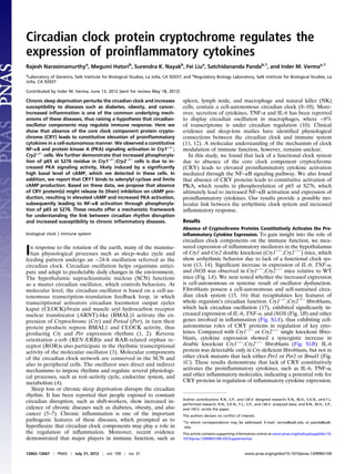
  • 5. CRY1 Expression Limits cAMP Production Likely by Binding to and Inhibiting the Function of Adenylyl Cyclase. On the basis of above results, we hypothesized that CRY regulates the intracellular cAMP levels to regulate NF–κB pathway. We addressed whether overexpression of CRY1 protein can suppress the cAMP production induced by either forskolin, prostaglandin E2 (PGE2), or isoproterenol (each 10 μM). Stimulation with forskolin directly activates adenylyl cyclase (30); whereas, induction with PGE2 and isoproterenol activates their cognate Gαs–coupled receptors, which ultimately activate adenylyl cyclase leading to transient increase in intracellular cAMP (31, 32). Dynamic changes in the intracellular cAMP levels were measured by a cAMP-sensitive luciferase reporter system (GloSensor cAMP assay; Promega) expressed in 293T cells. Overexpression of CRY1 resulted in a significant reduction in cAMP production induced by forskolin, PGE2, and also by isoproterenol (Fig. 5 A–C). Because we ob- served a substantial suppression in cAMP production induced by all three stimuli tested, we hypothesize that CRY1 may acts on adenylyl cyclase to block its function, thereby suppressing cAMP production. To test this proposal, lysates of 293T cells over- expressing Flag-tagged CRY1 or GFP were analyzed by immu- noprecipitation (IP). In 293T cell lysates, expressing Flag-CRY1 when adenylyl cyclase was immunoprecipitated with an antibody against adenylyl cyclase III (AC III), we detected Flag-CRY1 by immune blot (Fig. 5D). These results indicate that CRY1 bind to adenylyl cyclase to block its function and suppress the cAMP production. Discussion In summary, results presented here indicate that both PKA and IKK2–IκBα-dependent phosphorylation of NF–κB contributes to increased activation of inflammatory cytokines, for example IL-6, in Cry1−/− ;Cry2−/− fibroblasts. Here, we propose that circadian clock modulates NF–κB activity via CRY-mediated control of PKA activity. Circadian clock–controlled expression of CRY proteins regulate the production of cAMP, which in turn con- tributes to the observed circadian rhythms in cytosolic cAMP, which is a necessary component to core clock function in SCN neurons and in peripheral cell types (33). We presume that temporally gated oscillations of cAMP levels (33) and resultant PKA activity temporally modulates the basal activation state of the NF–κB pathway and may explain the well-described circa- dian rhythms in immune responses in mammals (34). Our data support the hypothesis that when CRY proteins are present they bind to adenylyl cyclase (Fig. 5D) and downregulate cAMP production (Fig. 5 A–C). In the absence of CRY, inhibition on adenylyl cyclase is relieved, which subsequently leads to in- creased cAMP production (Fig. 4F) and PKA activation (Fig. 4A) that cause increased phosphorylation of p65 at S276 (Fig. 4D), ultimately resulting in activation of its target genes such as IL-6 (Fig. 5E). We observed increased activation of TNF-α in Cry1−/− ;Cry2−/− fibroblasts and also in the hypothalamus of Cry1−/− ;Cry2−/− mice. Hashiramoto et al. has also previously shown high expression of TNF-α in Cry1−/− ;Cry2−/− fibroblasts (35). We also observed in- creased phospho-IKK2 in Cry1−/− ;Cry2−/− fibroblasts (Fig. 3D). We put forward that the most likely initiator of phosphorylation and activation of IKK2 signaling complex in Cry1−/− ;Cry2−/− cells is the increased activation of TNF-α (a well-known activator of IKK2 complex), which has been observed in these cells. This activated IKK2 complex subsequently leads to phosphorylation (Fig. 3C) and degradation of IκBα, which in turn cause increased p65 accumulation into the nucleus (Fig. 3E) and high level of DNA binding activity (Fig. 3F), ultimately resulting in increased expression of NF–κB target genes such as IL-6. Phosphorylation and degradation of IκBα is a critical step required to release p65 and PKA from IκBα, which allows PKA to phosphorylate p65 (36) and enter into nucleus, which is why overexpression of superrepressor mutant form of IκBα (IκBαM) dramatically suppressed up to 80% of IL-6 expression in Cry1−/− ;Cry2−/− fibroblasts (Fig. 3A). Recent reports have demonstrated the role of CRY in regu- lation of metabolism and immune function. CRY proteins are known to regulate glucose homeostasis by at least two different mechanisms. They bind to and repress glucocorticoid receptors (37), which regulate transcription of gluconeogenic genes. CRY proteins also bind to and inhibit Gαs function, thereby attenu- ating GPCR-activation-dependent increase in intracellular cAMP (38). However, we detected CRY1 in complex with ade- nylyl cyclase (Fig. 5D), and overexpression of CRY1 reduced AC III IgG GFP Flag-CRY1 AC III Tub Flag Lysate BA E DC IB IP Prostaglandin E2 Time (min) 0 6 12 18 24 30 Forskolin Foldstimulation 200 160 120 80 40 0 Vector/ Fsk CRY1/ Fsk Vector/ Vehicle Foldstimulation 200 160 120 Isoproterenol Vector/ ISP CRY1/ ISP Vector/ Vehicle Foldstimulation 240 200 160 80 40 0 120 Time (min) 0 6 12 18 24 30 Input cAMP ATP Forskolin GPCR IL-6 Ubiquitination & proteasomal degradation AC III IgG GFP Flag-CRY1RR AC III Tub Flag E IB IP Prostaglandin E2 200 160 120 Input cAMP ATPAA Forskolin IL-6 Ubiquitination & proteasomal degradation YCR G NEMO IKK1 P P IKK2 p65p50 p50 p65 CBP/p300 p50 p65 Ac P AKIP1 p65p50 P ADCY Sα P PKA P BακI Vector/ PGE2 CRY1/ PGE2 Vector/ Vehicle 80 40 0 Time (min) 0 6 12 18 24 30 Fig. 5. CRY1 inhibits the forskolin-, PGE2-, and isoproterenol-induced generation of intracellular cAMP, likely by binding to and inhibiting the function of adenylyl cyclase. (A–C) Luciferase assay performed to measure the kinetics of cAMP production after stimulation in the presence of 500 μM of IBMX in 293T cells are shown. Fold stimulation of cAMP production are shown after stimulation with either forskolin, PGE2, or isoproterenol (each 10 μM) in the absence (control vector) or presence of CRY1 expression. Data are mean ± SD (n = 3). (D) CRY1 interacts with adenylyl cyclase. All of the indicated im- munoprecipitate and lysate samples analyzed by Western blot for Flag tag (CRY1), adenylyl cyclase III and tubulin are shown. (E) The proposed model showing the likely function of CRY in binding to and suppressing the action of adenylyl cyclase in cAMP production and the pathways activated in the absence of CRY proteins. 12666 | www.pnas.org/cgi/doi/10.1073/pnas.1209965109 Narasimamurthy et al.
  • 6. cAMP production in response to PGE2, isoproterenol, and even the direct adenylyl cyclase activator, forskolin (30) (Fig. 5A). These results clearly suggest an additional cell-autonomous role of CRY in modulating cellular response to different stimuli, which contributes to our understanding of altered immune re- sponse under circadian disruption. It has previously been shown that p53 mutant mice lacking functional CRY proteins are more susceptible to TNF-α-initiated apoptosis through NF–κB acti- vation (39). Monje et al. had reported that depression like be- havior in mice induced by constant darkness is associated with increased levels of IL-6 in plasma and hippocampus mediated through NF–κB signaling pathway (40). Interestingly, NF–κB has been shown to play a role in metabolic adaptation both in normal and also in cancer cells by regulation of mitochondrial oxidative phosphorylation (41), and the current finding will suggest an additional CRY–NF–κB-mediated pathway in metabolic adap- tation of cancer cells. In this study, we analyzed the inflammatory response of mam- malian cells only lacking a functional circadian clock system, without any additional manipulations. Our results strongly indicate that an arrhythmic clock system, induced by the absence of CRY proteins, alone is sufficient to increase the stress levels of cells leading to constant expression of inflammatory cytokines and causing a low-grade chronic inflammatory status called metaflammation (42) and Cry-deficient cells could be a good model system to study this phenomenon. Compelling evidence has shown that low-grade constant inflammation could be the underlying cause for chronic diseases such as diabetes, obesity, and also cancer (43–45). Moreover, people exposed to frequent circadian clock disturbances, such as night-shift workers, are increasingly susceptible to these diseases. Therefore, further understanding connection between the circadian clock, me- tabolism, and inflammation will help to identify therapeu- tic drug targets to cure diseases like metabolic syndrome and cancer. Materials and Methods All animal experiments were carried out in accordance with the guidelines of the Institutional Animal Care and Use Committee of the Salk Institute. Male C57BL/6J and Cry1−/− ;Cry2−/− mice were used (12 wk old). TRIzol (Invitrogen) reagent was used to extract RNA from animal tissues by following the manufacturer’s protocol. Immortalized fibroblasts and 293T cells were cul- tured in Dulbecco-modified Eagle medium (Invitrogen) supplemented with 10% (vol/vol) FBS (Atlanta Biologicals) and 1% (vol/vol) of antibiotic–anti- mycotic (Invitrogen). The production of lentiviruses was performed accord- ing to the protocol described elsewhere (46). Additional details are in SI Materials and Methods. ACKNOWLEDGMENTS. This work was supported in part by grants from the National Institutes of Health (NIH), Ipsen/Biomeasure, Sanofi-aventis, and the H. N. and Frances C. Berger Foundation. The project described was supported by Grant R37AI048034 from the National Institute of Allergy and Infectious Diseases. R.N. was supported in part by a grant from the Swiss National Science Foundation (Bern, Switzerland). S.P. was supported by NIH Grant DK091618; M.H. was supported by Japan Society for Promotion of Science Fellowship. S.K.N. was supported by NIH Grant P30 CA014195-38. I.M.V. is an American Cancer Society Professor of Molecular Biology, and holds the Irwin and Joan Jacobs Chair in Exemplary Life Science. 1. Bass J, Takahashi JS (2010) Circadian integration of metabolism and energetics. Sci- ence 330:1349–1354. 2. Liu AC, Lewis WG, Kay SA (2007) Mammalian circadian signaling networks and therapeutic targets. Nat Chem Biol 3:630–639. 3. Cho H, et al. (2012) Regulation of circadian behaviour and metabolism by REV-ERB-α and REV-ERB-β. Nature 485:123–127. 4. Saini C, Suter DM, Liani A, Gos P, Schibler U (2011) The mammalian circadian timing system: Synchronization of peripheral clocks. Cold Spring Harb Symp Quant Biol 76:39–47. 5. Imeri L, Opp MR (2009) How (and why) the immune system makes us sleep. Nat Rev Neurosci 10:199–210. 6. Rajaratnam SM, Arendt J (2001) Health in a 24-h society. Lancet 358:999–1005. 7. Willyard C (2008) Hungry for sleep. Nat Med 14:477–480. 8. Arjona A, Sarkar DK (2005) Circadian oscillations of clock genes, cytolytic factors, and cytokines in rat NK cells. J Immunol 174:7618–7624. 9. Hayashi M, Shimba S, Tezuka M (2007) Characterization of the molecular clock in mouse peritoneal macrophages. Biol Pharm Bull 30:621–626. 10. Keller M, et al. (2009) A circadian clock in macrophages controls inflammatory im- mune responses. Proc Natl Acad Sci USA 106:21407–21412. 11. Bass J, Turek FW (2005) Sleepless in America: A pathway to obesity and the metabolic syndrome? Arch Intern Med 165:15–16. 12. Bollinger T, Bollinger A, Oster H, Solbach W (2010) Sleep, immunity, and circadian clocks: A mechanistic model. Gerontology 56:574–580. 13. van der Horst GT, et al. (1999) Mammalian Cry1 and Cry2 are essential for mainte- nance of circadian rhythms. Nature 398:627–630. 14. Vitaterna MH, et al. (1999) Differential regulation of mammalian period genes and circadian rhythmicity by cryptochromes 1 and 2. Proc Natl Acad Sci USA 96:12114–12119. 15. Nagoshi E, et al. (2004) Circadian gene expression in individual fibroblasts: Cell-au- tonomous and self-sustained oscillators pass time to daughter cells. Cell 119:693–705. 16. Welsh DK, Yoo SH, Liu AC, Takahashi JS, Kay SA (2004) Bioluminescence imaging of individual fibroblasts reveals persistent, independently phased circadian rhythms of clock gene expression. Curr Biol 14:2289–2295. 17. Yagita K, Tamanini F, van Der Horst GT, Okamura H (2001) Molecular mechanisms of the biological clock in cultured fibroblasts. Science 292:278–281. 18. Rey G, et al. (2011) Genome-wide and phase-specific DNA-binding rhythms of BMAL1 control circadian output functions in mouse liver. PLoS Biol 9:e1000595. 19. Li Q, Verma IM (2002) NF-kappaB regulation in the immune system. Nat Rev Immunol 2:725–734. 20. Vallabhapurapu S, Karin M (2009) Regulation and function of NF-kappaB transcrip- tion factors in the immune system. Annu Rev Immunol 27:693–733. 21. Hayden MS, Ghosh S (2008) Shared principles in NF-kappaB signaling. Cell 132: 344–362. 22. Hideshima T, et al. (2006) MLN120B, a novel IkappaB kinase beta inhibitor, blocks multiple myeloma cell growth in vitro and in vivo. Clin Cancer Res 12:5887–5894. 23. Chen LF, et al. (2005) NF-kappaB RelA phosphorylation regulates RelA acetylation. Mol Cell Biol 25:7966–7975. 24. Zhong H, Voll RE, Ghosh S (1998) Phosphorylation of NF-kappa B p65 by PKA stim- ulates transcriptional activity by promoting a novel bivalent interaction with the coactivator CBP/p300. Mol Cell 1:661–671. 25. Butt E, et al. (1994) cAMP- and cGMP-dependent protein kinase phosphorylation sites of the focal adhesion vasodilator-stimulated phosphoprotein (VASP) in vitro and in intact human platelets. J Biol Chem 269:14509–14517. 26. Clegg CH, Correll LA, Cadd GG, McKnight GS (1987) Inhibition of intracellular cAMP- dependent protein kinase using mutant genes of the regulatory type I subunit. J Biol Chem 262:13111–13119. 27. Sastri M, Barraclough DM, Carmichael PT, Taylor SS (2005) A-kinase-interacting pro- tein localizes protein kinase A in the nucleus. Proc Natl Acad Sci USA 102:349–354. 28. Gao N, Asamitsu K, Hibi Y, Ueno T, Okamoto T (2008) AKIP1 enhances NF-kappaB- dependent gene expression by promoting the nuclear retention and phosphorylation of p65. J Biol Chem 283:7834–7843. 29. Taylor SS, et al. (2005) Dynamics of signaling by PKA. Biochim Biophys Acta 1754: 25–37. 30. Seamon KB, Padgett W, Daly JW (1981) Forskolin: Unique diterpene activator of adenylate cyclase in membranes and in intact cells. Proc Natl Acad Sci USA 78: 3363–3367. 31. Furuyashiki T, Narumiya S (2011) Stress responses: The contribution of prostaglandin E(2) and its receptors. Nat Rev Endocrinol 7:163–175. 32. Kobilka BK (2007) G protein coupled receptor structure and activation. Biochim Bio- phys Acta 1768:794–807. 33. O’Neill JS, Maywood ES, Chesham JE, Takahashi JS, Hastings MH (2008) cAMP-de- pendent signaling as a core component of the mammalian circadian pacemaker. Science 320:949–953. 34. Hrushesky WJ, Langevin T, Kim YJ, Wood PA (1994) Circadian dynamics of tumor necrosis factor alpha (cachectin) lethality. J Exp Med 180:1059–1065. 35. Hashiramoto A, et al. (2010) Mammalian clock gene Cryptochrome regulates arthritis via proinflammatory cytokine TNF-alpha. J Immunol 184:1560–1565. 36. Zhong H, SuYang H, Erdjument-Bromage H, Tempst P, Ghosh S (1997) The tran- scriptional activity of NF-kappaB is regulated by the IkappaB-associated PKAc subunit through a cyclic AMP-independent mechanism. Cell 89:413–424. 37. Lamia KA, et al. (2011) Cryptochromes mediate rhythmic repression of the gluco- corticoid receptor. Nature 480:552–556. 38. Zhang EE, et al. (2010) Cryptochrome mediates circadian regulation of cAMP signal- ing and hepatic gluconeogenesis. Nat Med 16:1152–1156. 39. Lee JH, Sancar A (2011) Regulation of apoptosis by the circadian clock through NF- kappaB signaling. Proc Natl Acad Sci USA 108:12036–12041. 40. Monje FJ, et al. (2011) Constant darkness induces IL-6-dependent depression-like behavior through the NF-kappaB signaling pathway. J Neurosci 31:9075–9083. 41. Mauro C, et al. (2011) NF-κB controls energy homeostasis and metabolic adaptation by upregulating mitochondrial respiration. Nat Cell Biol 13:1272–1279. 42. Hotamisligil GS (2006) Inflammation and metabolic disorders. Nature 444:860–867. 43. Ben-Neriah Y, Karin M (2011) Inflammation meets cancer, with NF-κB as the match- maker. Nat Immunol 12:715–723. 44. Gregor MF, Hotamisligil GS (2011) Inflammatory mechanisms in obesity. Annu Rev Immunol 29:415–445. 45. Sun B, Karin M (2012) Obesity, inflammation, and liver cancer. J Hepatol 56:704–713. 46. Tiscornia G, Singer O, Verma IM (2006) Production and purification of lentiviral vectors. Nat Protoc 1:241–245. Narasimamurthy et al. PNAS | July 31, 2012 | vol. 109 | no. 31 | 12667 IMMUNOLOGY