Isolation and Screening of Starch Hydrolising Bacteria and its Effect of Diff...
Vaidyanathan JME 05
1. VECTOR/PATHOGEN/HOST INTERACTION, TRANSMISSION
Isolation of a Myoinhibitory Peptide from Leishmania major
(Kinetoplastida: Trypanosomatidae) and Its Function in the
Vector Sand Fly Phlebotomus papatasi (Diptera: Psychodidae)
RAJEEV VAIDYANATHAN1, 2
J. Med. Entomol. 42(2): 142Ð152 (2005)
ABSTRACT Protozoan parasites in the genus Leishmania are ingested by sand ßies with blood and
multiply in the gut until they are transmitted to a vertebrate host when the sand ßy blood feeds again.
Infections of the enzootic vector Phlebotomus papatasi Scopoli result in distended midguts with no
spontaneous gut contractions. Using a P. papatasi hindgut contraction bioassay, a paralytic factor
sensitive to trypsin, chymotrypsin, proteinase-K, and heating at 56ЊC was detected in crude lysates of
Leishmania major promastigotes. Application of parasite lysate to isolated hindguts resulted in re-
versible, dose-dependent inhibition of spontaneous contractions. Mean volume of isolated midguts
and hindguts increased by 50Ð60% after application of L. major lysate. L. major paralytic factor was
puriÞed 104
-fold over the total protein preparation and yielded a hydrophobic 12-kDa peptide.
Myoinhibitory activity eluted as a single peak in reverse phase-high-pressure liquid chromatography.
Tandem mass spectrometry resulted in 15 amino acid sequences, three of them sharing 45Ð73%
homology with short hypothetical gene products of undeÞned function from Pseudomonas, Halo-
bacterium, and Drosophila. This unique protozoan peptide mimics the function of endogenous insect
neuropeptides that control visceral muscle contractions. By this novel mechanism, parasites persist in
the expanded, relaxed midgut after blood meal and peritrophic matrix digestion. This allows time for
development and migration of infective forms, facilitating sand ßy vector competence and parasite
transmission.
KEY WORDS Leishmania major, Phlebotomus papatasi, vector competence, myoinhibition, hindgut
PROTOZOA IN THE GENUS Leishmania (Kinetoplastida:
Trypanosomatidae) are dimorphic parasites that al-
ternate between a ßagellated, extracellular promasti-
gote stage in the gut of a sand ßy vector and an
intracellular amastigote stage within the reticuloen-
dothelial cells of a mammal host (Peters and Killick-
Kendrick 1987, Dedet et al. 1999). Leishmania para-
sites cause a spectrum of diseases in humans, including
the clinically distinct forms of visceral, cutaneous, and
mucocutaneous leishmaniasis. Phlebotomus papatasi
Scopoli (Diptera: Psychodidae) is the most important
vector of cutaneous leishmaniasis in Israel, maintain-
ing enzootic transmission of Leishmania major among
fat sand rats, Psammomys obesus, and acting as sole
vector of L. major to humans (Schlein et al. 1982,
Janini et al. 1995).
Female sand ßies ingest blood that includes mac-
rophages containing Leishmania amastigotes when
feeding on an infected vertebrate host. In the midgut,
the blood is encased in a peritrophic matrix (PM),
a semipermeable chitinous sac produced by the gut
epithelium(Blackburnetal.1988).WithinthePM,the
amastigotes divide repeatedly and transform into a
uniform population of promastigotes. The PM disin-
tegrates at the end of blood digestion, and free para-
sites can be voided. Rapid loss of infection by excre-
tion indicates that susceptible parasites are not killed
outrightbutrathervoidedwiththePMandbloodmeal
remnants. Parasites might persist in the gut by an-
choring ßagella between midgut microvilli or by con-
formational changes in surface glycoconjugates (War-
burg et al. 1989; Pimenta et al. 1992; for summary, see
Sacks and Kamhawi 2001). However, a large popula-
tion of unattached parasites persists in the gut lumen
(Walters et al. 1987, 1989), and it has not been ex-
plained why these parasites are not excreted. Free
promastigotes transform into infective metacyclic
forms, which migrate forward and are transmitted
when the sand ßy blood feeds again.
Sand ßies heavily infected with L. major had dis-
tended midguts with no tonus and no peristalsis. Im-
mobility of the gut of infected ßies seemed to be a
mechanism by which parasites avoided expulsion, pre-
sumably by factors produced by L. major. The iden-
tiÞcation of this factor and deÞnition of its function
were the purpose of this study.
1 Department of Parasitology, Hebrew University of Jerusalem,
Hadassah Medical School, Ein Kerem, Jerusalem 91120 Israel.
2 Current address: Department of Entomology, University of Cal-
ifornia, Davis, CA 95616.
0022-2585/05/0142Ð0152$04.00/0 ᭧ 2005 Entomological Society of America
2. Materials and Methods
Parasite Preparation. L. major MHOM/IL/86/Blum
(Jordan Valley strain) was obtained from the World
Health Organization Leishmania Reference Center,
Department of Parasitology, Hebrew University,
Jerusalem, Israel. Reagents and protease inhibitors
were purchased from Sigma (Rehovot, Israel), unless
otherwise speciÞed. Parasites were grown in Dulbec-
coÕs modiÞed EagleÕs medium (Biological Industries,
Beit Haemek, Israel) with high glucose content, 10%
heat-inactivated fetal calf serum, 4 mM L-glutamine,
2 mM adenosine, and 2% (vol:vol) Þlter-sterilized
human urine. Cultures were maintained at 28ЊC and
passaged every 4 d.
Two other kinetoplastid parasites were used as con-
trols. Herpetomonas muscarum, an obligate parasite of
house ßies, was cultured identically to the L. major
cultures. Crithidia fasciculata, an obligate gut parasite
of mosquitoes, was cultured in brain-heart infusion at
28ЊC and passaged every day.
Late log-phase cultures of the three parasites at
high density (107
Ð108
parasites/ml) were spun at
2,000 ϫ g for 10 min at 8ЊC and washed twice with
Aedes aegypti L. buffered saline (ABS, Þnal concen-
tration 0.6 mM MgCl2, 4.0 mM KCl, 1.8 mM NaHCO3,
150 mM NaCl, 25 mM HEPES-NaOH, 1.7 mM CaCl2,
pH 7.4). A protease inhibitor cocktail was added to
wet parasite volume to inhibit autolysis (Þnal concen-
tration 1.0 mM AEBSF, 0.5 mM EDTA, 65 M bestatin,
7 M E-64, 0.5 M leupeptin, 0.15 M aprotinin). To
lyse parasites, cell pellets were snap-frozen in liquid
N2 and thawed three times at 30ЊC. Samples were
checked under phase-contrast microscope to verify
parasite lysis. Crude homogenates were frozen in liq-
uid N2 and stored at Ϫ70ЊC until use. Concentrations
of lysate proteins were assayed by the Bradford
method (Bradford 1976).
Crude homogenates were thawed on ice and spun
at 12,000 ϫ g for 30 min to precipitate particulates.
Both precipitate and supernatant were tested in the
sand ßy hindgut bioassay (below).
Sand Flies, Hindgut Bioassay, and Gut Distension
Measurements. The P. papatasi colony originated with
ßies from Kfar Adumim, 10 km east of Jerusalem. The
colony was maintained according to Modi and Tesh
(1983), and insectary conditions were 26 Ϯ 1ЊC, 80%
RH,andaphotoperiodof17:7(L:D)h.Two-to6-d-old
sugar-fed sand ßies were used for all experiments.
Whole guts from male and female sand ßies were
dissected into a watch glass with 90Ð99 l of oxygen-
ated ABS warmed to 30ЊC and allowed to recover until
hindgut contractions stabilized. Hindgut contractions
were counted for 5-min increments. Samples of par-
asite lysate proteins were added 5 min after the hind-
gut stabilized and contracted regularly. Preparations
were mixed with a pipette to distribute proteins. One
unit of paralytic activity is 1% inhibition of P. papatasi
hindgut contractions relative to the initial 5-min pe-
riod before addition of proteins. SpeciÞc activity is
deÞned as units of paralytic activity per milligram of
lysate proteins. To test whether inhibition was revers-
ible, the treated gut preparations were rinsed twice in
ABS, and returned to 100 l of warm, oxygenated ABS.
P. papatasi midguts and hindguts were measured on
an Olympus BH phase-contrast microscope. Whole
guts from unfed ßies were dissected into 98 l of
oxygenated ABS warmed to 30ЊC in glass-covered
watch glasses. Width and length of the midgut and the
hindgut were measured, 6.4 g of L. major proteins
was added (Þnal concentration 64 g/ml), and the
guts were kept in a humid chamber until they were
measured again at 5 and 30 min. Midgut and hindgut
volumes were estimated as cylinders for the calcula-
tions, as widest cross-sectional area multiplied by
length. Ten ßies (Þve males, Þve females) were used.
An equal concentration of H. muscarum lysate pro-
teins was used as a control.
SigniÞcant differences in speciÞc activity in the
sand ßy hindgut bioassay were tested using a two-
sample t-test assuming equal variances. Gut volume
calculations were tested for signiÞcant differences us-
ing the MannÐWhitney U test (Daniel 1991).
Protease and Heat Inactivation of the Paralytic
Factor. A 1-ml sample of L. major lysate proteins
(10.55 mg/ml) was prepared according to the above-
mentionedprotocol.Totestforthermalinactivationof
paralytic activity, lysate was heated at 56ЊC in a water
bath, gently agitated, and aliquots were removed at
0, 30, 60, 90, 120, 150, and 180 min. Each aliquot was
frozen in liquid N2 and stored at Ϫ70ЊC until tested on
the sand ßy hindgut bioassay.
L. major promastigote lysates were treated with
trypsin, chymotrypsin, and proteinase-K to test sen-
sitivity of the paralytic factor to proteases. Aliquots
of parasite proteins (3 mg/ml) were treated 1:1 with
1 mg/ml trypsin and chymotrypsin, or 10:1 with
0.5 mg/ml proteinase-K, heated for 30 min at 37ЊC,
and reactions halted with proteinase inhibitors, as
described above. Lysate samples treated with pro-
teases were frozen in liquid N2 and stored at Ϫ70ЊC
until tested on the sand ßy hindgut bioassay.
Purification of the Paralytic Factor. In brief, to pu-
rify the paralytic factor to apparent homogeneity,
cell proteins were precipitated with 85% (NH4)2SO4
at 0ЊC and subjected to two steps of hydrophobic
chromatography, size exclusion chromatography,
and Þnally reverse phase-high-pressure liquid chro-
matography (RP-HPLC). Chromatography, centrifu-
gation, and dialysis were performed at 0Ð4ЊC (Web-
ster and Prado 1970). RP-HPLC fractions were tested
for paralytic activity by using the sand ßy hindgut
bioassay. Active fractions were submitted to elec-
trospray ionization and tandem mass spectrometry
analysis to determine amino acid sequence (Na¨ssel
1999, Mann et al. 2001). These sequences, identiÞed
from peptide fragmentation data after mass spectrom-
etry, were matched to protein sequence databases.
Fraction 1 lysate proteins from freeze/thawed par-
asites,culturedandharvestedasdescribedabovewere
bulk precipitated with 85% (NH4)2SO4, pH adjusted
to 7.2 with NH4OH at 0ЊC (Englard and Seifter 1990).
Samples were centrifuged 5 ϫ 103
g for 30 min, and the
supernatant was discarded. Pellets were pooled, im-
March 2005 VAIDYANATHAN: L. major MYOINHIBITORY PEPTIDE 143
3. mediately frozen in liquid N2, and stored at Ϫ70ЊC
(fraction 2). Aliquots of fraction 1, fraction 2, and
supernatant were assayed using the sand ßy hindgut.
Fraction 2 and supernatant were Þrst dialyzed over-
night against 4.0 liters of ABSm (ABS modiÞed to
50 mM NaCl) to remove (NH4)2SO4.
Fraction 2 samples were thawed on ice, diluted with
ABSm to 1.5 M (NH4)2SO4, and centrifuged 5 ϫ 103
g for 30 min to remove insoluble particulates. In total,
2,878 mg of fraction 2 was loaded onto a Phenyl Sepha-
rose 6 Fast Flow column (6.5 by 23 cm) (Amersham
Biosciences Inc., Piscataway, NJ), equilibrated with
1.5 M (NH4)2SO4 in ABSm (Kennedy 1990). The col-
umn was eluted stepwise with the same buffer con-
taining 1.0, 0.75, 0.70, 0.65, 0.60, 0.55, 0.50, 0.25, and
0.0 M (NH4)2SO4. Aliquots of 100 l were dialyzed
against 4.0 liters of ABSm overnight and bioassayed.
Pooled active fractions were deÞned as fraction 3.
Fraction 3 (40 mg) was loaded onto a Fractogel
EMD Propyl Sepharose column (1.5 by 9 cm) (Merck,
Whitehouse Station, NJ), equilibrated with 1.5 M
(NH4)2SO4 inABSm.Samplewaselutedstepwisewith
the same buffer containing 1.25, 1.00, 0.75, 0.50, 0.25,
and 0.0 M (NH4)2SO4 (Kennedy 1990). Aliquots of
50 l were dialyzed against 4.0 liters of ABSm over-
night and tested on the sand ßy hindgut bioassay.
Active fractions from Propyl Sepharose chromatogra-
phy were deÞned as fraction 4. Before proceeding to
size exclusion chromatography, fraction 4 was con-
centrated on a 1-ml bed volume Phenyl Sepharose 6
Fast Flow column, equilibrated with 1.5 M (NH4)2SO4
in ABSm. The sample was eluted with the same buffer
at 1.5 and 0.0 M (NH4)2SO4, dialyzed, and bioassayed.
This concentrated sample was deÞned as fraction 5.
Protein recovery was conÞrmed by Bradford assay
(Bradford 1976), and fraction 5 was Þltered (0.22 m)
and loaded onto a Merck Fractogel EMD BioSEC (S)
size exclusion chromatography column, adapted for
HPLC(Merck,Darmstadt,Germany).Chromatography
wasperformedusingaVarian5000liquidchromatograph
and Rheodyne solvent delivery module (Cotati, CA).
FractionsweredetectedwithaVarianR1Ð3UVdetector
set at 280 nm. The following molecular weight markers
were run: -amylase (200 kDa), bovine serum albumin
(66 kDa), chymotrypsinogen (25 kDa), and aprotinin
(6.5 kDa). The column was equilibrated and washed
with ABS at a 0.5 ml/min ßow rate (Fischer 1980, Stell-
wagen 1990). Ninety 0.8-ml fractions were collected on
ice, and 10-l aliquots were bioassayed. Fractions with
paralytic activity were pooled and deÞned as fraction 6.
Fraction 6 was evaporated to dryness under vac-
uum, reconstituted in HPLC-grade triple-distilled
H2O, and Þltered (0.22 m) before loading onto a
Vydac 214 TP RP-C4HPLC column (0.46 by 25 cm).
Equipment was the same as for size exclusion chro-
matography, with the exception of UV detection at
220 nm. Column elution was performed with gradients
of solution A (5% acetonitrile, 95% H2O, and 0.1%
trißuoroacetic acid [TFA]) at 30 min, eluted using a
linear gradient with solution B (95% acetonitrile,
5% H2O, and 0.1% TFA) at a ßow rate of 0.5 ml/min
for 60 min. Ninety 0.5-ml fractions were collected
manually and immediately frozen in liquid N2; 2-l
aliquots were evaporated to dryness before reconsti-
tution in triple-distilled H2O and tested on sand ßy
hindgut (Veelaert et al. 1996, Duve et al. 1999).
Mass Spectrometric Identification of the Paralytic
Factor. Active Vydac fractions were combined and
vacuum-evaporated to Ϸ50 l. A further 25 l of
25mMNH4HCO3 wasaddedatpH8.0toactasabuffer
during trypsin addition. This 75 l was divided into
three aliquots. The Þrst aliquot was digested overnight
at 37ЊC with 10 l of trypsin (0.1 g/10 M
NH4HCO3). It was then eluted using a ZipTip C18 tip
(Millipore Corporation, Billerica, MA) and 75%
CH3CN, 1% formic acid (Merck). Part of this sample
was used as load, and the remaining part was digested
with Asp-N (see protocol below).
The second aliquot was digested overnight at 37ЊC
with 10 l of 0.04 g of Asp-N (Roche Diagnostics,
Mannheim, Germany) in 25 mM NH4HCO3 at pH 8.0.
After enzymatic digestion, the sample was eluted us-
ing a ZipTip C18 tip and 75% CH3CN, 1% formic acid.
The third aliquot underwent reduction alkylation.
It was treated with 10 l of 45 mM dithiothreitol for
30 min at 60ЊC and 10 l of 100 mM iodoacetamide for
30 min at 25ЊC. It was eluted with a ZipTip C18 tip
and 75% CH3CN, 1% formic acid. After alkylation
reduction, the sample was divided into two fractions.
The Þrst was treated with 10 l of 0.04 g of Asp-N in
25 mM NH4HCO3 at pH 8.0 overnight at 37ЊC and
eluted with a ZipTip C18 tip and 75% CH3CN, 1%
formic acid. The second was treated with 10 l of
trypsin (0.1 g/10 M NH4HCO3) overnight at 37ЊC
and eluted with a ZipTip C18 tip and 75% CH3CN, 1%
formic acid.
A load of 3 l of each eluted sample was injected
through a Long NanoES spray capillary (Protana Cor-
poration, Toronto, Canada) to an electrospray ioniza-
tion quadropole time of ßight (ESI QTOF2) (Micro-
mass London, London, United Kingdom) to ionize
protein fragments and to detect the mass-to-charge
ratio (m/z) of ionized peptides (Fenn et al. 1989).
Capillary voltage was 1,200 V, cone voltage was 25 V,
collision energy was 10.
Pumping the analyte at a low microliter per minute
ßow rate at high voltage causes electrostatic disper-
sion of micrometer-sized droplets that rapidly evap-
orate and impart their charge onto the analyte mole-
cules (Mann et al. 2001). Electrosprayed ions were
detected by tandem mass spectrometry (MS-MS)
spectra, with identical capillary and cone voltages as
described above, but with collision energy between 30
and 40 in the presence of argon (Hunt et al. 1986,
Biemann and Scoble 1987).
The MS-MS spectra were matched against nonre-
dundant database sequences; a sequence tag search
(www.matrixscience.com) and a full sequence search
(BLASTandFASTA)weredoneusingsmallidentiÞed
peptide fragments (Altschul et al. 1997, GrifÞn and
Aebersold 2001) against all recorded proteins in the
database.
144 JOURNAL OF MEDICAL ENTOMOLOGY Vol. 42, no. 2
4. Results
Inhibition of P. papatasi Hindgut Contractions.
Sand ßy hindguts in warmed ABS spontaneously con-
tracted for 1Ð6 h. Midgut activity was irregular; the
hindgutkeptasteadyrhythm.Activityofbothsections
was stopped by addition of L. major lysate; however,
only hindguts were suitable for measuring inhibitory
effect. Contractions of the rectum were not affected
by addition of parasite lysate.
A dose of 50Ð70 g/ml L. major lysate proteins
inhibited 60% of spontaneous P. papatasi hindgut con-
tractions within 5 min of application (Fig. 1). Hind-
guts exposed to parasite lysate for 20 min, rinsed in
ABS, and returned to fresh ABS resumed contracting
(Fig. 1). No effect was seen with equivalent concen-
trations of lysates prepared from H. muscarum or from
C. fasciculata (data not shown).
Increasing concentrations of L. major lysate pro-
teins inhibited hindgut contractions in a dose-depen-
dent manner (Fig. 2). An application of 12 or 24 g/ml
L.majorlysateproteinsinhibitedspontaneoushindgut
contractions by 20 and 34%, respectively. Distended
nodes formed along the length of the hindgut. An
application of 48 and 96 g/ml lysate proteins de-
creased hindgut contractions by 60 and 80%, respec-
tively. Hindguts completely distended within seconds
of a concentration of 48 g protein/ml or more. Hind-
gut lumen Þlled with liquid; gut and epithelial cells
were turgid.
Fig. 1. Inhibition of P. papatasi hindgut contractions with L. major lysate proteins (50Ð70 g/ml). Contractions were
monitored for 5 min. Hindguts in the “return” group were treated with L. major lysate proteins for 20 min, rinsed in ABS,
and immediately returned to fresh ABS to determine whether myoinhibition was reversible. Means and standard errors are
shown for 36 trials.
Fig. 2. Inhibition of hindgut contractions by different concentrations of L. major lysate proteins within 5 min. Means and
standard errors are shown for Þve trials.
March 2005 VAIDYANATHAN: L. major MYOINHIBITORY PEPTIDE 145
5. Distension of P. papatasi Midgut and Hindgut.
Based on observations of distended hindguts after
application of L. major lysate proteins, midgut and
hindgut distension was quantiÞed 5 and 30 min after
addition of 6.4 g of protein (64 g/ml) of L. major
and H. muscarum lysates (Fig. 3A and B).
P. papatasi midgut volume increased after 5-min
incubation with 64 g/ml L. major proteins, saline,
Fig. 3. Distension of P. papatasi midgut (A) and hindgut (B) volume 5 and 30 min after application of 64 g/ml L. major
or H. muscarum lysate proteins. Means and standard errors are shown for 10 trials (Þve males, Þve females).
Fig. 4. Heat inactivation of L. major lysate proteins tested on P. papatasi hindgut assays.
146 JOURNAL OF MEDICAL ENTOMOLOGY Vol. 42, no. 2
6. and 64 g/ml H. muscarum proteins by 19.5, 17.4, and
12.2%, respectively. The increase due to L. major
proteins was not signiÞcantly different from the saline
control or H. muscarum proteins (P Ͼ 0.05). However
the difference was signiÞcant (P Ͻ 0.01) after 30-min
incubation with L. major proteins (48.7%) versus
treatment with saline (19.2%) or H. muscarum pro-
teins (7.8%) (Fig. 3A).
Average hindgut volume increased 5 min after ap-
plication of 64 g/ml L. major proteins, saline, and
64 g/ml H. muscarum proteins by 20.7, 7.3, and
13.1%, respectively. After 30-min incubation, hindgut
volume increased by 57.1, 11.0, and 13.1%, respec-
tively. The increase in hindgut volume incubated with
L. major proteins was signiÞcantly greater than the
other two applications (P Ͻ 0.01) at both 5 and 30 min
(Fig. 3B).
Protease and Heat Inactivation of the Paralytic Fac-
tor. Heating of L. major lysate for 30 min at 56ЊC
resulted in a 42% loss of activity. Aliquots heated from
60 to 180 min resulted in 65Ð70% loss of activity, but
there was no difference among samples heated 60 min
or longer (Fig. 4).
Lysate samples treated with trypsin, chymotrypsin,
orproteinase-Knolongerinhibitedspontaneoushind-
gut contractions (data not shown). Based on these
results, the paralytic factor was considered to be a
protein.
Purification and Identification of the Paralytic Pro-
tein. A series of Þve chromatography columns were
used to purify the paralytic protein from L. major
lysate to apparent homogeneity. The puriÞcation
scheme was performed four times; results for the
Þnal trial are presented. Average values for activity
(percentage of inhibition per milliliter), protein con-
centration, and speciÞc activity are summarized in
Table 1.
After initial puriÞcation steps, more than one
(NH4)2SO4-precipitated fraction had myoinhibitory
activity on spontaneous contractions in P. papatasi
hindgut. The fraction with highest speciÞc activity
was puriÞed. Elution conditions for the active frac-
tions were: Phenyl Sepharose 6 Fast Flow, 0.65 M
(NH4)2SO4; EMD Propyl Sepharose, 1.0 M (NH4)2SO4;
Phenyl Sepharose 6 Fast ßow (to concentrate sam-
ple), plain ABSm; Fractogel EMD BioSEC, 134.9-min
elution time; and Vydac RP-C4HPLC, 33.5% acetoni-
trile. SpeciÞc activity increased 104
-fold from the orig-
inal crude proteins to the Þltered load injected onto
the Vydac RP-HPLC column (Table 1).
A preliminary estimation of the apparent native
protein mass could be deduced from the plotting of
Fig. 5. Elution sequence of molecular weight markers from Fractogel EMD BioSEC size exclusion column. Arrow denotes
point at which myoinhibitory activity eluted.
Table 1. Increase of myoinhibitory activity at different steps of purification of L. major proteins
Fraction
Vol
(ml)
Activity
(units/ml)
Total
activity
͓Protein͔
(mg/ml)
Total
protein
(mg)
SpeciÞc
activity
(units/
mg)
1 Original lysate 3,100 1.36 ϫ 104
4.21 ϫ 107
3.8 11,780 3.58 ϫ 103
2 AS precipitate 530 7.11 ϫ 104
3.77 ϫ 107
18.0 9540 3.95 ϫ 103
3 Phenyl Sepharose 1,300 1.73 ϫ 104
2.25 ϫ 107
0.15 195 1.15 ϫ 105
4 Propyl Sepharose 60 2.62 ϫ 105
1.57 ϫ 107
0.015 0.9 1.75 ϫ 107
6 Size exclusion 2.1 8.00 ϫ 104
1.68 ϫ 105
0.005 0.0105 1.60 ϫ 107
Final RP-HPLC 1.5 2.85 ϫ 105
4.28 ϫ 105
Ͻ0.005 Ͻ0.0075 Ͼ5.7 ϫ 107
Data are shown for active fractions only. AS, ammonium sulfate. Note that fraction 5 was a concentration step and is not shown.
March 2005 VAIDYANATHAN: L. major MYOINHIBITORY PEPTIDE 147
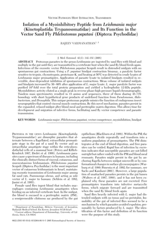
7. log molecular weight of the protein markers versus
their retention time on the preparative BioSEC col-
umn (Fig. 5). The paralytic activity eluted with a
retention time of 134.9 min, marked with an arrow
between chymotrypsinogen and aprotinin, indicating
an apparent mass of Ϸ12 kDa. A more accurate ana-
lytical measurement of the protein apparent mass has
yet to be conducted, based on both Stokes radii and
sedimentation measurements.
Myoinhibitory activity in the RP-HPLC step was
detected between 48 and 50 min (Fig. 6). The aceto-
nitrile gradient increased from 5% at 30 min (time 0)
to 95% at 90 min (that is, 60 min after the gradient
began). Using the linear regression equation y ϭ 1.5x
ϩ 5, the calculated acetonitrile concentration at
which myoinhibitory activity eluted is 33.5% (Fig. 7).
Active fractions from the RP-HPLC fractionation
step were subjected to enzymatic digestion, mass
Fig. 6. RP-HPLC fractionation on Vydac C4 column of a previous size exclusion chromatography fraction of Ϸ12 kDa.
Myoinhibitory activity eluted between 48 and 50 min (denoted by arrow).
Fig. 7. Increasing acetonitrile gradient on RP-HPLC Vydac C4 column during isocratic elution with solution B (95%
acetonitrile, 5% H2O, and 0.1% TFA). Arrow denotes point at which myoinhibitory activity eluted, 18Ð20 min after an initial
30-min void volume (corresponding to activity at 48Ð50 min in Fig. 6).
148 JOURNAL OF MEDICAL ENTOMOLOGY Vol. 42, no. 2
8. spectrometry, and MS-MS analysis (Fenn et al. 1989).
The m/z of ionized peptides was detected by a
QTOF2 Micromass mass spectrometer. The active my-
oinhibitory peptide was extremely difÞcult to frag-
ment, despite sequential trials of enzymatic digestion
with and without reduction alkylation. Peaks in the
MS spectra range of 1000 m/z (Fig. 8) are highly
charged, representing peptides Ϸ6,000 Da. Tandem
mass spectrometry of 10 major peaks and dozens of
minor peaks yielded m/z values, molecular weights,
and putative sequences for the myoinhibitory peptide.
Currently, sequence data are being used as templates
for RT-polymerase chain reaction and production of
recombinant peptide. Amino acid sequences may be
published once a more complete picture is obtained.
All the peptide sequences obtained from tandem
mass spectrometry were used to search for sequence
homologies in known proteins by using Mascot,
BLAST, and FASTA search programs (Altschul et al.
1997, GrifÞn and Aebersold 2001). By selecting entire
databases, three organisms yielded sequences of four
to seven amino acids with Ͼ45% homology to L. major
myoinhibitory peptide and low probability that the
search sequence was a random string (an expected
value, or E, Ͻ 1.0). A putative gene product of Pseudo-
monas aeruginosa had a 53% homology with some
Fig. 8. Tandem mass spectra data from electrospray ionization of active fractions from RP-HPLC. The 650Ð700 range is
expanded to show major peaks used for peptide sequence analysis. Each peak represents one peptide fragment; multiple
numbers for one peak represent amino acid modiÞcations within that fragment.
March 2005 VAIDYANATHAN: L. major MYOINHIBITORY PEPTIDE 149
9. identiÞed fragments and an E-value of 0.13 (Stover et
al. 2000). One hypothetical protein of a Halobacterium
sp. had 73% homology with one identiÞed fragment
and an E-value of 0.35. Three putative gene prod-
ucts in Drosophila melanogaster Meigen had 45% ho-
mology with identiÞed fragments and E-values of
0.84 (Adams et al. 2000). The functions for these pro-
teins in P. aeruginosa, Halobacterium, and D. melano-
gaster are unknown.
Discussion
Spontaneous contractions of P. papatasi gut prepa-
rationswereinhibitedbyL.majorlysateproteins;they
resumed regular activity after rinsing (Fig. 1). Lysates
from kinetoplastid parasites of other Diptera did not
affect P. papatasi hindgut contractions. Inhibition of
muscle activity was dose-dependent, although the ef-
fect was not strictly linear (Fig. 2). Similar lysate
preparations signiÞcantly increased midgut and hind-
gut volumes (Fig. 3). Paralytic activity in parasite
lysate was reduced when lysates were heated (Fig. 4)
and lost when lysates were treated with proteases,
indicating that the paralytic factor is a protein.
Leishmania promastigotes that are entirely in the
sand ßy gut beneÞt from decreased peristalsis and
increased gut volume. A distended gut retains more
sand ßy food, providing nutrients for parasites and
more room for multiplication. Immobility of the ex-
panded gut protects parasites from expulsion after PM
disintegration. This is coincident with development of
infective forms, or metacyclogenesis (Sacks and Per-
kins 1985). By inhibiting gut peristalsis and increasing
gut volume, parasites condition the midgut for meta-
cyclogenesis and transmission of infective forms.
Persistence of Leishmania promastigotes in the gut
by insertion of ßagella between midgut microvilli has
been studied by light microscopy (Adler and Theodor
1926) and later by electron microscopy (Killick-Ken-
drick et al. 1974, Warburg et al. 1986). Using isolated
ßagella, Warburg et al. (1989) found that a ßagellar
surface protein facilitated attachment to midgut epi-
thelial cells. Extensive studies have shown that pro-
cyclic promastigotes bind to gut epithelium by li-
pophosphoglycan (LPG), the most abundant cell
surface glycoconjugate. Other studies have detailed
differences in LPG structure and modiÞcations, cor-
relation with vector competence and speciÞcity, and
work with genetically modiÞed parasites (Pimenta et al.
1992; Sacks et al. 1994, 1995; Sacks and Kamhawi 2001).
However, infective metacyclic parasites with modiÞed
LPG and other morphotypes (Walters et al. 1987, 1989)
remain free in the gut lumen. Relaxation of the midgut
may protect the population of unattached parasites that
inhabit the midgut after PM disruption.
In the anterior midgut of infected ßies, a gelatinous
plug (Killick-Kendrick 1979) consisting of an elec-
tron-dense, Þlamentous precipitate surrounds many
parasites (Walters et al. 1987, Lawyer et al. 1990). The
gel plug has a framework of parasite-derived mucin-
like proteophosphoglycan that contains Leishmania-
secreted acid phosphatase. The gel plug presumably
enhances parasite transmission by impeding the in-
gestion of blood and causing repeated probing (Stier-
hof et al. 1999). Differentiation of promastigotes into
metacyclic forms also takes place in the plug (Rogers
et al. 2002). Assembly of the plug is cumulative; with-
out inhibition of gut peristalsis, gel components may
be expelled and plug assembly interrupted at an early
stage.
The factor responsible for inhibition of spontaneous
gut contractions was determined to be a protein be-
cause of its inactivation by heat and proteases. Based
on this observation, L. major lysate was precipitated
and fractionated to yield a 12-kDa peptide from size-
exclusion chromatography. A single peak from RP-
HPLC yielded a peptide with 104
-fold speciÞc activity
over the original crude protein extract. The peptide is
named stambhanin, from the Sanskrit verb stambh,
meaning “to hinder, suppress, paralyze, stupefy; to
become stiff or rigid” (Apte 1963).
SpeciÞc activity increased 100-fold from the Phenyl
to Propyl Sepharose column. A 100-fold increase in
speciÞc activity is unlikely by simple peptide isolation
and is most likely due to the removal of endogenous
inhibitors for the myoinhibitory peptide. Its elution
order in Phenyl and Propyl Sepharose chromatogra-
phy indicates that stambhanin is highly hydrophobic.
Myoinhibition was detected in more than one fraction
of Phenyl and Propyl Sepharose chromatography.
Several myoinhibiting peptides must be present in
L. major for activity to be present in more than one
fraction. PuriÞcation steps proceeded with fractions
of highest speciÞc activity and highest recovery
(Table 1).
Several peaks in the 650Ð700 range from the mass
spectrometric data represent the same peptide frag-
ment (Fig. 8). In these most likely sequences, hydro-
phobic amino acids (Gly, Ala, Val, Leu, Ile, Met, Phe,
Trp, and Pro) account for 64Ð73% of those identiÞed.
This explains the hydrophobic elution order for
active fractions in Phenyl and Propyl Sepharose chro-
matography. In addition, cysteine was detected in two
sequence fragments, possibly stabilizing internal pep-
tide conformation through disulÞde linkages. Cur-
rently, only fragmented sequence data are available.
Studies are focused on production of recombinant
peptide for further characterization.
A search of prokaryotic and eukaryotic genomes
yielded no homology with any arthropod-borne path-
ogen. Only three organisms expressed a protein with
Ͼ45% homology, and even these were for single frag-
ments. The three best-Þt sequences from P. aeruginosa,
Halobacterium sp., and D. melanogaster were putative
gene products with no known function. This means
either the sequences obtained are highly variable, or
stambhanin is a novel molecule that functions like
endogenous viscerotropic neuropeptides of insects.
The enteric nervous system in arthropods releases
endogenous neuropeptides and neurotransmitters
that regulate visceral muscle contraction (Coast and
Webster 1998). The Þrst inhibitory neuropeptide iso-
lated from an insect was the FMRF-amide-related
peptide leucomyosuppressin, which inhibits sponta-
150 JOURNAL OF MEDICAL ENTOMOLOGY Vol. 42, no. 2
10. neouscontractionsofthecockroachhindgut(Holman
et al. 1986). FLRF-amides of Locusta migratoria L.
inhibit locust heart rhythm, reduce spontaneous ovi-
duct contractions, and decrease amplitude of hindgut
contractions (Schoofs et al. 1993). Many of these pep-
tides, such as myosuppressins and locustamyoinhibit-
ing peptide, block voltage-gated and ligand-gated
Ca2ϩ
channels in the plasma membrane (Wilcox and
Lange 1995, Orchard et al. 1997). Blocking Ca2ϩ
chan-
nels and decreasing Ca2ϩ
-dependent action potentials
is a common mechanism shared by insect neuropep-
tides and may explain how stambhanin operates.
There are many examples of parasites modifying host
physiology to their own advantage. In this study, a pro-
tozoan produces a peptide that mimics the function of
myoinhibitory neuropeptides of insects. Amino acid se-
quences for fragments of stambhanin were compared
with proteins and protein fragments from several data-
bases,butitssequencesdidnotcorrespondtoanyknown
myoinhibiting agent. It seems to be a new protein that
reversibly inhibits visceral muscle contraction. Myoin-
hibition, in concert with modiÞcations in LPG, ßagellar
binding, and the gel plug, conditions the sand ßy gut for
development of infective forms and facilitates transmis-
sion of infective parasites.
Acknowledgments
This work was carried out under the guidance of Y. Schlein,
J. Shlomai, and A. Warburg (Department of Parasitology,
Hebrew University of Jerusalem, Hadassah Medical School,
Israel). Leishmania samples were provided by L. Schnur.
R. L. Jacobson helped with parasite cultures. O. Moshel (Blet-
terman Research Laboratory for Macromolecules and Mass
Spectrometry, Hebrew University of Jerusalem) performed the
mass spectrometric analysis.
References Cited
Adams, M. D., S. E. Celniker, R. A. Holt, C. A. Evans,
J. D. Gocayne, P. G. Amanatides, S. E. Scherer, P. W. Li,
R. A. Hoskins, R. F. Galle, et al. 2000. The genome se-
quence of Drosophila melanogaster. Science (Wash. DC)
287: 2185Ð2195.
Adler, S., and O. Theodor. 1926. Further observations on
the transmission of cutaneous leishmaniasis to man from
Phlebotomus papatasii. Ann. Trop. Med. Parasitol. 20: 175Ð
190.
Altschul, S. F., T. L. Madden, A. A. Scha¨ffer, J. Zhang,
Z. Zhang, W. Miller, and D. J. Lipman. 1997. Gapped
BLAST and PSI-BLAST: a new generation of protein
database search programs. Nucleic Acids Res. 25: 3389Ð
3402.
Apte, V. S. 1963. The studentÕs Sanskrit-English dictionary.
Motilal Banarsidass, Delhi, India.
Biemann, K., and A. Scoble. 1987. Characterization by tan-
dem mass spectrometry of structural modiÞcations in
proteins. Science (Wash. DC) 237: 992Ð998.
Blackburn, K., K. R. Wallbanks, D. H. Molyneux, and
D. R. Lavin. 1988. The peritrophic membrane of the
female sandßy Phlebotomus papatasi. Ann. Trop. Med.
Parasitol. 82: 613Ð619.
Bradford, M. M. 1976. A rapid and sensitive method for
the quantitation of microgram quantities of protein using
the principle of protein dye-binding. Anal. Biochem. 72:
248Ð254.
Coast, G. M., and S. G. Webster [eds]. 1998. Recent ad-
vances in arthropod endocrinology. SEB Seminar Series.
Cambridge University Press, Cambridge, United King-
dom.
Daniel, W. W. 1991. Biostatistics: a foundation for analysis
in the health sciences. Wiley, New York.
Dedet, J. P., F. Pratlong, G. Lanotte, and C. Ravel. 1999.
Cutaneous leishmaniasis: the parasite. Clin. Dermatol. 17:
261Ð268.
Duve, H., P. D. East, and A. Thorpe. 1999. Regulation of
lepidopteran foregut movement by allatostatins and al-
latotropinfromthefrontalganglion.J.Comp.Neurol.413:
405Ð416.
Englard, S., and S. Seifter. 1990. Precipitation techniques,
pp. 285Ð300. In M. P. Deutscher [ed.], Methods in enzy-
mology,vol.182.GuidetoproteinpuriÞcation.Academic,
San Diego, CA.
Fenn, J. B., M. Mann, C. K. Meng, S. F. Wong, and C. M.
Whitehouse. 1989. Electrospray ionization for mass
spectrometry of large biomolecules. Science (Wash. DC)
246: 64Ð71.
Fischer, L. 1980. Gel Þltration chromatography. Elsevier/
North-Holland Biomedical Press, Amsterdam, The Neth-
erlands.
Griffin, T. J., and R. Aebersold. 2001. Advances in pro-
teome analysis by mass spectrometry. J. Biol. Chem. 276:
45497Ð45500.
Holman, G. M., B. J. Cook, and R. J. Nachman. 1986. Isola-
tion, primary structure and synthesis of leucomyosup-
pressin, an insect neuropeptide that inhibits spontaneous
contractions of the cockroach hindgut. Comp. Biochem.
Physiol. 85C: 329Ð333.
Hunt, D. F., J. Shabanowitz, J. R. Yates, N.-Z. Zhu, and
D. H. Russell. 1986. Protein sequencing by tandem mass
spectrometry. Proc. Natl. Acad. Sci. U.S.A. 83: 6233Ð6237.
Janini, R., E. Saliba, S. Khoury, O. Oumeish, S. Adwan, and
S. Kamhawi. 1995. Incrimination of Phlebotomus pa-
patasi as vector of Leishmania major in the southern
Jordan Valley. Med. Vet. Entomol. 9: 420Ð422.
Kennedy, R. 1990. Hydrophobic chromatography, pp. 339Ð
342. In M. P. Deutscher [ed.], Methods in enzymology,
vol. 182. Guide to protein puriÞcation., ed. Academic,
San Diego, CA.
Killick-Kendrick, R. 1979. Recent advances and outstand-
ing problems in the biology of phlebotomine sandßies. A
review. Acta Trop. 35: 297Ð313.
Killick-Kendrick, R., D. H. Molyneux, and R. W. Ashford.
1974. Ultrastructural observations on the attachment of
Leishmania in the sandßy. Trans. R. Soc. Trop. Med. Hyg.
68: 269Ð276.
Lawyer, P. G., P. M. Ngumbi, C. O. Anjili, S. O. Odongo,
Y. B. Mebrahtu, J. I. Githure, D. K. Koech, and C. R.
Roberts. 1990. Development of Leishmania major in
Phlebotomus duboscqi and Sergentomyia schwetzi
(Diptera: Psychodidae). Am. J. Trop. Med. Hyg. 43: 31Ð
43.
Mann, M., R. C. Hendrickson, and A. Pandey. 2001. Analysis
of proteins and proteomes by mass spectrometry. Annu.
Rev. Biochem. 70: 437Ð473.
Modi, G., and R. Tesh. 1983. A simple technique for mass
rearing Lutzomyia longipalpis and Phlebotomus papatasi
(Diptera: Psychodidae) in the laboratory. J. Med.
Entomol. 20: 568Ð569.
Na¨ssel, D. R. 1999. Tachykinin-related peptides in inverte-
brates: a review. Peptides 20: 141Ð158.
March 2005 VAIDYANATHAN: L. major MYOINHIBITORY PEPTIDE 151
11. Orchard, I., B. C. Donly, M. Fuse´, A. B. Lange, S. S. Tobe, and
W. G. Bendena. 1997. FMRFamide-related peptides in
insects, with emphasis on the myosuppressins. Ann. N.Y.
Acad. Sci. 814: 307Ð309.
Peters, W., and R. Killick-Kendrick, eds. 1987. The leishman-
iases in biology and medicine, vols. 1 and 2. Academic,
New York.
Pimenta, P.F.P., S. J. Turco, M. J. McConville, P. G. Lawyer,
P. V. Perkins, and D. L. Sacks. 1992. Stage-speciÞc ad-
hesion of Leishmania promastigotes to the sand ßy mid-
gut. Science (Wash. DC) 256: 1812Ð1815.
Rogers, M. E., M. L. Chance, and P. A. Bates. 2002. The role
of promastigote secretory gel in the origin and transmis-
sion of the infective stage of Leishmania mexicana by the
sandßy Lutzomyia longipalpis. Parasitology 124: 495Ð507.
Sacks, D. L., and S. Kamhawi. 2001. Molecular aspects of
parasite-vector and vector-host interactions in leishman-
iasis. Annu. Rev. Microbiol. 55: 453Ð483.
Sacks, D. L., and P. V. Perkins. 1985. Development of in-
fective stage Leishmania promastigotes within phleboto-
mine sand ßies. Am. J. Trop. Med. Hyg. 34: 456Ð459.
Sacks, D. L., P. F. Pimenta, M. J. McConville, P. Schneider,
and S. J. Turco. 1995. Stage-speciÞc binding of Leishma-
nia donovani to the sand ßy vector midgut is regulated
by conformational changes in the abundant surface
lipophosphoglycan. J. Exp. Med. 181: 685Ð697.
Sacks, D. L., E. M. Saraiva, E. Rowton, S. J. Turco, and
P. F. Pimenta. 1994. The role of the lipophosphoglycan
of Leishmania in vector competence. Parasitology 108:
S55ÐS62.
Schlein, Y., A. Warburg, L. F. Schnur, and A. E. Gunders.
1982. Leishmaniasis in the Jordan Valley II. Sandßies and
transmission in the central endemic area. Trans. R. Soc.
Trop. Med. Hyg. 76: 582Ð586.
Schoofs, L., J. Vanden Broeck, and A. De Loof. 1993. The
myotropic peptides of Locusta migratoria: structures,
distribution, functions and receptors. Insect Biochem.
Mol. Biol. 23: 859Ð881.
Stellwagen, E. 1990. Gel Þltration, pp. 317Ð328. In M. P.
Deutscher [ed.], Methods in enzymology, vol. 182. Guide
to protein puriÞcation. Academic, San Diego, CA.
Stierhof, Y.-D., P. A. Bates, R. L. Jacobson, M. E. Rogers,
Y. Schlein, E. Handman, and T. Ilg. 1999. Filamentous
proteophosphoglycan secreted by Leishmania promastig-
otes forms gel-like three-dimensional networks that ob-
struct the digestive tract of infected sandßy vectors. Eur.
J. Cell Biol. 78: 675Ð689.
Stover, C. K., X. Q. Pham, A. L. Erwin, S. D. Mizoguchi,
P. Warrener, M. J. Hickey, F. S. Brinkman, W. O.
Hufnagle, D. J. Kowalik, M. Lagrou, et al. 2000. Com-
plete genome sequence of Pseudomonas aeruginosa PA01,
an opportunistic pathogen. Nature (Lond.) 406: 947Ð948.
Veelaert, D., B. Devreese, L. Schoofs, J. Van Beeumen,
J. Vanden Broeck, S. S. Tobe, and A. De Loof. 1996.
Isolation and characterization of eight myoinhibiting
peptides from the desert locust Schistocerca gregaria: new
members of the cockroach allatostatin family. Mol. Cell.
Endocrinol. 122: 183Ð190.
Walters, L. L., G. B. Modi, G. L. Chaplin, and R. B. Tesh.
1989. Ultrastructural development of Leishmania chagasi
in its vector, Lutzomyia longipalpis (Diptera: Psychodi-
dae). Am. J. Trop. Med. Hyg. 41: 295Ð317.
Walters, L. L., G. B. Modi, R. B. Tesh, and T. Burrage. 1987.
Host-parasite relationship of Leishmania mexicana mexi-
cana and Lutzomyia abonnenci (Diptera: Psychodidae).
Am. J. Trop. Med. Hyg. 36: 294Ð314.
Warburg, A., G. S. Hamada, Y. Schlein, and D. Shire. 1986.
Scanning electron microscopy of Leishmania major in
Phlebotomus papatasi. Z. Parasitenkd 72: 423Ð431.
Warburg, A., R. B. Tesh, and D. McMahon-Pratt. 1989.
Studies on the attachment of Leishmania ßagella to sand
ßy midgut epithelium. J. Protozool. 36: 613Ð617.
Webster, M. E., and E. S. Prado. 1970. Glandular kallikreins
from horse and human urine and from hog pancreas,
pp. 681Ð699. In G. E. Perlman and L. Lorand [eds.],
Methods in enzymology, vol. XIX. Proteolytic enzymes.
Academic, New York.
Wilcox, C. L., and A. B. Lange. 1995. Role of extracellular
and intracellular calcium on proctolin-induced contrac-
tions in an insect visceral muscle. Regul. Pept. 56: 49Ð59.
Received 8 June 2004; accepted 1 November 2004.
152 JOURNAL OF MEDICAL ENTOMOLOGY Vol. 42, no. 2