
Recommended
Recommended
More Related Content
Similar to Laboratory evaluation of prolonged APTT
Similar to Laboratory evaluation of prolonged APTT (20)
Recently uploaded
Recently uploaded (20)
Laboratory evaluation of prolonged APTT
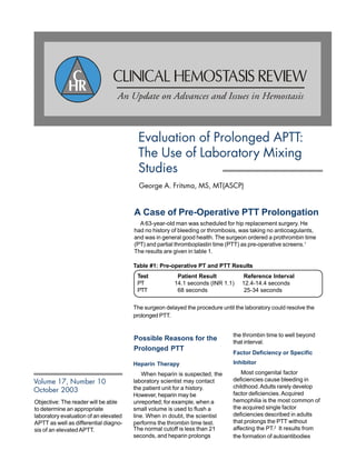
- 1. Objective: The reader will be able to determine an appropriate laboratory evaluation of an elevated APTT as well as differential diagno- sis of an elevatedAPTT. Evaluation of Prolonged APTT: The Use of Laboratory Mixing Studies George A. Fritsma, MS, MT(ASCP) Volume 17, Number 10 October 2003 A Case of Pre-Operative PTT Prolongation A 63-year-old man was scheduled for hip replacement surgery. He had no history of bleeding or thrombosis, was taking no anticoagulants, and was in general good health. The surgeon ordered a prothrombin time (PT) and partial thromboplastin time (PTT) as pre-operative screens.1 The results are given in table 1. The surgeon delayed the procedure until the laboratory could resolve the prolonged PTT. Possible Reasons for the Prolonged PTT Heparin Therapy When heparin is suspected, the laboratory scientist may contact the patient unit for a history. However, heparin may be unreported; for example, when a small volume is used to flush a line. When in doubt, the scientist performs the thrombin time test. Factor Deficiency or Specific Inhibitor Most congenital factor deficiencies cause bleeding in childhood.Adults rarely develop factor deficiencies.Acquired hemophilia is the most common of the acquired single factor deficiencies described in adults that prolongs the PTT without affecting the PT.2 It results from the formation of autoantibodies The normal cutoff is less than 21 seconds, and heparin prolongs the thrombin time to well beyond that interval. Table #1: Pre-operative PT and PTT Results Test Patient Result Reference Interval PT 14.1 seconds (INR 1.1) 12.4-14.4 seconds PTT 68 seconds 25-34 seconds
- 2. 2 CLINICAL HEMOSTASIS REVIEW / OCTOBER 2003 CLINICAL HEMOSTASIS REVIEW 3176 South Peoria Court Aurora, CO 80014 520.722.0797 chr@coagulation.com CLINICAL ADVISORS Dorothy M. Adcock, MD Alexander Duncan, MD, ChB H. James Day, MD, FACP Don W. Hill, MD, FACP CONTRIBUTOR Lynne Stevens, MT(ASCP) Clinical Hemostasis Review is published by Esoterix Coagulation, Inc. and is circu- lated to selected physicians and labora- torians. Copyright 2003. Esoterix Coagu- lation is part of the ESOTERIX, Inc. family of laboratories providing exoteric testing in numerous disease corridors. The opinions expressed in the articles are those of the author(s) and do not necessarily reflect the opinions or recommendations of the adver- tisers, editors, or publisher. The publisher reserves copyright and renewal on all pub- lished material and such material may not be reproduced in whole or in part without writ- ten permission from the publisher. Consult the full prescribing information on any drugs or devices discussed. All correspondence should be directed to the attention of the Editor, Clinical Hemo- stasis Review, 3176 South Peoria Court, Aurora, CO 80014. Subscription Rate: $65.00/year, 14 is- sues, prepaid. Outside the USA additional postage is required: Canada $20.00/year, all other destinations $50.00/year. Single copies $7.00. Subscriptions not accompa- nied by payment will be assessed a billing charge. To assure prompt delivery of your is- sues, please notify us of any address correc- tions. Six weeks are required to effect a change. Missing copies not received by mail will be provided free of charge if we are notified no later than two months after the issue date. After this deadline, we will charge subscribers $5.00 per issue. ISSN 0894-1025 specific for coagulation factor VIII. Anti-VIII autoantibodies may arise in pregnancy, autoimmune disorders, or in the elderly, causing severe, often life-threatening hemorrhage.3 Lupus Anticoagulant Lupus anticoagulants (LACs) are autoantibodies that react with phospholipid-bound proteins. LACs are present in 1-2% of unselected individuals.4 LACs cause prolongation of phospholipid- dependent laboratory assays, particularly the PTT, and may also be detected in immunoassay systems such as the anti-B-2 glycoprotein I, anti-prothrombin, or anti-cardiolipin antibody assay. Most LACs appear in response to inflammation, have no clinical effects, and disappear in 6-8 weeks. Chronic LACs, however, are associated with a risk of venous thrombosis, strokes, myocardial infarctions, peripheral artery occlusion, and recurring spontaneous abortions. LACs rarely cause bleeding. How to Decide: Mixing Studies Acute care laboratories must be equipped to perform PTT mixing studies. Mixing studies distinguish between factor deficiencies and the presence of inhibitors; also between specific inhibitors and LACs. The laboratory scientist prepares a 1:1 mixture of patient’s platelet-poor plasma and normal reagent platelet-poor plasma, and performs a PTT on the mixture. The normal reagent platelet-poor plasma may be purchased from a commercial distributor or prepared locally from a normal donor. Correction upon Immediate Testing If the mixture yields a PTT result within 10% of the normal reagent plasma’s PTT, the PTT is said to be “corrected.” In this instance, the patient may have a congenital factor deficiency. This is confirmed by performing a series of factor assays, first VIII, then IX, and then XI, the three most likely deficiencies. Most acute care hemostasis laboratories are equipped to measure these factors. Alternatively, the patient may have a temperature- and time-dependent anti-VIII inhibitor. The inhibitor may then be a warm reacting IgG antibody that requires one or two hour’s incubation at 37°C to be detected. If the incubated mixture does not correct, meaning if the PTT is at least 15% longer than the normal reagent plasma’s incubated PTT, anti-VIII is suspected. A low factor VIII activity level further suggests this possibility. The presence of a factor VIII inhibitor is confirmed by a Bethesda titer, usually performed by a specialty or reference hemostasis laboratory. No Correction Upon Immediate Testing If the PTT result fails to normalize or correct upon immediate testing, an inhibitor is present. The most likely possibility is LAC. Anti-VIII is still in the differential, as some VIII inhibitors can react immediately, however, anti-VIII is unlikely in the absence of bleeding. When chronic, LAC is associated with thrombosis.5 Lupus Anticoagulant Testing Many acute care facilities prefer to order LAC testing from specialty laboratories, as the diagnosis requires a complex series of reflexive assays and a pathologist’s interpretation. Two test systems are necessary, owing to the heterogeneity of LACs.6 The first uses a specially prepared PTT reagent that is sensitive to LAC. The second employs the dilute Russell’s viper venom time assay, also sensitive to LAC, even in instances where
- 3. 4 CLINICAL HEMOSTASIS REVIEW / OCTOBER 2003 the PTT system fails. Lack of correction in mixing studies of either abnormal test is presumptive evidence for LAC. These results are followed with neutralization studies using high phospholipid reagents. Correction by the neutralizing reagents confirms the presence of LAC. Lupus anticoagulants are part of a family of antibodies called anti- phospholipid antibodies. These may also be detected using a series of immunoassays: · Anticardiolipin IgG, IgM, or IgA antibody · Antiphosphatidyl serine IgG or IgM antibody · Anti-b2 glycoprotein I IgG or IgM antibody · Anti-prothrombin IgG or IgM antibody Like the selection of lupus anticoagulant assays, the selection of an anti-phospholipid antibody immunoassay is made in consultation with a specialty laboratory pathologist. Profiles consisting of several of these immunoassays are recommended. A Case of Pre-Operative PTT Prolongation: Conclusion Mixing studies were performed in follow-up to the prolonged PTT result as shown in Table 2. The thrombin time confirmed that the patient had received no heparin. The mixing study results indicate an inhibitor. LAC was first in the differential because the patient was experiencing no bleeding. The results from the reference laboratory confirmed the presence of LAC both in the PTT- and dRVVT-based assays. Surgery was successful and the patient was prescribed Coumadin® post-surgically to reduce the risk of thromboembolic disease.A PT and PTT performed after Coumadin® treatment was completed were normal, indicating that the LAC was transient. A Case of Post-Partum Hemorrhage A 31-year-old woman experienced vaginal bleeding, nosebleeds, and bruising one week after a normal delivery. She had no previous history of bleeding, no bleeding in her kindred, and was taking no anticoagulant drugs. Her gynecologist ordered a PT, PTT, and complete blood count. The results for the PT, PTT, and platelet count are shown in Table #3. As in the first case, the differential for this patient includes therapeutic heparin, factor deficiency, acquired factor inhibitor, or lupus anticoagulant. Disseminated intravascular coagulation and von Willebrand disease are further possibilities. Table #4: Expected results in DIC Table #3: PT, PTT, and platelet count results in a woman one week after delivery Table #2: Results of PTT Mixing Studies Disseminated intravascular coagulation Disseminated intravascular coagulation (DIC) is uncontrolled activation of the coagulation mechanism. DIC is the immediate cause of death in many cases of inflammation and infection, and the core hemostasis laboratory must be equipped to diagnose it and monitor its treatment.In DIC, platelets and coagulation factors are consumed, causing thrombocytopenia, prolonged PT and PTT, and reduced fibrinogen levels. Further, fibrinolysis becomes activated, producing a variety of peptides called fibrin degradation products. One such product is D-dimer. The laboratory must be equipped to measure D- dimer levels, either through the time-honored latex agglutination assay or the more recently available quantitative D-dimer methods. The recommended tests for DIC and their expected results are given in Table 4. Test Result Reference Interval Thrombin time 17.5 seconds < 21 seconds Patient PTT 68 seconds 25-34 seconds PTT reagent normal plasma 29 seconds Commercial platelet poor plasma 1:1 mixture 56 seconds No correction (> 15% above PTT reagent normal plasma, or > 33.4 seconds) Test Result Reference Interval PT 14.1 seconds (INR 1.1) 12.4-14.4 seconds PTT 82 seconds 25-34 seconds Platelet count 279 x 109 /L 140-450 x 109 /L Test Anticipated Value in DIC Platelet count < 150 x 109 /L D-dimer qualitative >1:2 D-dimer quantitative > 500 ng/mL Fibrinogen < 200 mg/dL PTT Prolonged
- 4. OCTOBER 2003 / CLINICAL HEMOSTASIS REVIEW 5 DIC treatment is monitored by serial platelet counts, D-dimer, and the fibrinogen assay. In the case of the 31 YO woman, DIC is unlikely as the PT and platelet count are normal. Nevertheless, because of the profound implications of DIC, a D-dimer and fibrinogen were performed as part of the follow-up laboratory assay. See table #5. Von Willebrand Disease Sometimes bleeding occurs when the PT and PTT are normal and the platelet count exceeds 50x109 /L. Von Willebrand disease (vWD), with a prevalence exceeding 0.1% worldwide, is the most likely explanation. Since von Willebrand factor is the carrier protein for factor VIII, low factor VIII activity, accompanied by a slightly prolonged PTT, may also point to vWD. When von Willebrand disease is suspected, the core laboratory should request a von Willebrand profile from a reference or specialty laboratory. In this case, the von Willebrand diagnosis is secondary, as the elevated estrogens in pregnancy raise the level of von Willebrand factor and reduce the bleeding symptoms. Further, because von Willebrand disease is inherited it is likely to appear with symptoms in childhood, and may be found in other members of the family. Nevertheless, a von Willebrand disease profile was ordered from a reference laboratory. The results demonstrated that the diagnosis was unlikely. A Case of Post-partum Hemorrhage: Conclusion Mixing studies were performed in follow-up to the prolonged PTT result as shown in Table # 5. The thrombin time confirmed that the patient had received no heparin. The normal fibrinogen and D-dimer levels rule out DIC. The mixing study results indicate an inhibitor. Factor VIII inhibitor was first in the differential because the patient was experiencing bleeding. The results of the incubated mixing study implied the presence of a warm- reacting antibody which was confirmed by the Bethesda titer. Acquired hemophilia is transient but often life-threatening. The patient was treated with an activated prothrombin complex concentrate, factor eight inhibitor bypassing activity (FEIBA®) and monitored with repeat factor VIII and Bethesda titers until the inhibitor could no longer be demonstrated. Conclusion and Further Recommendations • Mixing studies may be necessary any time there is a prolonged PTT in the absence of reported heparin therapy. • Mixing studies that include incubation are used to distinguish among single and multiple factor deficiencies, coagulation factor inhibitors, and lupus anticoagulant. • Mixing studies are effective when the patient plasma tested is platelet-poor plasma with a platelet count of less than 10 x 109 platelets per mL. To make platelet-poor plasma, centrifuge the specimen at 2500 x g for 10 minutes. Transfer the plasma with a plastic pipette into a plastic centrifuge tube, cap and centrifuge an additional 10 minutes at 2500 x g. Excessive numbers of plasma platelets will partially neutralize lupus anticoagulant in vitro, secrete excessive levels of stored coagulation factors, and partially neutralize heparin in vitro. The platelet poor plasma centrifugation method requires routine monitoring. • Mixing studies usually use equal portions of patient and normal platelet poor plasma. For increased sensitivity to weak inhibitors, a 4:1 ratio of patient to normal plasma may be used. Table #5: Final Laboratory workup of post-partum hemorrhage Test Result Reference Interval Thrombin time 18 seconds < 21 seconds Fibrinogen 324 mg/dL 226-467 mg/dL D-dimer quantitative 146 ng/mL < 240 ng/mL Patient PTT 82 seconds 25-34 seconds PTT reagent normal plasma 29 seconds Commercial platelet poor plasma 1:1 mixture 33 seconds Correction PTT reagent normal plasma 41 seconds Commercial platelet incubated 2 hours at 37ºC poor plasma 1:1 mixture incubated 67 seconds No correction 2 hours at 37ºC (> 47 seconds) Factor VIII < 1% Implies acquired hemophilia Bethesda titer 38 Bethesda units Autoimmune inhibitor to factor VIII
- 5. 6 CLINICAL HEMOSTASIS REVIEW / OCTOBER 2003 • Mixing studies may be employed any time the initial PTT result exceeds the upper limit of the reference interval.A mix is said to correct when the result of the mixture exceeds the result of the normal plasma by no more than 10%. A mix is uncorrected when the result exceeds the normal plasma result by more than 15%. Results in the 10-15% range are equivocal and require repeating. If a laboratory is getting a large number of equivocal results, it may be that the specimens are not sufficiently platelet poor. Separate cutoff limits are determined through empirical observation when a 4:1 or other mix is used. • Though less common, mixing studies may also be employed in the PT or thrombin time test system when appropriate. • All coagulation laboratories, large and small, should be equipped to perform mixing studies. References 1. Eisenberg J, Clarke JR, Sussman SA. Prothrombin and partial thromboplastin times as preoperative screening tests.Arch Surg 1982; 117:48. 2. Boggio LN, Green D.Acquired hemophilia. Rev Clin Exp Hematol 2001; 5: 889-404. 1185-1190. 3. Ludlam DA, Morrison AE, Kessler C. Treatment of acquired hemophilia. Semin Hematol 1994; 31 (Suppl 4) 16-19. 4. Bevers EM, et al. Lupus anticoagulant IgG’s are not directed to phospholipids only, but to a complex of lipid-bound human prothrombin. Thromb Haemostas 1991; 66: 629-632. 5. Goldsmith JC. Diagnosis of factor VIII versus nonspecific inhibitors. Semin Hematol 1993; 30 (Suppl 1): 3-6. 6. Brandt JT, et al. Criteria for the diagnosis of lupus anticoagulants: an update. Thromb Haemostas 1995; 74: 1185-1190.