SlideShare a Scribd company logo
1 of 19
Download to read offline
Internal Medicine
Harrison’s Principles of Internal Medicine
Rheumatology
Approach to Articular and MSK Disorders (Chapter 363)
 Rule out red flag diagnoses – suspected for acute onset, monoarticular
pattern or focal MSK pain
o Septic Arthritis
o Acute crystal-induced arthritis
o Fracture
o Vascular Ischemia
o Carpal Tunnel Syndrome
 Important questions:
o Articular or non-articular
o Acute (<6 weeks) or chronic (>6 weeks)
o Inflammatory or non-inflammatory
o Localized or widespread in distribution
Articular Non-articular
Deep or diffuse pain
Limited range of motion on active and
passive movement
Swelling – due to synovial proliferation,
effusion, bony enlargement
Crepitation
Instability
Locking
Deformity
Painful on active movement only
Site of pain is usually periarticular, or in
regions adjacent to the articular surfaces
(-) swelling
(-) crepitation
(-) instability
(-) deformity
Inflammatory Non-inflammatory
Erythema, pain, swelling, warmth
Systemic signs – fatigue, fever, rash,
weight loss
(+) ESR, CRP, thrombocytosis, anemia
of chronic disease, hypoalbuminemia
(+) prolonged morning stiffness
improving with activity
(+) gel phenomenon in OA only
Related to trauma
Repetitive use
Degeneration or ineffective repair
(+) intermittent gel phenomenon
Clinical History
Younger age group SLE, reactive arthritis
Middle age group Fibromyalgia
RA
Elderly age group OA
Polymyalgia rheumatica
Men Gout
Spondyloarthritis
Ankylosing spondylitis
Women RA
Fibromyalgia
Osteoporosis
SLE
African American Sarcoidosis
SLE
Whites Polymyalgia rheumatic
Giant cell arteritis
Granulomatosis with polyangiitis
Familial aggregation Ankylosing spondylitis
Gout
Heberdon nodes of OA
Onset
Abrupt onset Septic arthritis,Gout
Indolent onset OA, RA, fibromyalgia
Evolution
Chronic OA
Intermittent Crystal-induced, Lyme arthritis
Migratory RF, gonococcal, viral arthritis
Additive RA, Psoriatic arthritis
Evolution
Chronic (>6 weeks) OA, RA, fibromyalgia
Acute (<6 weeks) Infectious, crystal-induced,
reactive
Extent or Distribution
Mono articular (1 joint) Crystal-induced, infectious
Oligoarticular (2-3 joints)
Poly articular (4 or more joints) OA, RA (symmetric as well)
Asymmetric and polyarticular Spondyloarthritis, reactive arthritis,
gout, sarcoid
Symmetric or asymmetric
Polyarticular or oligoarticular
OA, psoriatic arthritis
Upper extremities RA, OA
Lower extremities Gout, reactive arthritis
Axial skeleton OA, ankylosing spondylitis
RA of the cervical spine
Diseases with MSK co-morbidities
Diabetes mellitus Carpal tunnel syndrome
Renal insufficiency Gout
Depression or insomnia Fibromyalgia
Myeloma Low back pain
Cancer Myositis
Osteoporosis Fracture
Glucocorticoids Osteonecrosis
Septic arthritis
Diuretics or chemotherapy Gout
Rheumatic Review of Systems
Fever SLE, infection
Rash SLE, psoriatic arthritis
Nail abnormalities Psoriatic arthritis, reactive arthritis
Myalgias Fibromyalgia, statin-induced,
drug-induced
Weakness Polymyositis, neuropathy
Eye involvement Behcet, sarcoidosis,
Spondyloarthritis
Gastrointestinal Scleroderma, IBD
Genitourinary Reactive arthritis, gonococcemia
Nervous system Lyme disease, vasculitis
Osteoarthritis (Chapter 364)
 Most common type of arthritis
 Joints commonly affected by arthritis:
Definition
 Defined as joint failure → hyaline articular cartilage loss present in a focal
and initially nonuniform manner → thickened subchondral bony plate,
outgrowth of osteophytes at the joint margin, stretching of the articular
capsule, snynovitis, weakness of the muscles bridging the joint
Joint Protective Mechanisms and their Failure
 Joint protectors – joint capsule, ligaments, muscles and tendons, afferent
sensory nerves, underlying bone
 Charcot arthropathy – severe and rapidly progressive OA developing when
minor injury occurs in the presence of posterior column peripheral
neuropathy
Joint capsule and ligaments Fixes range of motion
Synovial fluid lubrication Hyaluronic acid, lubricin
Bridges the joint Muscles and tendons
Cartilage and its Role in Joint Failure
 Two most important macromolecules in cartilage:
Type 2 collagen Provides cartilage tensile strength
Aggrecan Proteoglycan macromolecule linked with hyaluronic acid
molecule, with highly negatively charged glycosaminoglycans
Through electropulsion → gives cartilage compressive
stiffness
 OA cartilage
o (+) gradual depletion of aggrecan
o (+) unfurling of the tightly woven collagen matrix
o (+) loss of type 2 collagen
Risk Factors
 2 major factors contributing to OA development:
o Joint vulnerability
o Joint loading
Systemic Risk Factors
Age Most potent risk factor for osteoarthritis
 Rare in <40 yearsold
 >50% in >70 years old
Aging increases joint vulnerability – less responsive in matrix
synthesis, thinner cartilage with age, weaker bridging
muscles, slower nerve conduction, stretching of ligaments
Older women Especially in their sixth decade
Probably hormonal loss
Heritability and
genetics
Highly heritable disease, but joint specific
(+) polymorphism with GDF5 gene
Race Knee OA is more common among Chinese
Risk Factors in the Joint Environment
Joint anatomy 3 uncommon developmental abnormalities occurring in utero or in
childhood:
 Congenital dysplasia
 Legg-Perthes disease
 Slipped capital femoral epiphysis
Others: acetabular dysplasia, femoroacetabular impingement
Major injuries Fracture through the surface
Avascular necrosis
Tears of ligamentous and fibrocartilaginous structures
Meniscal tears
Joint misalignment
 Varus knees – cartilage loss in the medial or inner knee
 Valgus knees – cartilage loss in the lateral compartment
Weakness of the quadriceps – knee OA
Loading Factors
Obesity 3-6 times body weight is transmitted across the knee during a
single-leg stance
Well-recognized and potent risk factor for OA (esp. knee)
Precedes the development of disease
More potent in women
Adipokines may also be contributory
Repeated Use
of Joint and
Exercise
Seen in occupational use and leisure time physical activities
 Farmers – hip OA
 Miners – knee and spine OA
 Heavy carrying – knee OA
 Elite runners – knee and hip OA
 Recreational runners – hip OA
Pathology
 Non-uniform loss in cartilage in a focal disease
 Thickening of the subchondral bony plate
 Osteophyte formation – outgrowth of new cartilage that ossifies
o Important radiographic hallmark of OA
 Stretching of the capsule → edematous and fibrotic
Sources of Pain
 Likely occurs from structures outside the cartilage (aneural):
o Synovium, ligaments, joint capsule, muscles, subchondral
bone
 Severity of X-ray changes correlate poorly with pain severity
 Presence of MRI synovitis correlates with presence and severity of
knee pain
Clinical Features
 Joint pain
o During or just after joint use and then gradually resolves
o As disease progresses, pain becomes continuous and even
becomes bothersome at night
 Morning stiffness
o Usually brief (<30 minutes)
 Knee symptoms
o Most common cause of chronic knee pain in patients >45 years
o (+) knee buckling – from weakness of muscles crossing over
the joint
Buckling, catching, locking Anterior crucial ligament or meniscal tear
Needs to be evaluated after a knee injury
Pain during knee flexion (>35
degrees)
From the patellofemoral compartment
Prolonged morning stiffness
with many joints affected
Inflammatory arthritis
Pain medial and distal to the
knee
Anserine bursitis
Common cause of knee pain, responsive to
glucocorticoid injection
Prominent nocturnal pain in the
absence of end-stage OA
Merits diagnostic work-up
 Hip symptoms
o Can be detected by loss of internal rotation by passive
movement, pain isolated to an area lateral to the hip joint
(trochanteric bursitis)
Diagnostics
 No blood tests are routinely indicated for workup of patients with OA, unless
symptoms and signs suggest inflammatory arthritis
Examination of synovial fluid >1000 WBC/uL – presence of inflammatory
arthritis or crystal-induced arthritis (with presence
of crystals)
X-rays Indicated when joint pain and physical findings are
atypical of OA, or if pain persists after initiation of
pain treatment
Radiographs may be normal in early disease
as they are insensitive to cartilage loss and
early findings
MRI Not indicated as part of diagnostic workup
Almost never warrant change in therapy
Goal of Treatment
 Goal of treatment: To alleviate pain and minimize loss of physical function
 Most effective mode of treatment: nonpharmacological treatment
Non-pharmacological Treatment
 Goals of treatment:
o Avoiding painful activities – simplest treatment
o Improving strength and conditioning of muscles bridging the joints
o Unloading the joint via use of braces or splint to redistribute load or
unloading joint during weight bearing with a cane or crutch
 Exercise
o Inactivity leads to increased risk for CVD and obesity
o Weakness in muscles have multiple etiologies:
 Decline in strength with age
 Disuse muscle atrophy due to limitation of movement
 Diminishing muscle use due to alteration of gait
 Due to arthrogenous inhibition
o Weakness in a muscle makes joint more susceptible to further
damage and pain
o Exercise lessens pain and improves physical function
o Major challenge – adherence to program
o Strongest predictor of success – personal history of successful
exercise
Effective Exercise Non-effective Exercise
Aerobic exercises
Resistance training
Low-impact exercises (water
aerobics)
Tai chi – effective for knee OA
Exercise with caloric restriction
Range-of-motion exercises
Isometric exercises
 Correction of misalignment
o Via bracing for willing patients who can learn to put them on
correctly and on whom they do not slip
o Acupuncture
o Surgery
Pharmacological Treatment
 Serves as an important adjunct therapy for OA
Acetaminophen
(Paracetamol)
Up to 1 gram
TID
Initial analgesic of choice for patients with OA
Prolongs half-life of warfarin
NSAIDs Most popular drugs to treat osteoarthritic pain
Either topically or orally
30% greater pain alleviation than high-dose paracetamol
Important to take low-dose aspirin and iburprofen/naproxen at
different times to limit drug interaction – may increase bleeding
and GI complaints
Side effects: GI complaints (should be taken after meals)
If with high-risk, take PPIs
Others: edema, renal insufficiency (used with caution for CKD III)
NSAIDS with increased CVD risk – diclofenac, rofecoxib
Allowed NSAIDs – Naproxen (with high GI toxicity), salsalate,
ibuprofen, celecoxib
Topicals – lesser efficacy, less GI and systemic side effects but
may cause skin irritation
Intraarticular
injections –
glucocorticoids
May be used temporarily (less than 3 months) for pain alleviation
since synovitis is correlated with joint pain severity
Subsequent injections may cause minor amounts of cartilage
loss with unimportant clinical consequences
Hyaluronic acid
injections
Can be given as treatment but with controversy on efficacy over
placebo
Opioid
analgesics
Has modest efficacy
Duloxetine Has modest efficacy
Glucosamine
Chondroitin
Recommended against OA
Surgery
Arthroscopic
menisectomy
Indicated for acute meniscal tears in which symptoms such as
locking and acute pain are clearly related temporally to a knee
injury that produced the tear
High tibial
osteotomy
For patients with knee OA to the medial compartment
Total knee or
hip
arthroplasty
Highly efficacious operations that relieve pain and improve
function in majority of patients
Failure rates of 1% per year (higher in obese patients)
Success rates higher in hips > knees
Timing of knee or hip replacement is critical
Cartilage
regeneration,
abrasion
arthroplasty
(chondroplasty)
Not found to be efficacious in OA
May be efficacious when OA has not yet developed
Ineffective surgical plans
 Arthroscopic debridement and lavage
 Partial menisectomy
Gout and Other Crystal-induced Arthropathies (Chapter 365)
Gout
 Metabolic diseases mostly affecting
o Middle-aged to elderly men
o Postmenopausal women
 Due to increased body pool of urate with hyperuricemia → deposition of
MSU crystals in joints and CT tophi with risk for deposition in the
kidney interstitium or uric acid nephrolithiasis
Clinical Features
Involved
joints
MTP1, tarsal joints, ankles, knees, finger joints (usually in the
elderly)
First
episode
Begins at night with dramatic joint pain and swelling – mimicking
cellulitis
Precipitating
events
Dietary excess, trauma, surgery, excessive ethanol ingestion,
hypouricemic therapy, serious medical illnesses – MI and stroke
Presentation
in women
Represents 5-20% of the population
(+) osteoarthritis and arterial hypertension → mild renal
insufficiency (+) receiving diuretics
Laboratory Diagnosis
Needle aspiration of
acutely or chronically
involved joints or
tophaceous deposits
Confirms presumptive diagnosis
(+) negative birefrigent needle-shaped crystals in
compensated polarized light intracellularly and
extracellularly
Synovial fluid
leukocyte counts
More than 2,000 to 60,000/uL
Culture If suspected to have septic arthritis
Serum uric levels Can be normal or low at the time of an acute attack –
inflammatory cytokines can be uricosuric and effective
initiation of hypouricemic therapy can precipitate attacks
Serum urate levels Almost always elevated at some time
Important to use follow the course of hypouricemic
therapy
24-h uric acid Useful in assessing risk of stones, elucidating
overproducers or underexcretors of uric acid – to decide
if uricosuric therapy is appropriate
Excretion of >800 mg uric acid per day on regular diet –
suggests overproduction of purine
Other tests
Urinalysis
Serum creatinine
Hemoglobin
WBC count
Liver function tests
Serum lipids
To assess possible pathologic sequelae of gout and other
associated diseases requiring treatment and as baselines
because of possible adverse effects of gout treatment
Radiographic features
X-ray (+) cystic changes, well-defined erosions with sclerotic
margins (with overhanging bony edges)
(+) soft tissue masses – advanced chronic tophaceous
gout
Ultrasound (+) double contour sign overlying articular cartilage
Dual-energy CT (+) presence of urate crystals
Treatment
Acute Gouty Arthritis
 NSAIDs and colchicine – may be poorly tolerated and dangerous in the
elderly, especially in the presence of renal insufficiency and GI disorders
 Ice pack applications and rest of involved joints can be helpful
Oral colchicine Traditional and effective treatment if used early in an attack
Regimens: 0.6 mg given every 8 hours with subsequent tapering
1.2 mg followed by 0.6 mg in 1 hour with subsequent day dosing
depending on response
Temporarily discontinued promptly at the first sign of loose
stools, and symptomatic treatment of diarrhea must be given
Oral NSAIDs Effective in 90% of patients
With resolution of signs and symptoms after 5-8 days
Most effective are those with short-half-lives:
Celecoxib (800 mg – 400 mg 12 hour after, then 400 mg BID)
Diclofenac (50 mg TID)
Ibuprofen (800 mg TID)
Naproxen (500 mg BID)
Indomethacin (25-50 mg TID)
Intramuscular or
oral
glucocorticoids
Oral: prednisone 30-50 mg/day as initial dose → gradually
tapered with resolution of attack
May be effective in polyarticular gout
Intraarticular: triamcinolone acetonide 20-40 mg, or
methylprednisolone 25-50 mg
Anakinra IL-1 antagonist
Has an essential role on the inflammasome
Used when other treatments have failed or contraindicated
Hypouricemic Therapy
 Ultimate control – correction of hyperuricemial
 Normalized serum uric acid: <300-360 umol/L (5.0 – 6.0 mg/dL)
o To prevent recurrent gouty attacks and eliminate tophaceous
deposits
 Considered when hyperuricemia cannot be corrected by:
o Control of body weight
o Low-purine diet
o Increase in liquid intake
o Limitation of ethanol use
o Decreased use of fructose-containing foods and beverages
o Avoidance of diuretics
 Indications
o Increased number of acute gouty attacks
o Serum uric acid of >535 umol/L (>9.0 mg/dL)
o Willingness to commit to lifelong therapy
o Presence of uric acid stones
o Presence of tophi or chronic gouty arthritis
 Not initiated during acute attacks, but after patient is stable
 Low-dose colchicine has been initiated to decrease risk of flares
o Colchicine prophylaxis in doses of 0.6 mg one to two timed
daily – should be given along with Hypouricemic therapy until
patient is normouricemic and without gouty attacks for 6 months, or
as long as tophi are present
o Not used in patients in dialysis, given in lower doses to
patients with renal disease or with P glycoprotein or CYP3A4
inhibitors (clarithromycin)
Probenecid Uricosuric agent used in patients with good renal function
who underexcrete uric acid <600 mg/day
Urine volume is maintained by ingestion of 1500 mL water/day
Regimen
250 mg BID → gradually to 3 g per day
Not effective in those with serum creatinine levels of >2 mg/dL
- May require allopurinol or benzbromarone
Benzbromarone Uricosuric agent more effective for patients with CKD
Lesinurad New uricosuric agent that can be used as an adjuvant at 200
mg/day to a xanthise oxidase inhibitor
Others with mild
uricosuric
Losartan, amlodipine, fenofibrate
effects
Allopurinol Xanthine oxidase inhibitor
Most commonly used Hypouricemic agent
Best drug to lower serum urate in overproducers, urate stone
formers, patients with renal disease
Regimen
Single morning dose of 100 mg (up to 800 mg as needed)
Adjusted initial dose depending on creatinine clearance (10
mL/min = 100 mg)
Toxicity among:
(+) penicillin and ampicillin allergies
(+) thiazide diuretic users
Asians with HLA-B*58:01
Serious side effects: TEN, systemic vasculitis, bone marrow
suppression, granulomatous hepatitis, renal failure
With mild cutaneous reactions
Can shift to uricosuric agent, desentiziation to allopurinol, use of
febuxostat
Febuxostat Xanthine oxidase inhibitor (40-80 mg OD)
Does not require dose adjustments with mild-moderate renal
disease
Pegloticase Pegylated uricase
Available for patients who do not tolerate or fail full doses of
other treatment
IV 8mg every 2 weeks can dramatically lower serum uric acid to
up to 50%
Other Cyrstal-induced arthritis
Calcium
pyrophosphate
deposition
(CPPD)
disease
Definitive Diagnosis
(+) rhomboid or rodlike, weakly positive or nonbirefrigent with
polarized light
Others: (+) linear calcifications
Calcium
apatite
deposition
disease
(basic calcium
phosphate
disease)
Definitive Diagnosis:
(+) seen in EM – 1-20 um shiny intra or extracellular
nonbirefrigent globules or aggregates
(+) purplish in Wright’s stain
(+) bright red in alizarin red S.
Others: (+) intra or periarticular calcifications with or without
erosive, destructive, or hypertrophic changes
Calcium
oxalate
disease
Definitive Diagnosis:
(+) bipyramidal and small polymorphic crystals in light
microscopy
Primary oxalosis – rare metabolic disease
Secondary oxalosis – mainly due to chronic renal failure, more
common than primary
Rheumatoid Arthritis (Chapter 351)
 Most common form of chronic inflammatory arthritis
Clinical Features
 Increases between 25 and 55 years of age, plateaus until 75 years and
declines after
 Undifferentiated inflammatory arthritis – too few affected joints to be
classified as RA; if diagnosed later as RA:
o Higher number of tender and swollen joints
o Tests positive for RF and anti-CCP antibodies
o Higher scores for physical disability
Early morning stiffness Lasting more than 1 year, eases with physical activity
Earliest involved joints Small joints of the hands and feet:
Wrists, MCP, PIP joints
DIP joint involvement Manifestation of coexistent osteoarthritis
Flexor tendon
tenosynovitis
Frequent hallmark of RA → decreased range of
motion → reduced grip strength and trigger fingers
Ulnar deviation Subluxation of the MCP joints with subluxation of the
proximal phalanx to the volar side of the hand
Swan-neck deformity Hyperextension of the PIP
Flexion of the DIP
Boutonniere deformity Flexion of the PIP
Hyperextension of the DIP
Z-Line deformity Subluxation of the MCP1
Hyperextension of the IP1
Piano-key movement of
the ulnar styloid
Subluxation of the distal ulna – because of
inflammation of the ulnar styloid and tenosynovitis of
the extensor carpi ulnaris
Flat feet (pes planovalgus) Chronic inflammation of the ankle and midtarsal region
Atlantoaxial involvement May cause compressive myelopathy and neurologic
dysfunction
Rarely affects thoracic and lumbar spine
TMJ involvement Not associated with significant symptoms
Extraarticular Manifestations
 May develop in 40% of patients – even before onset of RA
 Most likely develop among patients:
o History of cigarette smoking
o Early onset of significant physical disability
o (+) RF and anti-CCP antibodies
Constitutional
symptoms
Weight loss, fatigue, malaise, depression, cachexia
Presence of fever >38.3 C – suspect for Vasculitis or
infection
Subcutaneous
nodules
Firm non-tender, adherent to the periosteum, tendonds or
bursae
More common (30-40%) in those with highest levels of disease
activity, disease-related shared epitope (SE), (+) RF,
radiographic evidence of joint erosions
Locations: forearm, sacral prominences, Achilles tendon
Others: lungs, pleura, pericardium, peritoneum
Sjogren
syndrome (10%)
(+) keratoconjunctivitis sicca (dry eyes)
(+) xerostomia (dry mouth)
Pulmonary
manifestations
Most common pulmonary manifestation - pleuritis
(+) pleuritic chest pain, dyspnea
(+) pleural friction rub, effusion (exudative)
Insterstitial lung disease – with high disease acitivity, smokers
– diagnosed via CT scan
 Usual interstitial pneumonia (UIP), non-specific
interstitial pneumonia (NSIP) – main histological and
radiologic patterns of ILD
 Poor prognosis (10% risk for mortality)
 Favors more than idiopathic ILD
 More responsive to immunosuppressive therapy
Caplan syndrome
(+) pulmonary nodulosis
(+) pneumoconiosis
(+) silica exposure
(+) rheumatoid arthritis
Cardiac Pericardium – most common site of involvement
MR – most common valvular abnormality in RA
Vasculitis Mostly seen in the following:
- Long-standing disease
- Positive for RF, anti-CCP
- Hypocomplementemia
Sensory polyneuropathies (Mononeuritis multiplex) may occur
Hematologic Normochromic, normocytic anemia – MC hematologic
abnormality
Felty’s syndrome (occurs in late stages of RA)
Neutropenia, splenomegaly, nodular RA
T-cell LGL
May have similar presentation with Felty
May occur early in disease
Lymphoma DLBCL – most common lymphoma
Associated Conditions
 CVD – most common cause of death in RA
o Due to chronic inflammation
 Osteoporosis – more common in RA patients
 Hypoandrogenisms
Epidemiology
TNF-alpha Major cytokine in RA
HLA-DRB1 HLA associations
PTPN22 Non-MHC gene with strong risk for RA
Smoking Most reproducible environmental risk factor for RA
Porphyromonas gingivalis
EBV
Associated infections with RA
Pathology
Pathologic hallmarks Synovial inflammation and proliferation (pannus)
Focal bone erosions
Thinning of articular cartilage
Most common inflammatory
infiltrate
T cells – driving chronic inflammatory response
Diagnosis
 Clinical diagnosis of RA is largely based on the following:
o Clinical presentation of chronic inflammatory arthritis
o Laboratory and radiographic results
Anti-CCP (ACPA) Has greater specificity for RA diagnosis than RF
Laboratory Features
Serum IgM RF Found in 75-80% of patients with RA
Also found in:
Primary Sjogren, SLE, type II mixed essential cryoglobulinemia
Subacute BE, hepatitis B and C
1-5% in healthy population
ACPA Same sensitivity with IgM RG
95% specificity (more useful in diagnosis)
Most predictive for predicting worse outcomes
SF analysis WBC count – 5,000-50,000 WBC/uL
(vs <2,000 in osteoarthritis)
Predominance of neutrophil
Purpose
Most useful in confirming an inflammatory athritis while
excluding infection or crystal-induced arthritis
Joint imaging Purpose
not only for diagnosis, but also for tracking progression of joint
damage
MRI, UTZ – preferred diagnostic modalities
X-ray Periarticular osteopenia – initial radiographic finding
Other findings:
ST swelling, symmetric joint space loss, subchondral erosions
(MCPs, PIPs, MTPs – lateral aspect of 5
th
MTP – most
common target)
MRI Offers greatest sensitivity for detecting Synovitis and joint
effusions, and early bone and bone marrow changes
UTZ Can detect more lesions than X-ray but operator-dependent
Clinical Course
 Natural history is mainly affected by:
o Age of onset, gender, genotype, phenotype, comorbid conditions
10% patients Will undergo spontaneous remission within 6 months
Especially Seronegative patients
IHD Most common cause of death
Shortening of
median life
expectancy
7 years less in men
3 year less in women
Risk factors
With systemic EA involvement, low functional capacity, low
socioeconomic status, low education, chronic prednisone use
Joint inflammation Main driver of joint damage, most important cause of functional
disability in earlier stages
Treatment of RA
 Development trends in management of RA
(1) Emergence of methotrexate as DMARD of first choice for early RA
(2) Development of novel biologicals that can be used alone or in
combination with methotrexate
(3) Proven superiority of combination DMARD regimens over methotrexate
alone
 Medications used in RA
o NSAIDS
o Glucocorticoids
o Conventional DMARDs
o Biological DMARDs
NSAIDs
 Presently considered as adjunctive agents for management of uncontrolled
symptoms by other measures
 Toxicities – gastritis, PUD
Glucocorticoids
 May be used in low-to-moderate doses to achieve rapid disease control
before onset of fully effective DMARD therapy
 1-2 week bursts of steroids for management of acute disease flares
 Chronic administration of prednisone – to control disease activity in patients
with inadequate response to DMARD therapy
Low-dose prednisone Can retard progression of joint disease
Must be balanced with side effects
Intraarticular injection of
intermediate steroid
(triamcinolone acetonide)
For rapid control of inflammation in a limited number
of affected joints
Must ensure that inflammation is not infectious
Osteoporosis Important long-term complication of steroids
Peptic ulcer No published guidelines for GI protection
Conventional DMARDs
 Hydroxychloroquine, sulfasalazine, methotrexate, Leflunomide
 Has delayed onset of action – 6-12 weeks
Methotrexate DMARD of choice for RA treatment
MOA – stimulates adenosine release → anti-inflammation
Leflunomide Inhibitor of pyrimidine synthesis
Similar clinical efficacy as methotrexate
Hydroxychloroquine Not a true DMARD; no retardation in RA progression
Sulfasalazine Has shown to retard RA progression Radiographically
Others Not used anymore
Minocycline, gold salts, penicillinamine, azathioprine, cylclosporine
Biological DMARDs
 Anti-TNF – first approved biological DMARDs for RA
Anti-TNF Infliximab – chimeric (mouse-human) monoclonal antibody
Adalimumab, golimumab – human monoclonal antibody
Certolizumab pegol – pegylated Fc-fragment of human monoclonal
antibody
Etanercept – soluble fusion protein (TNF receptor 2 in convalent
linkage with Fc portion of IgG1)
Can be used in combination therapy or monotherapy (latter 4)
Contraindication
Active infection
History of hypersensitivity with these agents
Chronic hepatitis B infection
Class III/IV CHF
Screening for latent TB is important (PPD, IFN-gamma)
Anakinra Recombinant form of naturally occurring IL-1 receptor antagonist
Has limited use in RA
Indications (for syndromes dependent on IL-1 production)
Neonatal-onset inflammatory disease
Muckle-Wells syndrome
Familial cold urticaria
Systemic juvenile-onset inflammatory arthritis
Adult-onset Still’s disease
Abatacept Soluble fusion protein consisting of extracellular domain of human
CTLA4 linked to modified human IgG portion
MOA – inhibits co-stimulation of T cells by blocking CD28-
CD80/CD86 inreractions
Can be used in combination therapy
Rituximab Chimeric monoclonal antibody against CD20
Indications
Refractory RA in combination with methotrexate
More effect on seropositive patients
Adverse effects
Increased risk for infection
PML
Tocilizumab Humanized monclonal antinody against IL-6
Adverse effects
Increased infection, increased LDL cholesterol
Small molecule inhibitors
 For RA resistant to conventional and biologic DMARDs
Tofacitinib Small-molecule inhibitor of JAK1 and JAK3
Adverse effects
Elevated liver enzymes, neutropenia, increased cholesterol, elevated
serum creatinine
Treatment of Extraarticular Manifestations
RA-ILD Challenging, some DMARDs (methotrexate, Leflunomide) can cause
pulmonary toxicity
Treatment
High-dose steroids + adjunctive immunosuppressants (azathioprine,
Mycophenolate mofetil, rituximab)
Management Considerations
Pregnancy
 Up to 75% of female RA patients – overall improvement during pregnancy,
but will flare after delivery
Flares during pregnancy Low-dose prednisone
Hydroxychloroquine
Sulfasalazine
Safest DMARDs to use during pregnancy
Elderly
 Conventional and biological DMARDs – equally effective and safe in younger
and older population
Methotrexate May need renal adjustments
Not prescribed when serum creatinine >2 mg/dL
SLE (Chapter 349)
 90% are women of child-bearing age
HLA
associations
HLA-DRB1
HLA-DR3
HLA-DQA2
HLA-CR2
HLA-FCGR2A/B
Genes for
lupus
nephritis
HLA-DR3, STAT4, APOL1 (African Americans)
FCGR2A, ITGAM, IRF5, IRF7, TNFSF4 (Ox40L), DNAse1
Pathology
Skin biopsy Deposition of Ig at dermal-epidermal junction (DEJ) (also in normal
skin)
Injury to basal keratinocytes
Inflammation by T cells in the DEJ and around blood vessels
Renal
biopsy
Pattern and severity of injury are important for diagnosis and selecting
best therapy
Requires aggressive immunosupression (steroids + another
drug)
Class III
Class IV
Class V accompanied by III and IV
Not required
Class I and II, or with extensive irreversible changes
Blood vessel
pathology
Leukocytoclastic vasculitis – most common pattern of vasculitis
Lymph
biopsy
Usually performed to rule out infection or malignancies
Nonspecific diffuse chronic inflmmation
Diagnosis
 Based on characteristic clinical features and autoantibodies
Classification of Nephritis (International Society of Nephrology and Renal Pathology
Society
Class I Minimal mesangial LN
Class II Mesangial proliferative LN
Class III Focal LN
Class IV Diffuse LN
Class V Membranous LN
Class VI Advanced Sclerotic LN
Manifestations
MSK Intermittent polyarthritis in hands, wrists, knees – most common
Joint pain – most common reason to increase dose of steroids
Ischemic necrosis of bone – diagnosed for persistence of pain in
single joint (treated with systemic steroids)
Myositis – usually drug-induced (steroids > antimalarials)
“rhupus” – combined SLE and RA presentation
Cutaneous Discoid lupus erythematosus (20%) – most common chronic
dermatitis in lupus
(+) roughly circular with slightly raised, scaly Hyperpigmented
erythematous rims
(+) depigmented atrophic centers (where all dermal appendages are
destroyed)
Treatment – topical or locally injected steroids + systemic
antimalarials
Photosensitive, slightly raised erythema, scaly on face – most
common acute SLE rash; “butterfly rash”
Subacute CLE – similar to psoriasis
(+) Ro (SS-A) antibodies
Renal Nephritis – most serious manifestation of SLE
Class IV – most severe, and most severe type of LN
DPGN – ESRD within 2 years (if untreated)
Can lead to atherosclerosis
CNS Cognitive dysfunction (memory difficulties, reasoning) - Most common
manifestation of diffuse CNS lupus
Seizures
Treatment – both antiseizure and immunosupression
Psychosis
Can be the predominant form of SLE
Must be distinguished from glucocorticoid-induced psychosis
Myelopathy
Tx: high-dose steroids
Pulmonary Pleuritis with or without pleural effusion – most common pulmonary
manifestation
Tx: NSAIDS → steroids if more severe
Life-threatening conditions
Interstitial fibrosis
Shrinking lung syndrome
Intraalveolar hemorrhage
Cardiac Pericarditis – most common cardiac manifestation
More serious manifestations – myocarditis, Libman-Sacks (MR, AR)
Vascular
occlusions
Cerebral event results from clotting
Tx: long-term anticoagulation if treatment of choice
Higher SLE MI events in women <49 years
Risk factors for atherosclerosis
Male, older, hypertension, dyslipidemia, diabetes, dysfunctional pro
inflammatory HDL, repeated high scores for disease activity, high
cumulative or daily doses of steroids, high levels of homocysteine and
leptin
Hematologic Normochromic, normocytic anemia – most common manifestation
Leukopenia (lymphopenia)
Thrombocytopenia
GI Vomiting, diarrhea, diffuse abdominal pain (autoimmune peritonitis,
and/or intestinal vasculitis), elevated liver enzymes
Tx: aggressive immunosuppression with high-dose steroids for short-
term control
Ocular Sicca syndrome, nonspecific conjunctivitis – common in SLE, rarely
threaten vision
Retinal vasculitis, optic neuritis – serious manifestations and blindness
can develop over days to weeks
Glaucoma, cataract – complications of steroids
Laboratory Tests
(1) To establish or rule out diagnosis
(2) Follow the course of disease
(3) Identify adverse effects of therapies
Autoantibody Testing
 ANA – most important autoantibodies to detect (positive in >95% patients)
Anti-Ro/SS-A Indicates increased risk for neonatal lupus, sicca syndrome, and
SCLE
Standard Tests for Diagnosis
 Screening using the following:
o CBC, PC, urinalysis
o May detect abnormalities contributing to diagnosis and
influence management decisions
Tests for Following Disease Course
UA, hemoglobin levels, PC,
serum creatinine, albumin
For hematuria, proteinuria
Identification of markers for
disease activity
See Table
Management of SLE
 Aim is to achieve low-level disease activity
o Mild symptoms on the lowest possible doses of medications
o Usually achieved in 30-50% of SLE patients within a year
Conservative Therapies for Management of Non-Life-Threatening Disease
 For patients with SLE autoantibodies, fatigue and pain without organ
involvement
Goal Management via suppression of symptoms
Anagesics, antimalarialas are mainstay
First issue with NSAIDs Increased risk for NSAID-induced aseptic
meningitis, elevated serum transaminases,
hypertension, renal dysfunction
Second issue with NSAIDs All NSAIDs (particularly COX-2 inhibitors), increases
MI risk
Issue with hydroxychloroquine Must have annual eye examination for potential
retinal toxicity
At least 750 ng/mL – blood level needed for optimal
response in active SLE
NSAIDs Arthritis/arthralgias
Antimalarials –
hydroxychloroquine,
chloroquine, quinacrine
Dermatitis
Arthritis
Fatigue
DHEA May reduce disease activity
Belimumab For fatigue, rash, and/or arthritis
Mostly effective with robust clinical activity:
SLEDA ≥10
Positive anti-DNA
Low serum complement
Topical sunscreens
Antimalarials
Topical steroids and/or
Tacrolimus
Lupus dermatitis
(systemic steroid with or without Mycophenolate
mofetil, azathioprine, belimumab – sever or
unresponse LD)
SLEDAI Widely used measure of SLE disease activitiy
>3 Clinically active disease
Life-threatening Management of SLE
Systemic Steroids
(0.5-1 mg/kg PO daily,
500-1000 mg IV daily)
Methylprednisolone sodium
succinate for 3 days → daily
(0.5-1 mg/kg) prednisone
Mainstay treatment for any inflammatory life-
threatening or organ-threatening manifestations of
SLE
Management of LN
(induction therapy)
Steroids +
Cylocphosphamide or mycophenolate mofetil
(azathioprine is effective, but associated with more
flares)
 African Americans, hispanics –
mycophenolate
 Whites, Asians – cyclophosphamide or
mycophenolate
Mycophenolate toxicity diarrhea
Cyclophosphamide toxicity Amenorrhea, leukopenia, nausea
Responses to treatment Steroid – within 24 hours
Cyclophosphamide, mycophenolate – 3-16 weeks
Management of LN
(maintenance therapy)
Mycophenolate > azathioprine
Cyclophosphamide – less safe
Cyclophosphamide toxicity Ovarian failure
Tx – GnRH agonist prior to each monhly
cyclophosphamide
Special Conditions
Crescentic LN
Treatment Either:
High-dose cyclophosphamide
High-dose mycophenolate
Membranous LN (INS V)
Treatment ACE-I or ABRs is recommended
But usually none, unless with nephrotic proteinuria
Pregnancy and Lupus
 Rates of fetal loss is higher in SLE
o High disease activity, antiphospholipid antibodies (lupus
anticoagulant), hypertension, and/or active nephritis
Mainstay for active flares Hydroxychloroquine + lowest effective doses of
prednisone/prednisolone + azathioprine (if failure to
suppress activity)
HCQ – reduces chance of subsequent fetus with heart
block
Dexamethasone – prevents progression of heart block
SLE + antiphospholipid
antibodies + prior fetal
losses
LMW heparin + low-dose aspirin
Warfarin is teratogenic
Adverse effects of prenatal
steroids (primarily
betamethasone)
LBW
CNS developmental abnormalities
Predilection to adult metabolic syndrome
Category A (no evidence of
teratogenicity in humans)
Steroids
Category C
(may be teratogenic in
animals, no good evidence
in humans)
Cyclosporine, Tacrolimus, rituximab
Category D
(benefits > risks of
teratogenicity)
Azathioprine, hydroxychloroquine, mycophenolate,
cyclophosphamide
Category X Methotrexate
Lupus and APAS
Treatment Warfarin
Target INR (2.0-2.5) for 1 episode of venous clotting
Target INR (3.0-3.5) for recurrent clots or arterial clotting (CNS)
Lupus (TTP, HUS)
Treatment Diagnosis
Identification of schistocytes, elevated LDH, anti-ADAMS13
Treatment
Plasmapheresis – life-saving
Concomitant steroid therapy – recommended by authorities
Lupus dermatitis
Mainstay treatment Topical steroids + antimalarials (hydroxychloroquine)
- Effective in reducing lesion severity and relatively safe
Systemic retinoic acid
- For recalcitrant cases
Extensive, pruritic,
bullous, ulcerating
Systemic steroids with tapering → (hydroxychloroquine,
retinoids, belimumab)
Reports of success
with
Tacrolimus
Dapsone or thalidomide
Tacrolimus Increased risk for malignancies
Thalidomide Extreme dangers for fetal deformities
Peripheral neuropathy
Associated Malignancies
 NHL
 Cancers of thyroid, lung, liver, vulvar/vaginal tissues
Death
Leading causes in 1
st
decade of disease Systemic disease activity
Renal failure
infections
After 1
st
decade of disease Thromboembolic events
Drug-induced Lupus
Definition (+) positive ANA associated with
Fever, malaise, arthritis/arthralgias, myalgias, serositis, and/or rash
Features Predominant in whites
Usually appear during therapy with certain medications, biologicals
Less female predilection
Rarely involved kidneys, brains
Rarely associated with anti-dsDNA
Commonly associated with anti-histones
Usually resolves after discontinuation of medication
Frequent suspects
Antiarryhtmias Procainamide, disopryamide, propafenone
Antihypertensive Hydralazine
Several ACE-I and beta blockers
Antithyroid PTU
Antipsychotics Lithium, chlorpromazine
Anticonvulsants Carbamazepine, phenytoin
Antibiotic Isoniazid, minocycline, nitrofurantoin
Antirheumatic Sulfasazaline
Diuretic HCT
Antihyperlipidemia Lovastastin, simvastatin
Biologics IFN and TNF inhibitors
PEARLS
Approach to MSK diseases
Focal, monoarthritis in acute onset Trauma/Fracture
Gout
Vascular ischemia
Carpal tunnel syndrome
Septic Arthritis
Osteoarhthritis
Most potent risk factor for OA Age
Polymorphism in this gene is involved in formation
of osteoarthritis
GDF5 gene
What joint is usually more involved in OA among
Chinese
Knees
Important radiographic hallmark of OA Osteophytes
MRI finding which correlates with severity of knee
pain
Presence of MRI synovitis
Most common cause of chronic knee pain in
patients >45 years
Knee OA
Amount of WBC in synovial fluid which accounts
the presence of inflammatory or crystal-induced
arthritis
>1000 WBC/uL
Never indicated as part of diagnostic workup
Furthermore, it almost never warrant change in
therapy
MRI
Mainstay treatment for osteoarthritis Non-pharmacological
Initial choice of treatment for OA Acetaminophen (high-dose)
The use of these agents were banned in the
treatment of OA
Chondroitin
Glucosamine
Gout and Other Crystal-Associated Arthritis
Excretion of this amount of uric acid per day on a
regular diet suggests overproduction of purine
>800 mg/day
Ultrasound manifestation of gout (+) double contour sign
Severe hyperuricemia requiring Hypouricemic
therapy
>9 mg/dL (>535 umol/L)
Underexcretor of uric acid <600 mg/day
Other medications with mild uricosuric effects Losartan
Amlodipine
Fenofibrate
Contraindications of colchicine use Dialysis patients
(+) P glycoprotein
(+) CYP3A4 inhibitors -
clarithromycin
Rheumatoid Arthritis
Temperature before one must suspect for
Vasculitis or infection in RA patients
>38.3 C
Main histological and radiologic patterns of ILD Usual interstitial pneumonia
Non-specific interstitial
pnuemonia
Most common cause of death in RA CVD
Associated conditions with RA CVD
Osteoporosis
hypoandrogenisms
Has greater specificity for RA diagnosis than RF ACPA
Initial radiographic finding in patients with RA Periarticular osteopenia
Delayed onset of actions of conventional
DMARDs
6-12 weeks
SLE
Most important autoantibody to test for SLE ANA
Achievement of low-level disease SLE activity 30-50% patients within a year
Rheumatology.pdf

More Related Content

Similar to Rheumatology.pdf

Osteoarthritis ppt
Osteoarthritis pptOsteoarthritis ppt
Osteoarthritis pptRupika Sodhi
 
DEGENERATIVE JOINT DISEASE.pptx
DEGENERATIVE JOINT DISEASE.pptxDEGENERATIVE JOINT DISEASE.pptx
DEGENERATIVE JOINT DISEASE.pptxMuhamadAfiqZul
 
بحث الدكتور سليم.pptx
بحث الدكتور سليم.pptxبحث الدكتور سليم.pptx
بحث الدكتور سليم.pptxHamdiAlaqal
 
31699186-History-Taking-in-Orthopaedics.ppt
31699186-History-Taking-in-Orthopaedics.ppt31699186-History-Taking-in-Orthopaedics.ppt
31699186-History-Taking-in-Orthopaedics.pptJeevan Mp
 
Visco supplymentation in osteoarthrosis of knee and autologous copy
Visco supplymentation in osteoarthrosis of knee and autologous   copyVisco supplymentation in osteoarthrosis of knee and autologous   copy
Visco supplymentation in osteoarthrosis of knee and autologous copypunithpc605
 
Knee pain.ppt by Dr havind Tandon.pptx
Knee pain.ppt by Dr havind Tandon.pptxKnee pain.ppt by Dr havind Tandon.pptx
Knee pain.ppt by Dr havind Tandon.pptxEmSophors1
 
Lower Back Pain - Part 2
Lower Back Pain - Part 2Lower Back Pain - Part 2
Lower Back Pain - Part 2cpppaincenter
 
Chronic pain syndromes
Chronic pain syndromes Chronic pain syndromes
Chronic pain syndromes Aftab Hussain
 
FracturesDescriptionA disruption or break in t.docx
FracturesDescriptionA disruption or break in t.docxFracturesDescriptionA disruption or break in t.docx
FracturesDescriptionA disruption or break in t.docxhanneloremccaffery
 
Arthritis and arthroplasty- dr. Mahmoud Abdel Kareem
Arthritis and arthroplasty- dr. Mahmoud Abdel KareemArthritis and arthroplasty- dr. Mahmoud Abdel Kareem
Arthritis and arthroplasty- dr. Mahmoud Abdel KareemAhmed-shedeed
 
Fractures medical surgical nursing .ppt
Fractures  medical surgical nursing .pptFractures  medical surgical nursing .ppt
Fractures medical surgical nursing .pptssuser47b89a
 

Similar to Rheumatology.pdf (20)

Osteoarthritis ppt
Osteoarthritis pptOsteoarthritis ppt
Osteoarthritis ppt
 
Osteoarthritis
Osteoarthritis Osteoarthritis
Osteoarthritis
 
Arthritis
ArthritisArthritis
Arthritis
 
DEGENERATIVE JOINT DISEASE.pptx
DEGENERATIVE JOINT DISEASE.pptxDEGENERATIVE JOINT DISEASE.pptx
DEGENERATIVE JOINT DISEASE.pptx
 
بحث الدكتور سليم.pptx
بحث الدكتور سليم.pptxبحث الدكتور سليم.pptx
بحث الدكتور سليم.pptx
 
OA for undergraduates: diagnosis & treatment.
OA for undergraduates: diagnosis & treatment.OA for undergraduates: diagnosis & treatment.
OA for undergraduates: diagnosis & treatment.
 
Seronegative arthropathies
Seronegative arthropathiesSeronegative arthropathies
Seronegative arthropathies
 
6256667.ppt
6256667.ppt6256667.ppt
6256667.ppt
 
31699186-History-Taking-in-Orthopaedics.ppt
31699186-History-Taking-in-Orthopaedics.ppt31699186-History-Taking-in-Orthopaedics.ppt
31699186-History-Taking-in-Orthopaedics.ppt
 
Visco supplymentation in osteoarthrosis of knee and autologous copy
Visco supplymentation in osteoarthrosis of knee and autologous   copyVisco supplymentation in osteoarthrosis of knee and autologous   copy
Visco supplymentation in osteoarthrosis of knee and autologous copy
 
Knee pain.ppt by Dr havind Tandon.pptx
Knee pain.ppt by Dr havind Tandon.pptxKnee pain.ppt by Dr havind Tandon.pptx
Knee pain.ppt by Dr havind Tandon.pptx
 
client care for arthritis.pptx
client care for arthritis.pptxclient care for arthritis.pptx
client care for arthritis.pptx
 
Orthopedics 5th year, 2nd lecture (Dr. Hamid)
Orthopedics 5th year, 2nd lecture (Dr. Hamid)Orthopedics 5th year, 2nd lecture (Dr. Hamid)
Orthopedics 5th year, 2nd lecture (Dr. Hamid)
 
Lower Back Pain - Part 2
Lower Back Pain - Part 2Lower Back Pain - Part 2
Lower Back Pain - Part 2
 
Chronic pain syndromes
Chronic pain syndromes Chronic pain syndromes
Chronic pain syndromes
 
FracturesDescriptionA disruption or break in t.docx
FracturesDescriptionA disruption or break in t.docxFracturesDescriptionA disruption or break in t.docx
FracturesDescriptionA disruption or break in t.docx
 
Arthritis and arthroplasty- dr. Mahmoud Abdel Kareem
Arthritis and arthroplasty- dr. Mahmoud Abdel KareemArthritis and arthroplasty- dr. Mahmoud Abdel Kareem
Arthritis and arthroplasty- dr. Mahmoud Abdel Kareem
 
Oesteoarthritis
OesteoarthritisOesteoarthritis
Oesteoarthritis
 
Fractures medical surgical nursing .ppt
Fractures  medical surgical nursing .pptFractures  medical surgical nursing .ppt
Fractures medical surgical nursing .ppt
 
Low back pain
Low back painLow back pain
Low back pain
 

Recently uploaded

Call Girl Number in Vashi Mumbai📲 9833363713 💞 Full Night Enjoy
Call Girl Number in Vashi Mumbai📲 9833363713 💞 Full Night EnjoyCall Girl Number in Vashi Mumbai📲 9833363713 💞 Full Night Enjoy
Call Girl Number in Vashi Mumbai📲 9833363713 💞 Full Night Enjoybabeytanya
 
Bangalore Call Girls Marathahalli 📞 9907093804 High Profile Service 100% Safe
Bangalore Call Girls Marathahalli 📞 9907093804 High Profile Service 100% SafeBangalore Call Girls Marathahalli 📞 9907093804 High Profile Service 100% Safe
Bangalore Call Girls Marathahalli 📞 9907093804 High Profile Service 100% Safenarwatsonia7
 
Call Girls In Andheri East Call 9920874524 Book Hot And Sexy Girls
Call Girls In Andheri East Call 9920874524 Book Hot And Sexy GirlsCall Girls In Andheri East Call 9920874524 Book Hot And Sexy Girls
Call Girls In Andheri East Call 9920874524 Book Hot And Sexy Girlsnehamumbai
 
💎VVIP Kolkata Call Girls Parganas🩱7001035870🩱Independent Girl ( Ac Rooms Avai...
💎VVIP Kolkata Call Girls Parganas🩱7001035870🩱Independent Girl ( Ac Rooms Avai...💎VVIP Kolkata Call Girls Parganas🩱7001035870🩱Independent Girl ( Ac Rooms Avai...
💎VVIP Kolkata Call Girls Parganas🩱7001035870🩱Independent Girl ( Ac Rooms Avai...Taniya Sharma
 
Call Girls Service Jaipur Grishma WhatsApp ❤8445551418 VIP Call Girls Jaipur
Call Girls Service Jaipur Grishma WhatsApp ❤8445551418 VIP Call Girls JaipurCall Girls Service Jaipur Grishma WhatsApp ❤8445551418 VIP Call Girls Jaipur
Call Girls Service Jaipur Grishma WhatsApp ❤8445551418 VIP Call Girls Jaipurparulsinha
 
College Call Girls Pune Mira 9907093804 Short 1500 Night 6000 Best call girls...
College Call Girls Pune Mira 9907093804 Short 1500 Night 6000 Best call girls...College Call Girls Pune Mira 9907093804 Short 1500 Night 6000 Best call girls...
College Call Girls Pune Mira 9907093804 Short 1500 Night 6000 Best call girls...Miss joya
 
Aspirin presentation slides by Dr. Rewas Ali
Aspirin presentation slides by Dr. Rewas AliAspirin presentation slides by Dr. Rewas Ali
Aspirin presentation slides by Dr. Rewas AliRewAs ALI
 
Call Girls Service Navi Mumbai Samaira 8617697112 Independent Escort Service ...
Call Girls Service Navi Mumbai Samaira 8617697112 Independent Escort Service ...Call Girls Service Navi Mumbai Samaira 8617697112 Independent Escort Service ...
Call Girls Service Navi Mumbai Samaira 8617697112 Independent Escort Service ...Call girls in Ahmedabad High profile
 
Low Rate Call Girls Pune Esha 9907093804 Short 1500 Night 6000 Best call girl...
Low Rate Call Girls Pune Esha 9907093804 Short 1500 Night 6000 Best call girl...Low Rate Call Girls Pune Esha 9907093804 Short 1500 Night 6000 Best call girl...
Low Rate Call Girls Pune Esha 9907093804 Short 1500 Night 6000 Best call girl...Miss joya
 
Call Girls Yelahanka Bangalore 📲 9907093804 💞 Full Night Enjoy
Call Girls Yelahanka Bangalore 📲 9907093804 💞 Full Night EnjoyCall Girls Yelahanka Bangalore 📲 9907093804 💞 Full Night Enjoy
Call Girls Yelahanka Bangalore 📲 9907093804 💞 Full Night Enjoynarwatsonia7
 
Russian Call Girls in Pune Tanvi 9907093804 Short 1500 Night 6000 Best call g...
Russian Call Girls in Pune Tanvi 9907093804 Short 1500 Night 6000 Best call g...Russian Call Girls in Pune Tanvi 9907093804 Short 1500 Night 6000 Best call g...
Russian Call Girls in Pune Tanvi 9907093804 Short 1500 Night 6000 Best call g...Miss joya
 
High Profile Call Girls Coimbatore Saanvi☎️ 8250192130 Independent Escort Se...
High Profile Call Girls Coimbatore Saanvi☎️  8250192130 Independent Escort Se...High Profile Call Girls Coimbatore Saanvi☎️  8250192130 Independent Escort Se...
High Profile Call Girls Coimbatore Saanvi☎️ 8250192130 Independent Escort Se...narwatsonia7
 
Call Girls Service Bellary Road Just Call 7001305949 Enjoy College Girls Service
Call Girls Service Bellary Road Just Call 7001305949 Enjoy College Girls ServiceCall Girls Service Bellary Road Just Call 7001305949 Enjoy College Girls Service
Call Girls Service Bellary Road Just Call 7001305949 Enjoy College Girls Servicenarwatsonia7
 
Call Girls Service Pune Vaishnavi 9907093804 Short 1500 Night 6000 Best call ...
Call Girls Service Pune Vaishnavi 9907093804 Short 1500 Night 6000 Best call ...Call Girls Service Pune Vaishnavi 9907093804 Short 1500 Night 6000 Best call ...
Call Girls Service Pune Vaishnavi 9907093804 Short 1500 Night 6000 Best call ...Miss joya
 
Vip Call Girls Anna Salai Chennai 👉 8250192130 ❣️💯 Top Class Girls Available
Vip Call Girls Anna Salai Chennai 👉 8250192130 ❣️💯 Top Class Girls AvailableVip Call Girls Anna Salai Chennai 👉 8250192130 ❣️💯 Top Class Girls Available
Vip Call Girls Anna Salai Chennai 👉 8250192130 ❣️💯 Top Class Girls AvailableNehru place Escorts
 
Call Girls Service Surat Samaira ❤️🍑 8250192130 👄 Independent Escort Service ...
Call Girls Service Surat Samaira ❤️🍑 8250192130 👄 Independent Escort Service ...Call Girls Service Surat Samaira ❤️🍑 8250192130 👄 Independent Escort Service ...
Call Girls Service Surat Samaira ❤️🍑 8250192130 👄 Independent Escort Service ...CALL GIRLS
 
VIP Call Girls Tirunelveli Aaradhya 8250192130 Independent Escort Service Tir...
VIP Call Girls Tirunelveli Aaradhya 8250192130 Independent Escort Service Tir...VIP Call Girls Tirunelveli Aaradhya 8250192130 Independent Escort Service Tir...
VIP Call Girls Tirunelveli Aaradhya 8250192130 Independent Escort Service Tir...narwatsonia7
 
Call Girls Service In Shyam Nagar Whatsapp 8445551418 Independent Escort Service
Call Girls Service In Shyam Nagar Whatsapp 8445551418 Independent Escort ServiceCall Girls Service In Shyam Nagar Whatsapp 8445551418 Independent Escort Service
Call Girls Service In Shyam Nagar Whatsapp 8445551418 Independent Escort Serviceparulsinha
 
Call Girl Coimbatore Prisha☎️ 8250192130 Independent Escort Service Coimbatore
Call Girl Coimbatore Prisha☎️  8250192130 Independent Escort Service CoimbatoreCall Girl Coimbatore Prisha☎️  8250192130 Independent Escort Service Coimbatore
Call Girl Coimbatore Prisha☎️ 8250192130 Independent Escort Service Coimbatorenarwatsonia7
 

Recently uploaded (20)

Russian Call Girls in Delhi Tanvi ➡️ 9711199012 💋📞 Independent Escort Service...
Russian Call Girls in Delhi Tanvi ➡️ 9711199012 💋📞 Independent Escort Service...Russian Call Girls in Delhi Tanvi ➡️ 9711199012 💋📞 Independent Escort Service...
Russian Call Girls in Delhi Tanvi ➡️ 9711199012 💋📞 Independent Escort Service...
 
Call Girl Number in Vashi Mumbai📲 9833363713 💞 Full Night Enjoy
Call Girl Number in Vashi Mumbai📲 9833363713 💞 Full Night EnjoyCall Girl Number in Vashi Mumbai📲 9833363713 💞 Full Night Enjoy
Call Girl Number in Vashi Mumbai📲 9833363713 💞 Full Night Enjoy
 
Bangalore Call Girls Marathahalli 📞 9907093804 High Profile Service 100% Safe
Bangalore Call Girls Marathahalli 📞 9907093804 High Profile Service 100% SafeBangalore Call Girls Marathahalli 📞 9907093804 High Profile Service 100% Safe
Bangalore Call Girls Marathahalli 📞 9907093804 High Profile Service 100% Safe
 
Call Girls In Andheri East Call 9920874524 Book Hot And Sexy Girls
Call Girls In Andheri East Call 9920874524 Book Hot And Sexy GirlsCall Girls In Andheri East Call 9920874524 Book Hot And Sexy Girls
Call Girls In Andheri East Call 9920874524 Book Hot And Sexy Girls
 
💎VVIP Kolkata Call Girls Parganas🩱7001035870🩱Independent Girl ( Ac Rooms Avai...
💎VVIP Kolkata Call Girls Parganas🩱7001035870🩱Independent Girl ( Ac Rooms Avai...💎VVIP Kolkata Call Girls Parganas🩱7001035870🩱Independent Girl ( Ac Rooms Avai...
💎VVIP Kolkata Call Girls Parganas🩱7001035870🩱Independent Girl ( Ac Rooms Avai...
 
Call Girls Service Jaipur Grishma WhatsApp ❤8445551418 VIP Call Girls Jaipur
Call Girls Service Jaipur Grishma WhatsApp ❤8445551418 VIP Call Girls JaipurCall Girls Service Jaipur Grishma WhatsApp ❤8445551418 VIP Call Girls Jaipur
Call Girls Service Jaipur Grishma WhatsApp ❤8445551418 VIP Call Girls Jaipur
 
College Call Girls Pune Mira 9907093804 Short 1500 Night 6000 Best call girls...
College Call Girls Pune Mira 9907093804 Short 1500 Night 6000 Best call girls...College Call Girls Pune Mira 9907093804 Short 1500 Night 6000 Best call girls...
College Call Girls Pune Mira 9907093804 Short 1500 Night 6000 Best call girls...
 
Aspirin presentation slides by Dr. Rewas Ali
Aspirin presentation slides by Dr. Rewas AliAspirin presentation slides by Dr. Rewas Ali
Aspirin presentation slides by Dr. Rewas Ali
 
Call Girls Service Navi Mumbai Samaira 8617697112 Independent Escort Service ...
Call Girls Service Navi Mumbai Samaira 8617697112 Independent Escort Service ...Call Girls Service Navi Mumbai Samaira 8617697112 Independent Escort Service ...
Call Girls Service Navi Mumbai Samaira 8617697112 Independent Escort Service ...
 
Low Rate Call Girls Pune Esha 9907093804 Short 1500 Night 6000 Best call girl...
Low Rate Call Girls Pune Esha 9907093804 Short 1500 Night 6000 Best call girl...Low Rate Call Girls Pune Esha 9907093804 Short 1500 Night 6000 Best call girl...
Low Rate Call Girls Pune Esha 9907093804 Short 1500 Night 6000 Best call girl...
 
Call Girls Yelahanka Bangalore 📲 9907093804 💞 Full Night Enjoy
Call Girls Yelahanka Bangalore 📲 9907093804 💞 Full Night EnjoyCall Girls Yelahanka Bangalore 📲 9907093804 💞 Full Night Enjoy
Call Girls Yelahanka Bangalore 📲 9907093804 💞 Full Night Enjoy
 
Russian Call Girls in Pune Tanvi 9907093804 Short 1500 Night 6000 Best call g...
Russian Call Girls in Pune Tanvi 9907093804 Short 1500 Night 6000 Best call g...Russian Call Girls in Pune Tanvi 9907093804 Short 1500 Night 6000 Best call g...
Russian Call Girls in Pune Tanvi 9907093804 Short 1500 Night 6000 Best call g...
 
High Profile Call Girls Coimbatore Saanvi☎️ 8250192130 Independent Escort Se...
High Profile Call Girls Coimbatore Saanvi☎️  8250192130 Independent Escort Se...High Profile Call Girls Coimbatore Saanvi☎️  8250192130 Independent Escort Se...
High Profile Call Girls Coimbatore Saanvi☎️ 8250192130 Independent Escort Se...
 
Call Girls Service Bellary Road Just Call 7001305949 Enjoy College Girls Service
Call Girls Service Bellary Road Just Call 7001305949 Enjoy College Girls ServiceCall Girls Service Bellary Road Just Call 7001305949 Enjoy College Girls Service
Call Girls Service Bellary Road Just Call 7001305949 Enjoy College Girls Service
 
Call Girls Service Pune Vaishnavi 9907093804 Short 1500 Night 6000 Best call ...
Call Girls Service Pune Vaishnavi 9907093804 Short 1500 Night 6000 Best call ...Call Girls Service Pune Vaishnavi 9907093804 Short 1500 Night 6000 Best call ...
Call Girls Service Pune Vaishnavi 9907093804 Short 1500 Night 6000 Best call ...
 
Vip Call Girls Anna Salai Chennai 👉 8250192130 ❣️💯 Top Class Girls Available
Vip Call Girls Anna Salai Chennai 👉 8250192130 ❣️💯 Top Class Girls AvailableVip Call Girls Anna Salai Chennai 👉 8250192130 ❣️💯 Top Class Girls Available
Vip Call Girls Anna Salai Chennai 👉 8250192130 ❣️💯 Top Class Girls Available
 
Call Girls Service Surat Samaira ❤️🍑 8250192130 👄 Independent Escort Service ...
Call Girls Service Surat Samaira ❤️🍑 8250192130 👄 Independent Escort Service ...Call Girls Service Surat Samaira ❤️🍑 8250192130 👄 Independent Escort Service ...
Call Girls Service Surat Samaira ❤️🍑 8250192130 👄 Independent Escort Service ...
 
VIP Call Girls Tirunelveli Aaradhya 8250192130 Independent Escort Service Tir...
VIP Call Girls Tirunelveli Aaradhya 8250192130 Independent Escort Service Tir...VIP Call Girls Tirunelveli Aaradhya 8250192130 Independent Escort Service Tir...
VIP Call Girls Tirunelveli Aaradhya 8250192130 Independent Escort Service Tir...
 
Call Girls Service In Shyam Nagar Whatsapp 8445551418 Independent Escort Service
Call Girls Service In Shyam Nagar Whatsapp 8445551418 Independent Escort ServiceCall Girls Service In Shyam Nagar Whatsapp 8445551418 Independent Escort Service
Call Girls Service In Shyam Nagar Whatsapp 8445551418 Independent Escort Service
 
Call Girl Coimbatore Prisha☎️ 8250192130 Independent Escort Service Coimbatore
Call Girl Coimbatore Prisha☎️  8250192130 Independent Escort Service CoimbatoreCall Girl Coimbatore Prisha☎️  8250192130 Independent Escort Service Coimbatore
Call Girl Coimbatore Prisha☎️ 8250192130 Independent Escort Service Coimbatore
 

Rheumatology.pdf

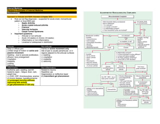
  • 1. Internal Medicine Harrison’s Principles of Internal Medicine Rheumatology Approach to Articular and MSK Disorders (Chapter 363)  Rule out red flag diagnoses – suspected for acute onset, monoarticular pattern or focal MSK pain o Septic Arthritis o Acute crystal-induced arthritis o Fracture o Vascular Ischemia o Carpal Tunnel Syndrome  Important questions: o Articular or non-articular o Acute (<6 weeks) or chronic (>6 weeks) o Inflammatory or non-inflammatory o Localized or widespread in distribution Articular Non-articular Deep or diffuse pain Limited range of motion on active and passive movement Swelling – due to synovial proliferation, effusion, bony enlargement Crepitation Instability Locking Deformity Painful on active movement only Site of pain is usually periarticular, or in regions adjacent to the articular surfaces (-) swelling (-) crepitation (-) instability (-) deformity Inflammatory Non-inflammatory Erythema, pain, swelling, warmth Systemic signs – fatigue, fever, rash, weight loss (+) ESR, CRP, thrombocytosis, anemia of chronic disease, hypoalbuminemia (+) prolonged morning stiffness improving with activity (+) gel phenomenon in OA only Related to trauma Repetitive use Degeneration or ineffective repair (+) intermittent gel phenomenon
  • 2. Clinical History Younger age group SLE, reactive arthritis Middle age group Fibromyalgia RA Elderly age group OA Polymyalgia rheumatica Men Gout Spondyloarthritis Ankylosing spondylitis Women RA Fibromyalgia Osteoporosis SLE African American Sarcoidosis SLE Whites Polymyalgia rheumatic Giant cell arteritis Granulomatosis with polyangiitis Familial aggregation Ankylosing spondylitis Gout Heberdon nodes of OA Onset Abrupt onset Septic arthritis,Gout Indolent onset OA, RA, fibromyalgia Evolution Chronic OA Intermittent Crystal-induced, Lyme arthritis Migratory RF, gonococcal, viral arthritis Additive RA, Psoriatic arthritis Evolution Chronic (>6 weeks) OA, RA, fibromyalgia Acute (<6 weeks) Infectious, crystal-induced, reactive Extent or Distribution Mono articular (1 joint) Crystal-induced, infectious Oligoarticular (2-3 joints) Poly articular (4 or more joints) OA, RA (symmetric as well) Asymmetric and polyarticular Spondyloarthritis, reactive arthritis, gout, sarcoid Symmetric or asymmetric Polyarticular or oligoarticular OA, psoriatic arthritis Upper extremities RA, OA Lower extremities Gout, reactive arthritis Axial skeleton OA, ankylosing spondylitis RA of the cervical spine Diseases with MSK co-morbidities Diabetes mellitus Carpal tunnel syndrome Renal insufficiency Gout Depression or insomnia Fibromyalgia Myeloma Low back pain Cancer Myositis Osteoporosis Fracture Glucocorticoids Osteonecrosis Septic arthritis Diuretics or chemotherapy Gout Rheumatic Review of Systems Fever SLE, infection Rash SLE, psoriatic arthritis Nail abnormalities Psoriatic arthritis, reactive arthritis Myalgias Fibromyalgia, statin-induced, drug-induced Weakness Polymyositis, neuropathy Eye involvement Behcet, sarcoidosis, Spondyloarthritis Gastrointestinal Scleroderma, IBD Genitourinary Reactive arthritis, gonococcemia Nervous system Lyme disease, vasculitis
  • 3.
  • 4.
  • 5. Osteoarthritis (Chapter 364)  Most common type of arthritis  Joints commonly affected by arthritis: Definition  Defined as joint failure → hyaline articular cartilage loss present in a focal and initially nonuniform manner → thickened subchondral bony plate, outgrowth of osteophytes at the joint margin, stretching of the articular capsule, snynovitis, weakness of the muscles bridging the joint Joint Protective Mechanisms and their Failure  Joint protectors – joint capsule, ligaments, muscles and tendons, afferent sensory nerves, underlying bone  Charcot arthropathy – severe and rapidly progressive OA developing when minor injury occurs in the presence of posterior column peripheral neuropathy Joint capsule and ligaments Fixes range of motion Synovial fluid lubrication Hyaluronic acid, lubricin Bridges the joint Muscles and tendons Cartilage and its Role in Joint Failure  Two most important macromolecules in cartilage: Type 2 collagen Provides cartilage tensile strength Aggrecan Proteoglycan macromolecule linked with hyaluronic acid molecule, with highly negatively charged glycosaminoglycans Through electropulsion → gives cartilage compressive stiffness  OA cartilage o (+) gradual depletion of aggrecan o (+) unfurling of the tightly woven collagen matrix o (+) loss of type 2 collagen Risk Factors  2 major factors contributing to OA development: o Joint vulnerability o Joint loading Systemic Risk Factors Age Most potent risk factor for osteoarthritis  Rare in <40 yearsold  >50% in >70 years old Aging increases joint vulnerability – less responsive in matrix synthesis, thinner cartilage with age, weaker bridging muscles, slower nerve conduction, stretching of ligaments Older women Especially in their sixth decade Probably hormonal loss Heritability and genetics Highly heritable disease, but joint specific (+) polymorphism with GDF5 gene Race Knee OA is more common among Chinese Risk Factors in the Joint Environment Joint anatomy 3 uncommon developmental abnormalities occurring in utero or in childhood:  Congenital dysplasia  Legg-Perthes disease  Slipped capital femoral epiphysis Others: acetabular dysplasia, femoroacetabular impingement Major injuries Fracture through the surface Avascular necrosis Tears of ligamentous and fibrocartilaginous structures Meniscal tears Joint misalignment
  • 6.  Varus knees – cartilage loss in the medial or inner knee  Valgus knees – cartilage loss in the lateral compartment Weakness of the quadriceps – knee OA Loading Factors Obesity 3-6 times body weight is transmitted across the knee during a single-leg stance Well-recognized and potent risk factor for OA (esp. knee) Precedes the development of disease More potent in women Adipokines may also be contributory Repeated Use of Joint and Exercise Seen in occupational use and leisure time physical activities  Farmers – hip OA  Miners – knee and spine OA  Heavy carrying – knee OA  Elite runners – knee and hip OA  Recreational runners – hip OA Pathology  Non-uniform loss in cartilage in a focal disease  Thickening of the subchondral bony plate  Osteophyte formation – outgrowth of new cartilage that ossifies o Important radiographic hallmark of OA  Stretching of the capsule → edematous and fibrotic Sources of Pain  Likely occurs from structures outside the cartilage (aneural): o Synovium, ligaments, joint capsule, muscles, subchondral bone  Severity of X-ray changes correlate poorly with pain severity  Presence of MRI synovitis correlates with presence and severity of knee pain Clinical Features  Joint pain o During or just after joint use and then gradually resolves o As disease progresses, pain becomes continuous and even becomes bothersome at night  Morning stiffness o Usually brief (<30 minutes)  Knee symptoms o Most common cause of chronic knee pain in patients >45 years o (+) knee buckling – from weakness of muscles crossing over the joint Buckling, catching, locking Anterior crucial ligament or meniscal tear Needs to be evaluated after a knee injury Pain during knee flexion (>35 degrees) From the patellofemoral compartment Prolonged morning stiffness with many joints affected Inflammatory arthritis Pain medial and distal to the knee Anserine bursitis Common cause of knee pain, responsive to glucocorticoid injection Prominent nocturnal pain in the absence of end-stage OA Merits diagnostic work-up  Hip symptoms o Can be detected by loss of internal rotation by passive movement, pain isolated to an area lateral to the hip joint (trochanteric bursitis) Diagnostics  No blood tests are routinely indicated for workup of patients with OA, unless symptoms and signs suggest inflammatory arthritis Examination of synovial fluid >1000 WBC/uL – presence of inflammatory arthritis or crystal-induced arthritis (with presence of crystals) X-rays Indicated when joint pain and physical findings are atypical of OA, or if pain persists after initiation of pain treatment Radiographs may be normal in early disease as they are insensitive to cartilage loss and early findings MRI Not indicated as part of diagnostic workup Almost never warrant change in therapy Goal of Treatment  Goal of treatment: To alleviate pain and minimize loss of physical function  Most effective mode of treatment: nonpharmacological treatment Non-pharmacological Treatment  Goals of treatment: o Avoiding painful activities – simplest treatment o Improving strength and conditioning of muscles bridging the joints o Unloading the joint via use of braces or splint to redistribute load or unloading joint during weight bearing with a cane or crutch  Exercise o Inactivity leads to increased risk for CVD and obesity o Weakness in muscles have multiple etiologies:  Decline in strength with age  Disuse muscle atrophy due to limitation of movement  Diminishing muscle use due to alteration of gait  Due to arthrogenous inhibition o Weakness in a muscle makes joint more susceptible to further damage and pain o Exercise lessens pain and improves physical function o Major challenge – adherence to program o Strongest predictor of success – personal history of successful exercise
  • 7. Effective Exercise Non-effective Exercise Aerobic exercises Resistance training Low-impact exercises (water aerobics) Tai chi – effective for knee OA Exercise with caloric restriction Range-of-motion exercises Isometric exercises  Correction of misalignment o Via bracing for willing patients who can learn to put them on correctly and on whom they do not slip o Acupuncture o Surgery Pharmacological Treatment  Serves as an important adjunct therapy for OA Acetaminophen (Paracetamol) Up to 1 gram TID Initial analgesic of choice for patients with OA Prolongs half-life of warfarin NSAIDs Most popular drugs to treat osteoarthritic pain Either topically or orally 30% greater pain alleviation than high-dose paracetamol Important to take low-dose aspirin and iburprofen/naproxen at different times to limit drug interaction – may increase bleeding and GI complaints Side effects: GI complaints (should be taken after meals) If with high-risk, take PPIs Others: edema, renal insufficiency (used with caution for CKD III) NSAIDS with increased CVD risk – diclofenac, rofecoxib Allowed NSAIDs – Naproxen (with high GI toxicity), salsalate, ibuprofen, celecoxib Topicals – lesser efficacy, less GI and systemic side effects but may cause skin irritation Intraarticular injections – glucocorticoids May be used temporarily (less than 3 months) for pain alleviation since synovitis is correlated with joint pain severity Subsequent injections may cause minor amounts of cartilage loss with unimportant clinical consequences Hyaluronic acid injections Can be given as treatment but with controversy on efficacy over placebo Opioid analgesics Has modest efficacy Duloxetine Has modest efficacy Glucosamine Chondroitin Recommended against OA Surgery Arthroscopic menisectomy Indicated for acute meniscal tears in which symptoms such as locking and acute pain are clearly related temporally to a knee injury that produced the tear High tibial osteotomy For patients with knee OA to the medial compartment Total knee or hip arthroplasty Highly efficacious operations that relieve pain and improve function in majority of patients Failure rates of 1% per year (higher in obese patients) Success rates higher in hips > knees Timing of knee or hip replacement is critical Cartilage regeneration, abrasion arthroplasty (chondroplasty) Not found to be efficacious in OA May be efficacious when OA has not yet developed Ineffective surgical plans  Arthroscopic debridement and lavage  Partial menisectomy
  • 8. Gout and Other Crystal-induced Arthropathies (Chapter 365) Gout  Metabolic diseases mostly affecting o Middle-aged to elderly men o Postmenopausal women  Due to increased body pool of urate with hyperuricemia → deposition of MSU crystals in joints and CT tophi with risk for deposition in the kidney interstitium or uric acid nephrolithiasis Clinical Features Involved joints MTP1, tarsal joints, ankles, knees, finger joints (usually in the elderly) First episode Begins at night with dramatic joint pain and swelling – mimicking cellulitis Precipitating events Dietary excess, trauma, surgery, excessive ethanol ingestion, hypouricemic therapy, serious medical illnesses – MI and stroke Presentation in women Represents 5-20% of the population (+) osteoarthritis and arterial hypertension → mild renal insufficiency (+) receiving diuretics Laboratory Diagnosis Needle aspiration of acutely or chronically involved joints or tophaceous deposits Confirms presumptive diagnosis (+) negative birefrigent needle-shaped crystals in compensated polarized light intracellularly and extracellularly Synovial fluid leukocyte counts More than 2,000 to 60,000/uL Culture If suspected to have septic arthritis Serum uric levels Can be normal or low at the time of an acute attack – inflammatory cytokines can be uricosuric and effective initiation of hypouricemic therapy can precipitate attacks Serum urate levels Almost always elevated at some time Important to use follow the course of hypouricemic therapy 24-h uric acid Useful in assessing risk of stones, elucidating overproducers or underexcretors of uric acid – to decide if uricosuric therapy is appropriate Excretion of >800 mg uric acid per day on regular diet – suggests overproduction of purine Other tests Urinalysis Serum creatinine Hemoglobin WBC count Liver function tests Serum lipids To assess possible pathologic sequelae of gout and other associated diseases requiring treatment and as baselines because of possible adverse effects of gout treatment Radiographic features X-ray (+) cystic changes, well-defined erosions with sclerotic margins (with overhanging bony edges) (+) soft tissue masses – advanced chronic tophaceous gout Ultrasound (+) double contour sign overlying articular cartilage Dual-energy CT (+) presence of urate crystals Treatment Acute Gouty Arthritis  NSAIDs and colchicine – may be poorly tolerated and dangerous in the elderly, especially in the presence of renal insufficiency and GI disorders  Ice pack applications and rest of involved joints can be helpful Oral colchicine Traditional and effective treatment if used early in an attack Regimens: 0.6 mg given every 8 hours with subsequent tapering 1.2 mg followed by 0.6 mg in 1 hour with subsequent day dosing depending on response Temporarily discontinued promptly at the first sign of loose stools, and symptomatic treatment of diarrhea must be given Oral NSAIDs Effective in 90% of patients With resolution of signs and symptoms after 5-8 days Most effective are those with short-half-lives: Celecoxib (800 mg – 400 mg 12 hour after, then 400 mg BID) Diclofenac (50 mg TID) Ibuprofen (800 mg TID) Naproxen (500 mg BID) Indomethacin (25-50 mg TID)
  • 9. Intramuscular or oral glucocorticoids Oral: prednisone 30-50 mg/day as initial dose → gradually tapered with resolution of attack May be effective in polyarticular gout Intraarticular: triamcinolone acetonide 20-40 mg, or methylprednisolone 25-50 mg Anakinra IL-1 antagonist Has an essential role on the inflammasome Used when other treatments have failed or contraindicated Hypouricemic Therapy  Ultimate control – correction of hyperuricemial  Normalized serum uric acid: <300-360 umol/L (5.0 – 6.0 mg/dL) o To prevent recurrent gouty attacks and eliminate tophaceous deposits  Considered when hyperuricemia cannot be corrected by: o Control of body weight o Low-purine diet o Increase in liquid intake o Limitation of ethanol use o Decreased use of fructose-containing foods and beverages o Avoidance of diuretics  Indications o Increased number of acute gouty attacks o Serum uric acid of >535 umol/L (>9.0 mg/dL) o Willingness to commit to lifelong therapy o Presence of uric acid stones o Presence of tophi or chronic gouty arthritis  Not initiated during acute attacks, but after patient is stable  Low-dose colchicine has been initiated to decrease risk of flares o Colchicine prophylaxis in doses of 0.6 mg one to two timed daily – should be given along with Hypouricemic therapy until patient is normouricemic and without gouty attacks for 6 months, or as long as tophi are present o Not used in patients in dialysis, given in lower doses to patients with renal disease or with P glycoprotein or CYP3A4 inhibitors (clarithromycin) Probenecid Uricosuric agent used in patients with good renal function who underexcrete uric acid <600 mg/day Urine volume is maintained by ingestion of 1500 mL water/day Regimen 250 mg BID → gradually to 3 g per day Not effective in those with serum creatinine levels of >2 mg/dL - May require allopurinol or benzbromarone Benzbromarone Uricosuric agent more effective for patients with CKD Lesinurad New uricosuric agent that can be used as an adjuvant at 200 mg/day to a xanthise oxidase inhibitor Others with mild uricosuric Losartan, amlodipine, fenofibrate effects Allopurinol Xanthine oxidase inhibitor Most commonly used Hypouricemic agent Best drug to lower serum urate in overproducers, urate stone formers, patients with renal disease Regimen Single morning dose of 100 mg (up to 800 mg as needed) Adjusted initial dose depending on creatinine clearance (10 mL/min = 100 mg) Toxicity among: (+) penicillin and ampicillin allergies (+) thiazide diuretic users Asians with HLA-B*58:01 Serious side effects: TEN, systemic vasculitis, bone marrow suppression, granulomatous hepatitis, renal failure With mild cutaneous reactions Can shift to uricosuric agent, desentiziation to allopurinol, use of febuxostat Febuxostat Xanthine oxidase inhibitor (40-80 mg OD) Does not require dose adjustments with mild-moderate renal disease Pegloticase Pegylated uricase Available for patients who do not tolerate or fail full doses of other treatment IV 8mg every 2 weeks can dramatically lower serum uric acid to up to 50% Other Cyrstal-induced arthritis Calcium pyrophosphate deposition (CPPD) disease Definitive Diagnosis (+) rhomboid or rodlike, weakly positive or nonbirefrigent with polarized light Others: (+) linear calcifications Calcium apatite deposition disease (basic calcium phosphate disease) Definitive Diagnosis: (+) seen in EM – 1-20 um shiny intra or extracellular nonbirefrigent globules or aggregates (+) purplish in Wright’s stain (+) bright red in alizarin red S. Others: (+) intra or periarticular calcifications with or without erosive, destructive, or hypertrophic changes Calcium oxalate disease Definitive Diagnosis: (+) bipyramidal and small polymorphic crystals in light microscopy Primary oxalosis – rare metabolic disease Secondary oxalosis – mainly due to chronic renal failure, more common than primary
  • 10. Rheumatoid Arthritis (Chapter 351)  Most common form of chronic inflammatory arthritis Clinical Features  Increases between 25 and 55 years of age, plateaus until 75 years and declines after  Undifferentiated inflammatory arthritis – too few affected joints to be classified as RA; if diagnosed later as RA: o Higher number of tender and swollen joints o Tests positive for RF and anti-CCP antibodies o Higher scores for physical disability Early morning stiffness Lasting more than 1 year, eases with physical activity Earliest involved joints Small joints of the hands and feet: Wrists, MCP, PIP joints DIP joint involvement Manifestation of coexistent osteoarthritis Flexor tendon tenosynovitis Frequent hallmark of RA → decreased range of motion → reduced grip strength and trigger fingers Ulnar deviation Subluxation of the MCP joints with subluxation of the proximal phalanx to the volar side of the hand Swan-neck deformity Hyperextension of the PIP Flexion of the DIP Boutonniere deformity Flexion of the PIP Hyperextension of the DIP Z-Line deformity Subluxation of the MCP1 Hyperextension of the IP1 Piano-key movement of the ulnar styloid Subluxation of the distal ulna – because of inflammation of the ulnar styloid and tenosynovitis of the extensor carpi ulnaris Flat feet (pes planovalgus) Chronic inflammation of the ankle and midtarsal region Atlantoaxial involvement May cause compressive myelopathy and neurologic dysfunction Rarely affects thoracic and lumbar spine TMJ involvement Not associated with significant symptoms Extraarticular Manifestations  May develop in 40% of patients – even before onset of RA  Most likely develop among patients: o History of cigarette smoking o Early onset of significant physical disability o (+) RF and anti-CCP antibodies Constitutional symptoms Weight loss, fatigue, malaise, depression, cachexia Presence of fever >38.3 C – suspect for Vasculitis or infection Subcutaneous nodules Firm non-tender, adherent to the periosteum, tendonds or bursae More common (30-40%) in those with highest levels of disease activity, disease-related shared epitope (SE), (+) RF, radiographic evidence of joint erosions Locations: forearm, sacral prominences, Achilles tendon Others: lungs, pleura, pericardium, peritoneum Sjogren syndrome (10%) (+) keratoconjunctivitis sicca (dry eyes) (+) xerostomia (dry mouth) Pulmonary manifestations Most common pulmonary manifestation - pleuritis (+) pleuritic chest pain, dyspnea (+) pleural friction rub, effusion (exudative) Insterstitial lung disease – with high disease acitivity, smokers – diagnosed via CT scan  Usual interstitial pneumonia (UIP), non-specific interstitial pneumonia (NSIP) – main histological and radiologic patterns of ILD  Poor prognosis (10% risk for mortality)  Favors more than idiopathic ILD  More responsive to immunosuppressive therapy Caplan syndrome (+) pulmonary nodulosis (+) pneumoconiosis (+) silica exposure (+) rheumatoid arthritis Cardiac Pericardium – most common site of involvement MR – most common valvular abnormality in RA Vasculitis Mostly seen in the following: - Long-standing disease - Positive for RF, anti-CCP - Hypocomplementemia Sensory polyneuropathies (Mononeuritis multiplex) may occur Hematologic Normochromic, normocytic anemia – MC hematologic abnormality Felty’s syndrome (occurs in late stages of RA) Neutropenia, splenomegaly, nodular RA T-cell LGL May have similar presentation with Felty May occur early in disease Lymphoma DLBCL – most common lymphoma
  • 11. Associated Conditions  CVD – most common cause of death in RA o Due to chronic inflammation  Osteoporosis – more common in RA patients  Hypoandrogenisms Epidemiology TNF-alpha Major cytokine in RA HLA-DRB1 HLA associations PTPN22 Non-MHC gene with strong risk for RA Smoking Most reproducible environmental risk factor for RA Porphyromonas gingivalis EBV Associated infections with RA Pathology Pathologic hallmarks Synovial inflammation and proliferation (pannus) Focal bone erosions Thinning of articular cartilage Most common inflammatory infiltrate T cells – driving chronic inflammatory response Diagnosis  Clinical diagnosis of RA is largely based on the following: o Clinical presentation of chronic inflammatory arthritis o Laboratory and radiographic results Anti-CCP (ACPA) Has greater specificity for RA diagnosis than RF Laboratory Features Serum IgM RF Found in 75-80% of patients with RA Also found in: Primary Sjogren, SLE, type II mixed essential cryoglobulinemia Subacute BE, hepatitis B and C 1-5% in healthy population ACPA Same sensitivity with IgM RG 95% specificity (more useful in diagnosis) Most predictive for predicting worse outcomes SF analysis WBC count – 5,000-50,000 WBC/uL (vs <2,000 in osteoarthritis) Predominance of neutrophil Purpose Most useful in confirming an inflammatory athritis while excluding infection or crystal-induced arthritis Joint imaging Purpose not only for diagnosis, but also for tracking progression of joint damage MRI, UTZ – preferred diagnostic modalities X-ray Periarticular osteopenia – initial radiographic finding Other findings: ST swelling, symmetric joint space loss, subchondral erosions (MCPs, PIPs, MTPs – lateral aspect of 5 th MTP – most common target) MRI Offers greatest sensitivity for detecting Synovitis and joint effusions, and early bone and bone marrow changes UTZ Can detect more lesions than X-ray but operator-dependent Clinical Course  Natural history is mainly affected by: o Age of onset, gender, genotype, phenotype, comorbid conditions 10% patients Will undergo spontaneous remission within 6 months Especially Seronegative patients IHD Most common cause of death Shortening of median life expectancy 7 years less in men 3 year less in women Risk factors With systemic EA involvement, low functional capacity, low socioeconomic status, low education, chronic prednisone use Joint inflammation Main driver of joint damage, most important cause of functional disability in earlier stages
  • 12. Treatment of RA  Development trends in management of RA (1) Emergence of methotrexate as DMARD of first choice for early RA (2) Development of novel biologicals that can be used alone or in combination with methotrexate (3) Proven superiority of combination DMARD regimens over methotrexate alone  Medications used in RA o NSAIDS o Glucocorticoids o Conventional DMARDs o Biological DMARDs NSAIDs  Presently considered as adjunctive agents for management of uncontrolled symptoms by other measures  Toxicities – gastritis, PUD Glucocorticoids  May be used in low-to-moderate doses to achieve rapid disease control before onset of fully effective DMARD therapy  1-2 week bursts of steroids for management of acute disease flares  Chronic administration of prednisone – to control disease activity in patients with inadequate response to DMARD therapy Low-dose prednisone Can retard progression of joint disease Must be balanced with side effects Intraarticular injection of intermediate steroid (triamcinolone acetonide) For rapid control of inflammation in a limited number of affected joints Must ensure that inflammation is not infectious Osteoporosis Important long-term complication of steroids Peptic ulcer No published guidelines for GI protection Conventional DMARDs  Hydroxychloroquine, sulfasalazine, methotrexate, Leflunomide  Has delayed onset of action – 6-12 weeks Methotrexate DMARD of choice for RA treatment MOA – stimulates adenosine release → anti-inflammation Leflunomide Inhibitor of pyrimidine synthesis Similar clinical efficacy as methotrexate Hydroxychloroquine Not a true DMARD; no retardation in RA progression Sulfasalazine Has shown to retard RA progression Radiographically Others Not used anymore Minocycline, gold salts, penicillinamine, azathioprine, cylclosporine Biological DMARDs  Anti-TNF – first approved biological DMARDs for RA Anti-TNF Infliximab – chimeric (mouse-human) monoclonal antibody Adalimumab, golimumab – human monoclonal antibody Certolizumab pegol – pegylated Fc-fragment of human monoclonal antibody Etanercept – soluble fusion protein (TNF receptor 2 in convalent linkage with Fc portion of IgG1) Can be used in combination therapy or monotherapy (latter 4) Contraindication Active infection History of hypersensitivity with these agents Chronic hepatitis B infection Class III/IV CHF Screening for latent TB is important (PPD, IFN-gamma) Anakinra Recombinant form of naturally occurring IL-1 receptor antagonist Has limited use in RA Indications (for syndromes dependent on IL-1 production) Neonatal-onset inflammatory disease Muckle-Wells syndrome Familial cold urticaria Systemic juvenile-onset inflammatory arthritis Adult-onset Still’s disease Abatacept Soluble fusion protein consisting of extracellular domain of human CTLA4 linked to modified human IgG portion MOA – inhibits co-stimulation of T cells by blocking CD28- CD80/CD86 inreractions Can be used in combination therapy Rituximab Chimeric monoclonal antibody against CD20 Indications Refractory RA in combination with methotrexate More effect on seropositive patients Adverse effects Increased risk for infection PML Tocilizumab Humanized monclonal antinody against IL-6 Adverse effects Increased infection, increased LDL cholesterol
  • 13. Small molecule inhibitors  For RA resistant to conventional and biologic DMARDs Tofacitinib Small-molecule inhibitor of JAK1 and JAK3 Adverse effects Elevated liver enzymes, neutropenia, increased cholesterol, elevated serum creatinine Treatment of Extraarticular Manifestations RA-ILD Challenging, some DMARDs (methotrexate, Leflunomide) can cause pulmonary toxicity Treatment High-dose steroids + adjunctive immunosuppressants (azathioprine, Mycophenolate mofetil, rituximab) Management Considerations Pregnancy  Up to 75% of female RA patients – overall improvement during pregnancy, but will flare after delivery Flares during pregnancy Low-dose prednisone Hydroxychloroquine Sulfasalazine Safest DMARDs to use during pregnancy Elderly  Conventional and biological DMARDs – equally effective and safe in younger and older population Methotrexate May need renal adjustments Not prescribed when serum creatinine >2 mg/dL SLE (Chapter 349)  90% are women of child-bearing age HLA associations HLA-DRB1 HLA-DR3 HLA-DQA2 HLA-CR2 HLA-FCGR2A/B Genes for lupus nephritis HLA-DR3, STAT4, APOL1 (African Americans) FCGR2A, ITGAM, IRF5, IRF7, TNFSF4 (Ox40L), DNAse1 Pathology Skin biopsy Deposition of Ig at dermal-epidermal junction (DEJ) (also in normal skin) Injury to basal keratinocytes Inflammation by T cells in the DEJ and around blood vessels Renal biopsy Pattern and severity of injury are important for diagnosis and selecting best therapy Requires aggressive immunosupression (steroids + another drug) Class III Class IV Class V accompanied by III and IV Not required Class I and II, or with extensive irreversible changes Blood vessel pathology Leukocytoclastic vasculitis – most common pattern of vasculitis Lymph biopsy Usually performed to rule out infection or malignancies Nonspecific diffuse chronic inflmmation Diagnosis  Based on characteristic clinical features and autoantibodies Classification of Nephritis (International Society of Nephrology and Renal Pathology Society Class I Minimal mesangial LN Class II Mesangial proliferative LN Class III Focal LN Class IV Diffuse LN Class V Membranous LN Class VI Advanced Sclerotic LN
  • 14. Manifestations MSK Intermittent polyarthritis in hands, wrists, knees – most common Joint pain – most common reason to increase dose of steroids Ischemic necrosis of bone – diagnosed for persistence of pain in single joint (treated with systemic steroids) Myositis – usually drug-induced (steroids > antimalarials) “rhupus” – combined SLE and RA presentation Cutaneous Discoid lupus erythematosus (20%) – most common chronic dermatitis in lupus (+) roughly circular with slightly raised, scaly Hyperpigmented erythematous rims (+) depigmented atrophic centers (where all dermal appendages are destroyed) Treatment – topical or locally injected steroids + systemic antimalarials Photosensitive, slightly raised erythema, scaly on face – most common acute SLE rash; “butterfly rash” Subacute CLE – similar to psoriasis (+) Ro (SS-A) antibodies Renal Nephritis – most serious manifestation of SLE Class IV – most severe, and most severe type of LN DPGN – ESRD within 2 years (if untreated) Can lead to atherosclerosis CNS Cognitive dysfunction (memory difficulties, reasoning) - Most common manifestation of diffuse CNS lupus Seizures Treatment – both antiseizure and immunosupression Psychosis Can be the predominant form of SLE Must be distinguished from glucocorticoid-induced psychosis Myelopathy Tx: high-dose steroids Pulmonary Pleuritis with or without pleural effusion – most common pulmonary manifestation Tx: NSAIDS → steroids if more severe Life-threatening conditions Interstitial fibrosis Shrinking lung syndrome Intraalveolar hemorrhage Cardiac Pericarditis – most common cardiac manifestation More serious manifestations – myocarditis, Libman-Sacks (MR, AR) Vascular occlusions Cerebral event results from clotting Tx: long-term anticoagulation if treatment of choice Higher SLE MI events in women <49 years Risk factors for atherosclerosis Male, older, hypertension, dyslipidemia, diabetes, dysfunctional pro inflammatory HDL, repeated high scores for disease activity, high cumulative or daily doses of steroids, high levels of homocysteine and leptin Hematologic Normochromic, normocytic anemia – most common manifestation Leukopenia (lymphopenia) Thrombocytopenia GI Vomiting, diarrhea, diffuse abdominal pain (autoimmune peritonitis, and/or intestinal vasculitis), elevated liver enzymes Tx: aggressive immunosuppression with high-dose steroids for short- term control Ocular Sicca syndrome, nonspecific conjunctivitis – common in SLE, rarely threaten vision
  • 15. Retinal vasculitis, optic neuritis – serious manifestations and blindness can develop over days to weeks Glaucoma, cataract – complications of steroids Laboratory Tests (1) To establish or rule out diagnosis (2) Follow the course of disease (3) Identify adverse effects of therapies Autoantibody Testing  ANA – most important autoantibodies to detect (positive in >95% patients) Anti-Ro/SS-A Indicates increased risk for neonatal lupus, sicca syndrome, and SCLE Standard Tests for Diagnosis  Screening using the following: o CBC, PC, urinalysis o May detect abnormalities contributing to diagnosis and influence management decisions Tests for Following Disease Course UA, hemoglobin levels, PC, serum creatinine, albumin For hematuria, proteinuria Identification of markers for disease activity See Table Management of SLE  Aim is to achieve low-level disease activity o Mild symptoms on the lowest possible doses of medications o Usually achieved in 30-50% of SLE patients within a year Conservative Therapies for Management of Non-Life-Threatening Disease  For patients with SLE autoantibodies, fatigue and pain without organ involvement Goal Management via suppression of symptoms Anagesics, antimalarialas are mainstay First issue with NSAIDs Increased risk for NSAID-induced aseptic meningitis, elevated serum transaminases, hypertension, renal dysfunction Second issue with NSAIDs All NSAIDs (particularly COX-2 inhibitors), increases MI risk Issue with hydroxychloroquine Must have annual eye examination for potential retinal toxicity At least 750 ng/mL – blood level needed for optimal response in active SLE NSAIDs Arthritis/arthralgias Antimalarials – hydroxychloroquine, chloroquine, quinacrine Dermatitis Arthritis Fatigue DHEA May reduce disease activity Belimumab For fatigue, rash, and/or arthritis Mostly effective with robust clinical activity: SLEDA ≥10 Positive anti-DNA Low serum complement Topical sunscreens Antimalarials Topical steroids and/or Tacrolimus Lupus dermatitis (systemic steroid with or without Mycophenolate mofetil, azathioprine, belimumab – sever or unresponse LD)
  • 16. SLEDAI Widely used measure of SLE disease activitiy >3 Clinically active disease Life-threatening Management of SLE Systemic Steroids (0.5-1 mg/kg PO daily, 500-1000 mg IV daily) Methylprednisolone sodium succinate for 3 days → daily (0.5-1 mg/kg) prednisone Mainstay treatment for any inflammatory life- threatening or organ-threatening manifestations of SLE Management of LN (induction therapy) Steroids + Cylocphosphamide or mycophenolate mofetil (azathioprine is effective, but associated with more flares)  African Americans, hispanics – mycophenolate  Whites, Asians – cyclophosphamide or mycophenolate Mycophenolate toxicity diarrhea Cyclophosphamide toxicity Amenorrhea, leukopenia, nausea Responses to treatment Steroid – within 24 hours Cyclophosphamide, mycophenolate – 3-16 weeks Management of LN (maintenance therapy) Mycophenolate > azathioprine Cyclophosphamide – less safe Cyclophosphamide toxicity Ovarian failure Tx – GnRH agonist prior to each monhly cyclophosphamide Special Conditions Crescentic LN Treatment Either: High-dose cyclophosphamide High-dose mycophenolate Membranous LN (INS V) Treatment ACE-I or ABRs is recommended But usually none, unless with nephrotic proteinuria Pregnancy and Lupus  Rates of fetal loss is higher in SLE o High disease activity, antiphospholipid antibodies (lupus anticoagulant), hypertension, and/or active nephritis Mainstay for active flares Hydroxychloroquine + lowest effective doses of prednisone/prednisolone + azathioprine (if failure to suppress activity) HCQ – reduces chance of subsequent fetus with heart block Dexamethasone – prevents progression of heart block SLE + antiphospholipid antibodies + prior fetal losses LMW heparin + low-dose aspirin Warfarin is teratogenic Adverse effects of prenatal steroids (primarily betamethasone) LBW CNS developmental abnormalities Predilection to adult metabolic syndrome Category A (no evidence of teratogenicity in humans) Steroids Category C (may be teratogenic in animals, no good evidence in humans) Cyclosporine, Tacrolimus, rituximab Category D (benefits > risks of teratogenicity) Azathioprine, hydroxychloroquine, mycophenolate, cyclophosphamide Category X Methotrexate Lupus and APAS Treatment Warfarin Target INR (2.0-2.5) for 1 episode of venous clotting Target INR (3.0-3.5) for recurrent clots or arterial clotting (CNS) Lupus (TTP, HUS) Treatment Diagnosis Identification of schistocytes, elevated LDH, anti-ADAMS13 Treatment Plasmapheresis – life-saving Concomitant steroid therapy – recommended by authorities
  • 17. Lupus dermatitis Mainstay treatment Topical steroids + antimalarials (hydroxychloroquine) - Effective in reducing lesion severity and relatively safe Systemic retinoic acid - For recalcitrant cases Extensive, pruritic, bullous, ulcerating Systemic steroids with tapering → (hydroxychloroquine, retinoids, belimumab) Reports of success with Tacrolimus Dapsone or thalidomide Tacrolimus Increased risk for malignancies Thalidomide Extreme dangers for fetal deformities Peripheral neuropathy Associated Malignancies  NHL  Cancers of thyroid, lung, liver, vulvar/vaginal tissues Death Leading causes in 1 st decade of disease Systemic disease activity Renal failure infections After 1 st decade of disease Thromboembolic events Drug-induced Lupus Definition (+) positive ANA associated with Fever, malaise, arthritis/arthralgias, myalgias, serositis, and/or rash Features Predominant in whites Usually appear during therapy with certain medications, biologicals Less female predilection Rarely involved kidneys, brains Rarely associated with anti-dsDNA Commonly associated with anti-histones Usually resolves after discontinuation of medication Frequent suspects Antiarryhtmias Procainamide, disopryamide, propafenone Antihypertensive Hydralazine Several ACE-I and beta blockers Antithyroid PTU Antipsychotics Lithium, chlorpromazine Anticonvulsants Carbamazepine, phenytoin Antibiotic Isoniazid, minocycline, nitrofurantoin Antirheumatic Sulfasazaline Diuretic HCT Antihyperlipidemia Lovastastin, simvastatin Biologics IFN and TNF inhibitors PEARLS Approach to MSK diseases Focal, monoarthritis in acute onset Trauma/Fracture Gout Vascular ischemia Carpal tunnel syndrome Septic Arthritis Osteoarhthritis Most potent risk factor for OA Age Polymorphism in this gene is involved in formation of osteoarthritis GDF5 gene What joint is usually more involved in OA among Chinese Knees Important radiographic hallmark of OA Osteophytes MRI finding which correlates with severity of knee pain Presence of MRI synovitis Most common cause of chronic knee pain in patients >45 years Knee OA Amount of WBC in synovial fluid which accounts the presence of inflammatory or crystal-induced arthritis >1000 WBC/uL Never indicated as part of diagnostic workup Furthermore, it almost never warrant change in therapy MRI Mainstay treatment for osteoarthritis Non-pharmacological Initial choice of treatment for OA Acetaminophen (high-dose) The use of these agents were banned in the treatment of OA Chondroitin Glucosamine Gout and Other Crystal-Associated Arthritis Excretion of this amount of uric acid per day on a regular diet suggests overproduction of purine >800 mg/day Ultrasound manifestation of gout (+) double contour sign Severe hyperuricemia requiring Hypouricemic therapy >9 mg/dL (>535 umol/L) Underexcretor of uric acid <600 mg/day Other medications with mild uricosuric effects Losartan Amlodipine Fenofibrate Contraindications of colchicine use Dialysis patients (+) P glycoprotein (+) CYP3A4 inhibitors - clarithromycin Rheumatoid Arthritis Temperature before one must suspect for Vasculitis or infection in RA patients >38.3 C Main histological and radiologic patterns of ILD Usual interstitial pneumonia Non-specific interstitial pnuemonia Most common cause of death in RA CVD Associated conditions with RA CVD
  • 18. Osteoporosis hypoandrogenisms Has greater specificity for RA diagnosis than RF ACPA Initial radiographic finding in patients with RA Periarticular osteopenia Delayed onset of actions of conventional DMARDs 6-12 weeks SLE Most important autoantibody to test for SLE ANA Achievement of low-level disease SLE activity 30-50% patients within a year