
Recommended
More Related Content
Similar to The Lymphatic System.pdf
Similar to The Lymphatic System.pdf (20)
More from Pritamjit3
Recently uploaded
Recently uploaded (20)
The Lymphatic System.pdf
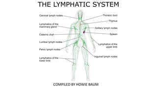
- 1. THE LYMPHATIC SYSTEM COMPILED BY HOWIE BAUM
- 2. THE LYMPHATIC SYSTEM – ALSO CALLED THE LYMPH OR IMMUNE SYSTEM Protects the body from invasion by bacteria or other germs. The active part of the system is lymph fluid. It drains into networks of tiny capillaries in tissue spaces that unite to form larger vessels called lymphatics. Lymph nodes (lymph glands) are the filtering and storage areas of the system, and they are scattered along the routes of the lymphatics. Unlike blood, lymph is not pumped; instead it moves passively as lymphatics are compressed by contraction of surrounding muscles during movement. Lymph fluid contains large numbers of specialized white blood cells, particularly lymphocytes, which protect the body against non-self material such as invading microorganisms. https://www.youtube.com/watch?v=JRkLDKrDtDY
- 5. THE AUXILIARY IMMUNE SYSTEM Many organs have a role in protecting the body against invading microbes. Together they can be termed the auxiliary immune system, since they supplement the true internal immune system. Physical mechanisms of protection are provided by structures such as the skin and microscopic hairs, while defenders such as gastric enzymes and useful bacteria protect by chemical means.
- 6. REMOVING AND DESTROYING PATHOGENS: HOW THE LYMPHATIC SYSTEM PROTECTS THE BODY The lymphatic system includes several components; it’s a network of vessels, ducts, and nodes, as well as organs and diffuse tissue. These structures work to filter unwanted substances out of the bloodstream and body tissues. How does the body destroy harmful pathogens?
- 7. Immunity is the body’s defense system against infection and disease. White blood cells play a key role. Some rush to attack any harmful microbes that invade the body. Other white blood cells become specialists, adapted to fight particular pathogens. All of them work to keep the body as healthy as possible. IMMUNCE CELLS EATING BACTERIA 1.5 MIN https://www.youtube.com/watch?v=iZYLeIJwe4w
- 8. White blood cells, also called leukocytes, defend the body against disease. They normally account for only 1% of circulating blood volume but increase during infection or inflammation. Neutrophils are the most common type, comprising 60% to 70% of all white blood cells. Neutrophils are phagocytes, cells that consume invading pathogens. A Macrophage is a large phagocytic cell found in stationary form in the tissues or as a mobile white blood cell, especially at sites of infection https://www.youtube.com/watch?v=w0-0Bqoge2E (USE LOW SPEED)
- 9. Lymphocytes, the second most common type of white blood cell are produced in red bone marrow and move through the organs and tissues of the lymphatic system. They include several sub-types: B cells produce antibodies T cells target virus or fungal-infected cells, cancer cells, and transplanted cells. Natural killer (NK) cells attack and destroy foreign microbes.
- 10. Phagocytes of humans and other animals are called "professional" or "non-professional" depending on how effective they are at phagocytosis. The professional phagocytes include many types of white blood cells (such as neutrophils, monocytes, macrophages, mast cells, and dendritic cells). The main difference between professional and non- professional phagocytes is that the professional phagocytes have molecules called receptors on their surfaces that can detect harmful objects, such as bacteria, that are not normally found in the body The process begins when chemicals from a pathogen, or damaged tissue, attract a phagocyte. The phagocyte binds to the microbe, envelopes it, and then eats it. Enzymes within the phagocyte kill and digest the pathogen.
- 11. The video shows a human polymorphonuclear leukocyte (neutrophil) crawling among red blood cells, notable for their dark color and principally spherical shape. The neutrophil is "chasing" Staphylococcus aureus (Staph) microorganisms. https://www.youtube.com/watch?v=Z_mXDvZQ6dU (make loud at end)
- 12. INNATE IMMUNITY When an infection occurs, fever elevates body temperature to accelerate the immune response. The reaction can happen relatively quickly. Blood vessels dilate around the injury site, inflaming the area. The vessel dilation allows more white blood cells to leave the bloodstream and enter the infected tissues. Phagocytes then do the job of consuming invading microbes. This rapid response by the body to an infection is an example of innate immunity.
- 13. Activated B cells multiply to produce large numbers of clones, most of which become plasma cells. Plasma cells produce antibodies that recognize antigens on foreign microbes. The antibodies act as tags to identify the invaders. This is called an antibody- mediated response. T cells, activated by antigens presented by phagocytes, multiply then seek out and destroy infected cells. This is called a cell-mediated response.
- 14. LYMPH NODES The lymph nodes (or “glands”) are vital to the body’s defense system – they produce and hold immune cells (lymphocytes) that protect the body from disease. Lymph nodes are scattered throughout the body and also concentrated in groups. Each node is a mass of lymphatic tissue divided into compartments by partitions of connective tissue known as trabeculae. Lymph fluid from most tissues or organs flows through one or more lymph nodes, where it is filtered and cleaned, before draining into the venous bloodstream. Lymph nodes vary in diameter from 1 to 25mm ( 1/25 to 1in), although they can swell during infection or illness. Covered in a fibrous capsule, they contain sinuses, where many scavenging white blood cells, called macrophages, ingest bacteria as well as other foreign matter and debris.
- 15. WHITE CELL TYPES There are numerous types of white blood cell, known by the general name of leucocytes. Some grow and mature into other types. All are derived from bone marrow. Monocyte Largest cell in the blood, with big and rounded, or indented, nucleus Engulfs pathogens.
- 16. Lymphocyte - Chief immune cell, with large nucleus that almost fills the cell; either B or T, depending on development.
- 17. Neutrophil - Granulocyte (having many small particles in the cytoplasm) with a multi-lobed nucleus; engulfs pathogens.
- 18. Basophil - Circulating granulocyte with lobed nucleus; involved in allergic reactions.
- 19. Eosinophil - Granulocyte that is important during allergic reactions; B-shaped nucleus; destroys antigen– antibody complexes.
- 20. LOCAL INFECTION If harmful microbes enter body tissues, both the inflammatory and immune responses act swiftly to limit their spread. The infection may be confined to a naturally defined site, such as the boundary between two sets of tissues. White blood cells and invaders, living and dead, accumulate, along with fluids, toxins, and general debris. The resulting mixture is known as pus, and if it gathers in a localized area, this is an abscess. As the pus collects, it puts pressure on surrounding structures. This may cause discomfort and pain, especially if the surroundings have no flexibility, as in a tooth abscess. Microbes gain access through a decayed region of enamel and dentine, infect the pulp, and spread into the root, where pus collects. As pus presses on the pulp nerves, it causes the pain of toothache.
- 21. RESPONSE Once an inflammatory response is triggered, blood flow to the damaged area is increased. The blood vessels, especially capillaries, widen and the capillary walls become thinner and more permeable, allowing plasma and fluid to leak into the space between the cells. Next, white blood cells, such as neutrophils, start to arrive. The neutrophils leave the blood and enter the tissue, drawn to the damaged area by chemicals released by the disrupted cells.
- 22. PHAGO CYTOSIS Various kinds of white blood cells can surround, engulf, and ingest smaller items, such as bacteria and cellular debris, in a process known as phagocytosis (“cell eating”). The cell exploits its ability to change shape and move, using the intracellular components of microtubules and microfilaments that form its flexible, mobile internal scaffolding. The ingestion usually takes less than one second, and the consumed material is gradually broken down by enzymes and other chemicals within the cell. The phagocyte binds to the microbe, envelopes it, and then eats it. Enzymes within the phagocyte kill and digest the pathogen. https://www.youtube.com/watch?v=ohFQEl4z0Yc
- 23. ENGULFING STAGE The white cell extends pseudopods (“false feet”) towards and around the unwanted items – here a bacterium. The pseudopods merge to engulf them.
- 24. LYSIS STAGE The items are trapped in phagocytic vesicles, which with enzyme-containing lysosomes form phagolysosomes, where lysis (breaking down) occurs.
- 25. Exocytosis stage Harmless products of cell eating are expelled through the white blood cell’s membrane, or in tiny membrane- bound exocytic vesicles to the extracellular fluid.
- 26. Defensive cells Various types of white blood cells (leucocytes) become involved in inflammation, including neutrophils and monocytes. The latter are immature leaving the blood vessels and enter the tissues, but rapidly develop into, active cells called macrophages that replace neutrophils. A single macrophage (“big eater”) can consume up to 100 bacteria or similar- sized items before dying.
- 27. FIGHTING INFECTIONS IN LYMPH AND IMMUNITY An infection occurs when microscopic organisms gain entry into the body, survive, multiply, and disrupt normal cell function. The infection may be localized, such as in a patch of skin or in a wound, or systemic, in which the organisms are carried around the body by the blood and lymph to invade many parts. Flu Attack! How A Virus Invades Your Body https://www.youtube.com/watch?v=Rpj0emEGShQ
- 28. VIRUSES - An important group of harmful microorganisms, or pathogenic microbes (commonly known as “germs” or “bugs”), is the viruses. They are the smallest of all the microbes; millions would cover the head of a pin. Many viruses can remain inactive for long periods and survive freezing, boiling, and chemical attack, yet they can activate suddenly when they have the opportunity of invading a living cell. VIRUS SHAPES There are thousands of different types of virus, with shapes such as balls, boxes, polygons, sausages, golf balls, spirals, and even tiny “space rockets”. Viruses are classified by their size, shape, and symmetry as well as by the groups of diseases they cause. The protein coat is corkscrew-like with the DNA or RNA genetic material inside. Examples include myxoviruses and paramyxoviruses.
- 29. Image used from article: https://www.assignmentexpert.com/blog/wp-content/uploads/2015/06/Picture-structure-1024x460.png SHAPES OF DIFFERENT TYPES OF VIRUS
- 30. Icosahedral Twenty equal-sided triangles connect to form a faceted container. Examples include adenoviruses and herpes viruses. Contains DNA or RNA genetic material so it can replicate itself once its in the body.
- 31. Complex Like a tiny rocket with “landing legs” that settle on the host cell. They only attack bacteria, so they are important in health terms when they attack pathogenic bacteria in the human body. Examples include the T4 bacteriophage.
- 32. Bacteria The microorganisms known as bacteria are present almost everywhere – in soil, water, air, food, drink, and on and in our own bodies. Many types of bacteria are harmless; indeed, those present naturally in the human intestines, the “gut flora”, have a beneficial effect in helping to extract nutrients from food. However, hundreds of types of bacteria cause infections, ranging from mild to lethal. Bacteria are simpler than other single-cell organisms in that their genetic material (DNA) is free in the cell, rather than contained in a membrane- A typical rod-shaped bacterium (bacillus) has a cell membrane enclosing cytoplasm and organelles, such as ribosomes, which are distributed in it. Unlike animal cells, it has a semirigid cell wall outside its cell membrane.
- 33. Bacterial shapes - There are several typical shapes for bacteria, and these, along with the way they are colored by laboratory stains, are important for classification and working out their origins and relationships. Many thousands of bacterial types are known, with more discovered each year. Cocci Generally spherical (shown here in the process of dividing). Examples include Staphylococcus and Streptococcus.
- 34. Bacilli Oval, or rod-like, with or without surface hairs (pili) or whip-like flagella. Examples are Streptobacillus and Clostridium.
- 35. Spirilla Spiral or, more accurately, helical (corkscrew-like) in shape, as open or tight coils. Examples include Leptospira and Treponema.
- 36. Resistance to antibiotics - Many bacteria are able to develop resistance to antibiotics by changing (mutating) into new strains. Their most effective mechanism is the rapid transfer of plasmids – small loop-like packages of the bacterial genetic material, DNA – between bacterial populations. The gene for antibiotic resistance crops up by accident, and the bacterium possessing it is able to pass the gene to others by the process of conjugation, or “bacterial sex”, in which plasmid genetic material is donated or exchanged. 1) Role of plasmids - Plasmids may cause the bacterium to make enzymes against antibiotic drugs, or alter its surface receptor sites, where antibiotics bind. Then, the plasmid duplicates itself.
- 37. 2) Plasmid transfer Plasmid transfer takes place during a process known as conjugation. The plasmid copy is passed from the donor, through a pilus, to the recipient bacterium.
- 38. 3) Drug-resistant strains Recipient bacteria inherit the resistant gene. Plasmid transfer produces populations of bacteria resistant to a range of antibiotics.
- 39. ALLERGIES - The immune system normally defends the body against infections, cancer, injuries, and damaging substances such as toxic chemicals. Sometimes, however, it over-reacts, attacking a foreign substance that is normally harmless. This reaction is an allergic response. Such responses can vary from mild conditions to life-threatening disorders. Allergic Response - An allergy develops if the immune system becomes sensitized to a foreign substance (an allergen). When first exposed to an allergen, such as pollen, nuts, or penicillin, the immune system makes antibodies to fight it. The antibodies coat the surface of mast cells, found in the skin, stomach lining, lungs, and upper airways. If the allergen enters the body again, these cells mount an allergic response.
- 40. 1) Exposure to an allergen - Antibodies bind to the surfaces of mast cells. A mast cell (also known as a mastocyte or a labrocyte) is a migrant cell of connective tissue that contains many granules rich in histamine and heparin.
- 41. 2) Antibodies triggered Allergens come into contact with the antibodies. If they link two or more antibodies, they cause the cell to burst.
- 42. 3) Histamine released Granules inside the mast cell release histamine as the cell bursts. Histamine causes an inflammatory response that irritates body tissues and produces the symptoms of an allergy.
- 43. Allergic Rhinitis Airborne allergens that irritate the lining of the nose and throat cause allergic rhinitis; this allergy may be seasonal or occur all year. In allergic rhinitis, the lining of the nose and throat becomes inflamed after contact with an airborne allergen. One form is hay fever, which is brought on by pollen grains in the spring and summer.
- 44. Another form, perennial rhinitis, may be caused by house dust mites, bird feathers, or animal fur or skin flakes (dander) and may occur at any time of year. Both forms can cause sneezing, a blocked, runny nose, and itchy, watery eyes, although symptoms tend to be more severe in hay fever. Often, the cause of rhinitis is easy to identify. If a person cannot avoid contact with the allergen, ant-iallergy drugs taken before or during an attack may relieve itchy eyes or a blocked nose. Drugs can be applied directly to the inside of the nose or eyes or taken orally.
- 45. Food Allergies - Some allergies are caused by an excessive immune response to certain foods, most commonly nuts, seafood, eggs, Gluten from wheat, and milk. Gluten is a substance present in cereal grains, especially wheat, that is responsible for the elastic texture of dough. A mixture of two proteins, it causes illness in people with Celiac disease. Symptoms of food allergies may appear as soon as the food is eaten or develop over a few hours. Some affect the digestive system, causing swelling and itching in the mouth and throat, nausea and vomiting, and diarrhea. Others affect the whole body, causing skin rashes, swollen tissues, and shortness of breath. The only effective treatment is to avoid the problem food.
- 46. THE END