Micro-Osteoperforation Accelerating Orthodontic Tooth Movement
•
2 likes•662 views

This research paper examines the use of micro-osteoperforation (MOP) to accelerate orthodontic tooth movement. MOP is a minimally invasive procedure that involves creating controlled microtraumas in the bone using specialized devices. This stimulates an inflammatory response and increases the rate of alveolar bone remodeling compared to conventional orthodontics. Several clinical studies showed MOP resulted in 41-50% faster canine retraction and higher levels of inflammatory markers associated with bone remodeling. While MOP reduces treatment time, future research is still needed to further validate its efficacy.

Recommended
Recommended
More Related Content
What's hot
What's hot (20)
Similar to Micro-Osteoperforation Accelerating Orthodontic Tooth Movement
Similar to Micro-Osteoperforation Accelerating Orthodontic Tooth Movement (20)
More from Dr. Prathamesh Fulsundar
More from Dr. Prathamesh Fulsundar (20)
Recently uploaded
Recently uploaded (20)
Micro-Osteoperforation Accelerating Orthodontic Tooth Movement
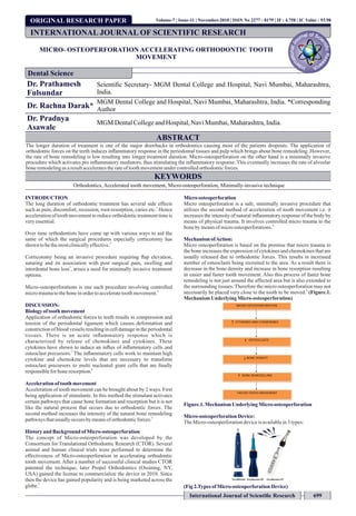
- 1. ORIGINAL RESEARCH PAPER MICRO- OSTEOPERFORATION ACCELERATING ORTHODONTIC TOOTH MOVEMENT Dr. Prathamesh Fulsundar Scientific Secretary- MGM Dental College and Hospital, Navi Mumbai, Maharashtra, India. Dr. Rachna Darak* MGM Dental College and Hospital, Navi Mumbai, Maharashtra, India. *Corresponding Author Dr. Pradnya Asawale MGM DentalCollegeandHospital,NaviMumbai,Maharashtra,India. ABSTRACT The longer duration of treatment is one of the major drawbacks in orthodontics causing most of the patients dropouts. The application of orthodontic forces on the teeth induces inflammatory response in the periodontal tissues and pulp which brings about bone remodeling. However, the rate of bone remodeling is low resulting into longer treatment duration. Micro-osteoperforation on the other hand is a minimally invasive procedure which activates pro inflammatory mediators, thus stimulating the inflammatory response. This eventually increases the rate of alveolar boneremodelingasaresultacceleratestherateoftoothmovementundercontrolledorthodonticforces. KEYWORDS Orthodontics, Accelerated tooth movement, Micro-osteoperforation, Minimally-invasive technique INTRODUCTION The long duration of orthodontic treatment has several side effects 1 such as pain, discomfort, recession, root-resorption, caries etc. Hence accelerationof toothmovementtoreduceorthodontictreatmenttimeis veryessential. Over time orthodontists have come up with various ways to aid the same of which the surgical procedures especially corticotomy has 2 shown tobethemostclinicallyeffective. Corticotomy being an invasive procedure requiring flap elevation, suturing and its association with post surgical pain, swelling and 3 interdental bone loss , arises a need for minimally invasive treatment options. Micro-osteoperforations is one such procedure involving controlled 4 microtraumatotheboneinordertoacceleratetoothmovement. DISCUSSION: Biologyof tooth movement Application of orthodontic forces to teeth results in compression and tension of the periodontal ligament which causes deformation and constriction of blood vessels resulting in cell damage in the periodontal tissues. There is an acute inflammatory response which is characterized by release of chemokines and cytokines. These cytokines have shown to induce an influx of inflammatory cells and 5 osteoclast precursors. The inflammatory cells work to maintain high cytokine and chemokine levels that are necessary to transform osteoclast precursors to multi nucleated giant cells that are finally 6 responsiblefor boneresorption. Accelerationoftooth movement Acceleration of tooth movement can be brought about by 2 ways. First being application of stimulants. In this method the stimulant activates certain pathways that cause bone formation and resorption but it is not like the natural process that occurs due to orthodontic forces. The second method increases the intensity of the natural bone remodeling 4 pathwaysthatusuallyoccursby meansoforthodonticforces. Historyand Background ofMicro-osteoperforation The concept of Micro-osteoperforation was developed by the Consortium for Translational Orthodontic Research (CTOR). Several animal and human clinical trials were performed to determine the effectiveness of Micro-osteoperforation in accelerating orthodontic tooth movement. After a number of successful clinical studies CTOR patented the technique, later Propel Orthodontics (Ossining, NY, USA) gained the license to commercialize the device in 2010. Since then the device has gained popularity and is being marketed across the 7 globe. Micro osteoperforation Micro osteoperforation is a safe, minimally invasive procedure that utilizes the second method of acceleration of tooth movement i.e. it increases the intensity of natural inflammatory response of the body by means of physical trauma. It involves controlled micro trauma to the 4 bonebymeansofmicroosteoperforations. MechanismofAction: Micro osteoperforation is based on the premise that micro trauma to the bone increases the expression of cytokines and chemokines that are usually released due to orthodontic forces. This results in increased number of osteoclasts being recruited to the area. As a result there is decrease in the bone density and increase in bone resorption resulting in easier and faster tooth movement. Also this process of faster bone remodeling is not just around the affected area but is also extended to the surrounding tissues. Therefore the micro osteoperforation may not 8 necessarily be placed very close to the tooth to be moved. (Figure.1. MechanismUnderlyingMicro-osteoperforation) Figure.1.MechanismUnderlyingMicro-osteoperforation Micro-osteoperforationDevice: TheMicro-osteoperforationdeviceisavailablein3types: (Fig2.Types ofMicro-osteoperforationDevice) INTERNATIONAL JOURNAL OF SCIENTIFIC RESEARCH Dental Science Volume-7 | Issue-11 | November-2018 | ISSN No 2277 - 8179 | IF : 4.758 | IC Value : 93.98 International Journal of Scientific Research 699
- 2. ISSN No 2277 - 8179 | IF : 4.758 | IC Value : 93.98Volume-7 | Issue-11 | November-2018 700 International Journal of Scientific Research 1- Excellerator: it comprise of single use tip and a fine grip handle as a single unit. It has an adjustable depth dial with the markings of 0mm, 3mm, 5mm and 7mm which can be fixed at a desired depth by rotating in clockwise direction. The LED light depth indicator adds to its user friendly design. However the device is meant for single use and cannot be sterilized which adds on to the cost of the treatment. 2- Excellerator RT kit: It comprises of one large handle, 2 closed tips and one open tip.The handle comes with a textured grip and can be sterilized. The 2 disposable closed tips have depth graduations at 3mm, 5mm and 7mm; while the open tip do not show depth graduations. 3- Excellerator PT kit: it comprises of a powered handpiece, charging unit, a contra-angled head attachment and disposable tips. The handpiece has a digital display which shows the battery level, speed in rpm, torque and the forward or reverse mode. The accessibility in entire mouth is made easy with the help of contra- angled head attachment. Greater rotational speed drives the tips easily through the cortical bone without the need of pressure application. MethodofUse: The patient is advised to use Chlorhexidine mouth wash twice daily, one day prior to the procedure. Immediately before the procedure the patient is asked to rinse for 1 minute using 0.12 % Chlorhexidine digluconate. The placement and depth of micro-osteoperforations should be determined. Local anesthesia should be achieved using local infiltration. The depth of 3mm, 5mm or 7 mm should be set on the device by rotating the depth dial in clockwise direction for anterior, premolar and molar region respectively.The tissues are held taught and the device is positioned such that the tip is perpendicular at the point of contact, gentle pressure is applied. The LED indicator turns red once the desired depth is obtained, rotating counterclockwise the device should be removed. One to three micro-osteoperforations should be done either on the buccal or the lingual aspect of the interdental bone to obtainoptimalresults. Indications: Micro osteoperforations can be successfully used to increase the rate of tooth movement around teeth that require to be moved along long distances such as ectopic canines, forced eruption, edentulous space closingetc. Contraindications: Micro osteoperforation should not be used nearby anchorage devices such as temporary anchorage devices in form of implants and anchor teeth since it causes reduction in bone density in the involved area and hence can cause loss of stability of the device and loss of anchorage. Also it should not be used in patients not having a healthy periodontal status or medical conditions such as seizures, hematological disorders, diabetesetc. Advantages: It increases the rate of bone remodeling, thus reduces the orthodontic treatment time. The procedure of micro osteoperforation is performed under infiltration local anesthesia hence there is no pain during the procedure. Also it is shown that there is no additional pain or discomfort experienced by patients undergoing micro osteoperforation compared with usual orthodontic patients. Although this procedure causes a reduction in bone density but it does not cause external apical root resorption. The procedure is easy, efficient and safe. Disadvantages: The increase in cytokine activity due to micro osteoperforation decreases after a period of two months. Therefore the procedure has to be repeated in every one or two months. The micro osteoperforation deviceisexpensivewhichincreasesthecostof thetreatment. ClinicalStudies: 1- Astudy conducted by Zamora EY et al. on 10 patients undergoing canine distalization following extraction of first premolars; mini- implants were placed to provide anchorage. Micro- osteoperforation was performed on one side of the arch while the other side receiving conventional retraction served as control. The results showed 41% faster space closure on the side where micro- osteoperforationwas performed.9 2- Study was performed by Alikhani M et al. on 20 patients with Angles Class 2 Division 1 malocclusion undergoing canine retraction. Micro-osteoperforation was performed on either right or left side of maxilla in the experimental group. The canine retraction and the activity of inflammatory markers in the GCF were measured after 28 days. The results showed increased rate of tooth movement and higher levels of inflammatory markers in the experimentalgroup.10 3- A study was conducted on 15 patients undergoing canine retraction following first premolar extractions. The study group received one to four Micro osteoperforations between along the retraction site on one side of maxilla. The level of IL 1 alpha in GCF was measured using ELISA test one day prior to force application then measured one week and one month after application of force respectively. The study group showed higher levels of IL1alpha and greater tooth movement as compared to the controlgroup.4 FutureScope: With long treatment time in orthodontics predisposing patients to a variety of other dental problems and increasing number of adult patients opting for orthodontic treatment, need for reducing the treatment time becomes utmost important. Micro osteoperforation has proved to be effective in accelerating the tooth movement and thus reducing the treatment time for orthodontics. It can be used in routine orthodontics at different stages of treatment. However further studies, researchandclinicaltrialsarerequiredtodemonstrateitsefficacy. REFERENCES: 1. Accelerated orthodontics-a review. S Shenava, KUS Nayak, V Bhaskar - International Journalof ScientificStudy, February2014,Vol1,Issue 5. 2. Frost, H. M. The regional acceleratory phenomenon: a review. Henry Ford Hospital MedicalJournal.1983;31(1):3. 3. Ali H Hassan, Ahmad A Al-Fraidi, and Samar H Al-Saeed. Corticotomy-Assisted OrthodonticTreatment:Review;Open DentJ. 2010;4:159–164. 4. Alikhani et al, Micro-osteoperforations: Minimally invasive accelerated tooth movement.SeminOrthod 2015;21:162–169 5. Teixeira CC, Khoo E, Tran J, et al. Cytokine expression and accelerated tooth movement.J DentRes.2010;89(10):1135–1141. 6. Alhashimi et al, Orthodontic tooth movement and de novo synthesis of proinflammatory cytokines;Volume119,Issue 3,March2001,Pages307-312. 7. Chou MY, Alikhani M. A successful story of translational orthodontic research: Micro- osteoperforation-from experiments to clinical practice.APOS Trends Orthod 2017;7:6- 11 8. Alikhani M, Raptis M, Zoldan B, et al. Effect of microosteoperforations. Authors’ response.AmJ Orthod DentofacialOrthop. 2014;145(3):273–274. 9. Zamora EY et al. Micro-osteoperforations for accelerating tooth movement during canine distalization, split-mouth study Revista Mexicana de Ortodoncia 2017;5 (4): e201-e209 10. Alikhani et al. Effect of micro osteoperforations on the rate of tooth movement, American journal of orthodontics and dentofacial orthopaedics, Nov 2013; 144 (5): 639- 648