SlideShare a Scribd company logo
1 of 45
Retinitis Pigmentosa (RP) Dr. Pradeep Bastola, MBBS, MD, MPH,
Professor, Department of Ophthalmology,
Chitwan Medical College Teaching Hospital,
Nepal
References
• American Academy of Ophthalmology
• Wolff’s Ocular Anatomy
• Myrin Yanoff
• Ryan’s Retina 5th Edition
• Clinical Ophthalmology by Jack J. Kanski
• The Massachusetts Eye and Ear
Infirmary Illustrated Manual of
Ophthalmology
• AIOS Ready Reckoner in Ophthalmology
• Duke Elder Book of Ophthalmology
• Various articles from Internet
• Presentations of Dr. Pradeep Bastola
Differential diagnosis
of Night Blindness
(Nyctalopia)
• Xerophthalmia
• Retinitis pigmentosa
• Primary open angle glaucoma/Advanced glaucoma
• High myopia
• Post pan-retinal photocoagulation
• Congenital rod dystrophy
• Congenital stationary night blindness
• Familial night blindness
• Pharmacologically constricted pupil
• Pontine hemorrhage
• Opioid poisoning cases
• Cuneiform type of cortical cataract
Retinitis pigmentosa (RP) is a clinically and genetically heterogeneous group of
inherited retinal disorders characterized by diffuse progressive dysfunction of
predominantly rod photoreceptors with subsequent degeneration of cone
photoreceptors and the retinal pigment epithelium (RPE).
Visual impairment usually manifests as night blindness and progressive visual field
loss.
Its prevalence is 1:3000 to 1:5000
RP may be seen in isolation (Typical RP), in association with systemic disease or with atypical
presentation (Atypical RP).
Inheritance:
Sporadic (23%)
Autosomal recessive (20%)
Autosomal dominant (43%)
X Linked (8%)
Undetermined Inheritance (6%)
Prevalence: 1 in 5000, appears in childhood and slowly progresses
No racial predilection, M:F::3:2, usually always bilateral
Primary retinal dystrophy affecting the rods more than cones and is usually hereditary
ICD-10
H35.5
Risk Factors
• There are no known risk factors for RP other than genetic predisposition.
• RP may occur as an isolated sporadic disorder or be inherited as autosomal dominant,
autosomal recessive, or X-linked.
• Mutation of the rhodopsin gene has been reported in many RP cases.
• RP is also associated with certain systemic disorders which are usually autosomal recessive.
• Many genes associated with RP have been identified and patients can undergo genetic testing.
• Association with RPE65 is important as there is now effective gene therapy available
for these patients.
Molecular genetics of RP
• More than 100 gene loci that cause RP have been mapped or identified.
• Genes that cause RP can be categorized into those that affect the phototransduction cascade, the retinoid
cycle, photoreceptor structure, or other biological function of photoreceptor and retinal pigment
epithelium.
• The most frequent known causes are mutations in the rhodopsin (phototransduction cascade), USH2A
(photoreceptor structure), or RPGR (maintenance of cilia or ciliated cells with a possible role in trafficking)
genes.
• Patients with the same gene defect can have variable severity of disease at a given age.
• Despite recent advances, about 50% of cases still have an unknown molecular genetic basis.
There is a genetic treatment for RPE65 defects.
Clinical features
Symptoms/Signs
 Night blindness (Nyctalopia): Diminution of vision in dim light
 Diminution of vision (Painless gradual, progression depends upon the type of
inheritance, AD with best prognosis and AR with worst prognosis)
 Dark adaption (affected lately)
 Peripheral visual field loss leading to tubular vision in advanced cases
Fundus changes (Important) – Triad of pigments, attenuated arterioles and waxy
pale optic disc
• Retinal pigmentary changes: Typically, perivascular resembling bone corpuscles
in shape found in equatorial region but later spread both anteriorly and
posteriorly.
• Retinal arterioles: Attenuated and later become thread like
• Optic disc: Waxy pale but consecutive type of optic atrophy
• Presence of Colloid bodies, choroidal sclerosis, cystoid macular edema, atrophic
or cellophane maculopathy are also been seen in retina
Visual field changes
• Ring shaped scotoma later the scotoma increase
anteriorly and posteriorly and finally a central tubular
vision is only left.
• Eventually it is also lost, and the patient becomes blind.
ERG/EOG: Sub normal eventually extinguished
Progression of retinitis pigmentosa
Fine dust like pigmentation Perivascular ‘bone-spicule’
pigmentation
Initially mid-peripheral
Unmasking of large choroidal vessels
Optic disc pallor
Maculopathy
Arteriolar attenuation
Anterior and peripheral
spread
Associations of retinitis pigmentosa (RP)
Ocular: Myopia, primary open angle glaucoma, microphthalmos, conical cornea and
posterior sub capsular cataract
Systemic: 25% patients have systemic association
Laurence Moon Bardet Biedl syndrome: RP, obesity, hypogonadism, polydactyly, mental
deficiency
Bassen-Kornzweig syndrome: AR, abetalipoproteinemia, RP, diarrhea, Microsomal TG
Protein (MTP) Deficiency disease.
Ocular associations of retinitis pigmentosa
Cataract
(very common)
Keratoconus
(uncommon)
Optic disc drusen
(uncommon)
Vitreous degeneration
(common)
Open-angle glaucoma
(uncommon
Myopia
(common)
Cockayne’s syndrome: RP, Infantile deafness,
dwarfism, mental retardation, nystagmus,
ataxia
Refsum’s disease: RP, Peripheral neuropathy,
cerebellar ataxia
Usher’s syndrome: RP and labyrinthine
deafness
Hallgren’s syndrome: RP, Vestibulo cerebellar
ataxia, congenital deafness, mental
deficiency
Kearns- Sayre Syndrome: RP, AV conduction
defects, Mitochondrial transmission,
progressive external Ophthalmoplegia,
premature death
Atypical RP
• Retinitis pigmetosa sine pigmento: All features of RP but no visible pigments
• Sectoral RP: Only a sector of retina involved
• Pericentric RP: Pigments around the macula, rest all features present
• Retinitis Punctata albescens: Presence of innumerable while dots scattered all
over the retina other features include attenuated vessels, night blindness and
visual field changes.
• Pigmented paravenous chorio-retinal atrophy: Retinal pigments along the veins,
usually sporadic, etiology unknown
Retinitis punctata albescens
Presents - usually under 30 years of ages
• Scattered white dots extending from posterior pole
to periphery
• Subsequent development of ‘bone-spicule’
pigmentation
Probably variant of retinitis
pigmentosa
Prognosis - poor
ERG - reduced
Fundus albipunctatus
Inheritance - recessive
Multitude of tiny yellow-white spots
Extend from posterior pole to mid-periphery
Congenital stationary night blindness
Prognosis - excellent
ERG - reduced
Fovea spared
Atypical retinitis pigmentosa
Sectorial
Quadrantic
Paravenous
Pericentric
Diagnosis
The diagnosis of retinitis pigmentosa is established when the following findings are
present.
• Bilateral involvement (can be asymmetric)
• Impairment of night vision and loss of peripheral vision
• Rod dysfunction evidenced by elevated rod final threshold on dark adaptation and/or
rod responses on ERG testing that are either reduced in b-wave amplitude and
prolonged in implicit time or are essentially non-detectable (extinguished ERG)
• Progressive loss in photoreceptor function
Investigations
• Visual fields: Defects leading to tubular vision in
advanced RP
• Electroretinogram (ERG): Reduction in a and b wave
amplitudes
• Dark adaptometry: Prolonged in RP
• Electrooculogram (EOG): Abnormal in RP
• Optical coherence tomography (OCT): Macular edema
detection
• Fundus fluorescein angiography (FFA): Detects early
deterioration of RPE
Treatment
• Vitamin A, Vit E, and Placental extracts to stop progression
• Correct refractive error
• Acetazolamide, steroids, anti VEGF for cystoid macular edema (CME)
• Low vision aid
• Rehabilitation
• Genetic counselling, and gene therapy
• FDA approved Humanitarian Device, called the ARGUS II implant, which may help patients with end-stage
RP, only indicated in RP patients with a visual acuity (VA) of perception of light to NPL.
CurrentlythereisnocureforRetinitisPigmentosa
Prognosis
• Autosomal dominant RP has the best prognosis, with most patients
under 30 years having a visual acuity of 20/30 or better.
• X-linked is the most severe form with appreciable impairment of central
visual acuity to 20/200 or less by the fifth decade of life.
• Autosomal recessive and sporadic cases were intermediate in severity.
Further reading
Boyd K, Vemulakonda GA. Retinitis Pigmentosa. American Academy of
Ophthalmology. EyeSmart® Eye health. https://www.aao.org/eye-
health/diseases/retinitis-pigmentosa-list. Accessed March 25, 2019.
XEROPHTHALMIA
Definition (World
Health
Organization,
1976)
• Xerophthalmia refers to the spectrum of ocular disease
caused by severe Vitamin A deficiency (VAD).
• Vitamin A serves several essential functions in the eye, and
deficiency can lead to a constellation of ocular signs and
symptoms that affect the conjunctiva, cornea, and retina.
• Xerophthalmia continues to be a leading cause of
preventable blindness in developing countries.
• Xerophthalmia signifies a severity of VAD that causes
significant morbidity and mortality from malnutrition and
increased susceptibility to mucosal infections.
Epidemiology
• VAD is among the leading causes of blindness worldwide, estimated to blind half
a million children each year
• The World Health Organization (WHO) estimated that 228 million children have
VAD, causing 1-3 million childhood deaths and 5-10 million cases of eye disease.
• VAD is especially prevalent in Africa and South-East Asia, where young children
and pregnant women are disproportionally affected
VAD usually involves a malabsorptive process, such as inflammatory bowel disease or post-
gastric bypass surgery, or a severely restrictive diet.
VAD is a public Health problem in more than half of all the countries.
Vitamin A deficiency affects approximately 21 percent of the developing world's preschool-
aged children and leads to the deaths of over 800,000 women and children each year.
Prevalence of VAD very low in developed world, in the US (2013), 0.3%
According to Nepal Blindness Survey (NBS, 1981), nutritional cause (0.9%) was also a
leading cause of blindness, Xerophthalmia was the major nutritional deficiency noted then.
Prevalence of VAD in Nepal before Nepal National Vitamin A program was 2-8%.
Nepal National Vitamin A Program
• Nepal Government has initiated National Vitamin A Program (NVAP) in 1993 to improve
Vitamin A status of the children aged 6‐59 months and reduce child mortality.
• This program has been recognized as a global success story of public health interventions
with consistent coverage over 85%.
• It initially covered 8 districts and was scaled up nationwide by 2002.
• FCHVs distribute the capsules to the targeted children twice a year through a campaign
style activity.
• Biannual deworming tablets distribution: Family Welfare Division, DoHS
has been implemented biannual deworming tablets distribution to the
children aged 12-59 months aiming to reduce childhood anemia with
control or parasitic infestation through public health measures.
• This activity is integrated with biannual Vitamin A supplementation to the
children aged 6-59 months, which takes place nationally in every ward on
first week of Baisakh and Kartik each year.
Government’s
initiations to control
of vitamin A
deficiency in Nepal
• The biannual supplementation of high dose vitamin A capsules to 6–59 months
children.
• Postpartum vitamin A supplementation for mothers within 42 days of delivery.
• Strengthen implementation of vitamin A treatment protocol for severe
malnutrition, persistent diarrhoea, measles and xerophthalmia.
• Nutrition education to promote dietary diversification and consumption of
vitamin A rich foods.
• Ensuring the availability of vitamin, A capsules at health facilities.
• Increase awareness of importance of vitamin A supplementation.
• The biannual distribution of vitamin A capsules to 6- to 59 months children
through FCHVs.
• Advocate for increased home production, consumption and preservation of
vitamin A rich foods.
• Strengthen the use of the vitamin A Treatment protocol.
Etiology of Xerophthalmia (VAD)
Reduced intake of Vitamin A Impaired absorption of Vitamin A Reduced Storage of Vitamin A
• Inadequate food supply
• Chronic alcoholism
• Highly selective dieting
• Dysphagia
• Mental illness
• Crohn's disease
• Celiac disease
• Pancreatic insufficiency
• Short bowel syndrome
• Chronic diarrhea
• Inflammatory bowel disease
• Upper gastrointestinal surgery
• Giardiasis
• Abetalipoproteinemia
• Liver disease
• Cystic fibrosis
Recommended daily intake of Vitamin A
• The recommended dietary allowance of vitamin A is 700ug/day in females and
900ug/day in the males.
• For children and pregnant or lactating women, the recommended amount is 300-900,
770, and 1300ug/day, respectively.
• Children aged 1-5 years old require a minimum of 200ug/day to prevent symptomatic
VAD.
• Fat soluble Vitamin, 80-90% stored in the Liver
Dietary sources of preformed vitamin A include dark leafy greens, orange-colored vegetables, fish liver oils, liver,
egg yolks, butter, and vitamin A-fortified dairy products.
Risk factors
• Living in an endemic area
• Low socioeconomic status
• Malnutrition
• Maternal malnourishment (affects Vitamin A concentration in breastmilk)
• Zinc deficiency: inadequate zinc can depress the hepatic synthesis of retinol-binding protein (RBP),
which is required for metabolism of retinol from the liver. Zinc may also play a role in the conversion
of beta-carotene to retinol via the enzyme 15-15’-dioxygenase.
• Co-existing measles or other respiratory/diarrheal illness
• Vitamin A has several essential functions in the body, including cell development, metabolism,
immune function, vision, and reproductive function.
Diagnosis (WHO Classification of Xerophthalmia, 1976 based on
clinical signs)
Grade of xerophthalmia Peak age group (years) Type of deficiency
XN: Night blindness 2-6; adult women Longstanding. Not blinding
X1A: Conjunctival xerosis 3-6 Longstanding. Not blinding
X1B: Bitot's spots 3-6 Longstanding. Not blinding
X2: Corneal xerosis 1-4 Acute deficiency. Can be blinding
X3A: Corneal ulcer <1/3 cornea 1-4 Severe acute deficiency. Blinding
X3B: Corneal
ulcer/keratomalacia1/3 cornea or
greater
1-4 Severe acute deficiency. Blinding
XS: Corneal scarring (from X3) >2 Consequence of corneal ulceration
XF: Xerophthalmos fundus Adults Longstanding. Not blinding. Rare
Night blindness (Grade XN): Defective vision in dim light or night blindness is one of the
most common manifestations of VAD, especially in children aged 2-6 years or pregnant or
lactating women.
Although it is considered one of the earliest manifestations, children with VAD may
develop one of the more severe signs, such as corneal ulcers, after infection or diarrhoea
without any of the classically early signs.
Children may not be able to verbalize their symptoms, and parents need to be asked if they
have noticed their children behaving differently in the dark, e.g., becoming less active or
fearful. Night blindness is both a sensitive and specific indicator for serum retinol levels.
Conjunctival xerosis (Grade X1A): Conjunctival xerosis is characterized by a dull and dry
appearance of the conjunctiva with slight wrinkling. It is caused by the loss of goblet cells
and insufficient mucin secretion, and it can be subtle and difficult to detect clinically.
Bitot spots (Grade X1B): Bitot spots are collections of desquamated, keratinized epithelial
cells mixed with the gas-forming bacteria Corynebacterium xerosis.
• They appear as triangular patches of foamy, whitish, opaque deposits, typically located on the
bulbar conjunctiva near the limbus at the 3 and 9 o’clock positions. They are more common
temporally.
Corneal xerosis (Grade X2): Conjunctival xerosis is characterized by a dull and hazy
appearance of the cornea that is caused by drying of the cornea secondary to conjunctival gland
dysfunction.
• It may initially present with bilateral punctate corneal epithelial erosions, and it can quickly
progress to the stage of corneal ulceration.
• Up to this stage, high-dose Vitamin A supplementation can result in the full preservation of vision.
Corneal ulceration (Grade X3A and B): Corneal xerosis can lead corneal ulceration and melting if
not treated urgently.
Keratomalacia, the melting away of the cornea by liquefactive necrosis, is the most severe form of
xerophthalmia, which can perforate and destroy the cornea in just a matter of days.
A child who appears relatively healthy but develops keratomalacia should be questioned about
a recent history of measles or diarrhea, which could rapidly deplete already deficient vitamin A
stores.
Corneal scar (Grade XS): Corneal scarring due to VAD is often symmetric and bilateral. Other
causes of corneal scarring must be ruled out.
Xerophthalmic fundus (Grade XF):
Prolonged VAD can lead to structural
changes in the retina.
Fundus changes appear as small, white,
deep retinal lesions scattered
throughout the posterior pole.
Work up/Investigations
• History, and physical examination
• Clinical signs
• Serum vitamin A/retinol (reference range: 20-60 mcg/dL).
(VAD - related ocular symptoms have been shown to develop at concentrations
<10mcg/dL)
• Serum retinol binding protein (reference range: 30-75 ug/ml).
• Serum zinc (reference range: 75-120 mcg/dL)
• Dark adaptometry and night vision threshold tests -
Prolonged
• Electroretinogram (ERG): Retinopathy from VAD is
associated with decreased amplitude
• Impression cytology: Conjunctival specimens can be
viewed for the presence of goblet cells, a decrease in
normal amount is considered an effective
measurement of VAD
• Liver biopsy: Considered the gold standard for
evaluating total body vitamin A, not practiced except
for research
Differential Diagnosis
• Parasitic eye disease, e.g., Acanthamoeba keratitis or Onchocerciasis
• Trachoma
• Allergic conjunctivitis
• Viral conjunctivitis
• Dry eye syndrome
• Retinitis pigmentosa and other retinal dystrophies that can present with night blindness
• Pinguecula/pterygium (may resemble Bitot spots)
Treatment (Oral or Intramuscular Vitamin A supplement)
Keratomalacia should be treated as a medical emergency, as it is an indicator of
very severe VAD
Vitamin A Dosage International Units (IU),
(IM dose half of oral dose)
Young infants 0-5 months1 50,000
Older infants 6-11 months (less than 8kgs)1 100,000
Children (males: 12 months or more; females 12 months
to 12 year and 50 y or more)1 200,000
Women (13-49 y) with night blindness and/or Bitot's
spots
10,000 every day or 25,000 every week for at least 3 mo
Women (13-49 y) with active corneal lesions 200,000 on days 1, 2, and 14
Vitamin A treatment regimens. 1 Schedule: severe malnutrition, day 1; measles, days 1 and 2; xerophthalmia,
days 1, 2, and 14.
Patients with concomitant zinc deficiency should also undergo zinc supplementation.
Keratomalacia in Xerophthalmia is treated in line with the Bacterial Corneal Ulcer.
VAD associated with malabsorptive, or other processes is treated based on the severity of deficiency and provider
discretion, usually involving daily treatment.
Localized ocular treatment includes intense lubrication, topical retinoid acid, and management of perforation.
Patient education is a large component of management, and patients and their families should be educated on
dietary sources of vitamin A and well-balanced, nutrient-rich diets.
Alcohol abuse should be addressed if applicable.
In resource poor countries, large-scale vitamin A supplementation programs are in place eg, Nepal, India, Sub
Saharan Africa
Prognosis
Nearly 2/3 of children with keratomalacia die within months.
Mortality in children with night blindness is triple the mortality found in children with subclinical
VAD, and mortality in children with both Bitot spots and night blindness is reported to be nine
times that of children with subclinical VAD.
With prompt treatment of high-dose vitamin A, the early Ophthalmologic signs, such as a rod
function, conjunctival xerosis, and night blindness, can resolve completely within about 2
months of supplementation, without long-term sequelae.
Corneal xerosis and ulceration may also improve; however, they can lead to scarring and
permanent vision loss.
Retinitis Pigmetosa_Xerophthalmia_Dr. Pradeep Bastola.pptx

More Related Content

What's hot

DISORDERS OF THE CRYSTALLINE LENS
DISORDERS OF THE CRYSTALLINE LENSDISORDERS OF THE CRYSTALLINE LENS
DISORDERS OF THE CRYSTALLINE LENSHossein Mirzaie
 
Subretinal hemorrhage
Subretinal hemorrhageSubretinal hemorrhage
Subretinal hemorrhageSujay Chauhan
 
Optic neuropathy
Optic neuropathyOptic neuropathy
Optic neuropathyNiwar Ameen
 
Superior oblique palsy
Superior oblique palsySuperior oblique palsy
Superior oblique palsysiraj safi
 
Hypertensive retinopathy converted
Hypertensive retinopathy  convertedHypertensive retinopathy  converted
Hypertensive retinopathy convertedVinitkumar MJ
 
Acute retinal necrosis syndrome
Acute retinal necrosis syndromeAcute retinal necrosis syndrome
Acute retinal necrosis syndromePavanShroff
 
Diabetic retinopathy
Diabetic retinopathyDiabetic retinopathy
Diabetic retinopathySujay Chauhan
 
Pigment epithelial detachment (PED)
Pigment epithelial detachment (PED)Pigment epithelial detachment (PED)
Pigment epithelial detachment (PED)Md Riyaj Ali
 
Differential Diagnosis of Disc Edema
Differential Diagnosis of Disc EdemaDifferential Diagnosis of Disc Edema
Differential Diagnosis of Disc EdemaSahil Thakur
 
Iridocorneal endothelial syndrome
Iridocorneal endothelial syndromeIridocorneal endothelial syndrome
Iridocorneal endothelial syndromeSSSIHMS-PG
 
Sudden loss of vision IN A PATIENT ACUTELY
Sudden loss of vision IN A PATIENT ACUTELYSudden loss of vision IN A PATIENT ACUTELY
Sudden loss of vision IN A PATIENT ACUTELYAjayDudani1
 
Duanes retraction syndrome ..
Duanes retraction syndrome ..Duanes retraction syndrome ..
Duanes retraction syndrome ..SSSIHMS-PG
 
Retinal vascular disease: Diabetic Retinopathy, Branch Retinal Artery Occlusi...
Retinal vascular disease: Diabetic Retinopathy, Branch Retinal Artery Occlusi...Retinal vascular disease: Diabetic Retinopathy, Branch Retinal Artery Occlusi...
Retinal vascular disease: Diabetic Retinopathy, Branch Retinal Artery Occlusi...RabindraAdhikary
 
Cystoid macular oedema
Cystoid macular oedemaCystoid macular oedema
Cystoid macular oedemaArushi Prakash
 
Age related macular degeneration
Age related macular degenerationAge related macular degeneration
Age related macular degenerationNikita Jaiswal
 

What's hot (20)

DISORDERS OF THE CRYSTALLINE LENS
DISORDERS OF THE CRYSTALLINE LENSDISORDERS OF THE CRYSTALLINE LENS
DISORDERS OF THE CRYSTALLINE LENS
 
Subretinal hemorrhage
Subretinal hemorrhageSubretinal hemorrhage
Subretinal hemorrhage
 
Optic neuropathy
Optic neuropathyOptic neuropathy
Optic neuropathy
 
Superior oblique palsy
Superior oblique palsySuperior oblique palsy
Superior oblique palsy
 
Meibography
MeibographyMeibography
Meibography
 
Hypertensive retinopathy converted
Hypertensive retinopathy  convertedHypertensive retinopathy  converted
Hypertensive retinopathy converted
 
Acute retinal necrosis syndrome
Acute retinal necrosis syndromeAcute retinal necrosis syndrome
Acute retinal necrosis syndrome
 
Diabetic retinopathy
Diabetic retinopathyDiabetic retinopathy
Diabetic retinopathy
 
Pigment epithelial detachment (PED)
Pigment epithelial detachment (PED)Pigment epithelial detachment (PED)
Pigment epithelial detachment (PED)
 
Differential Diagnosis of Disc Edema
Differential Diagnosis of Disc EdemaDifferential Diagnosis of Disc Edema
Differential Diagnosis of Disc Edema
 
Disc edema
Disc edemaDisc edema
Disc edema
 
Retinal Vein Occlusion
Retinal Vein OcclusionRetinal Vein Occlusion
Retinal Vein Occlusion
 
Macular hole
Macular holeMacular hole
Macular hole
 
6 th nerve palsy
6 th nerve palsy6 th nerve palsy
6 th nerve palsy
 
Iridocorneal endothelial syndrome
Iridocorneal endothelial syndromeIridocorneal endothelial syndrome
Iridocorneal endothelial syndrome
 
Sudden loss of vision IN A PATIENT ACUTELY
Sudden loss of vision IN A PATIENT ACUTELYSudden loss of vision IN A PATIENT ACUTELY
Sudden loss of vision IN A PATIENT ACUTELY
 
Duanes retraction syndrome ..
Duanes retraction syndrome ..Duanes retraction syndrome ..
Duanes retraction syndrome ..
 
Retinal vascular disease: Diabetic Retinopathy, Branch Retinal Artery Occlusi...
Retinal vascular disease: Diabetic Retinopathy, Branch Retinal Artery Occlusi...Retinal vascular disease: Diabetic Retinopathy, Branch Retinal Artery Occlusi...
Retinal vascular disease: Diabetic Retinopathy, Branch Retinal Artery Occlusi...
 
Cystoid macular oedema
Cystoid macular oedemaCystoid macular oedema
Cystoid macular oedema
 
Age related macular degeneration
Age related macular degenerationAge related macular degeneration
Age related macular degeneration
 

Similar to Retinitis Pigmetosa_Xerophthalmia_Dr. Pradeep Bastola.pptx

Retinal Dystrophy 1. Farhad.pptx
Retinal Dystrophy 1. Farhad.pptxRetinal Dystrophy 1. Farhad.pptx
Retinal Dystrophy 1. Farhad.pptxFarhad Ahmed Dipu
 
Nightblindness and xerophthalmia
Nightblindness and xerophthalmiaNightblindness and xerophthalmia
Nightblindness and xerophthalmiaRaju Kaiti
 
Retinal dystrophy
Retinal dystrophyRetinal dystrophy
Retinal dystrophydipusarkar2
 
Hereditary macular dystrophies
Hereditary macular  dystrophiesHereditary macular  dystrophies
Hereditary macular dystrophiesShruti Laddha
 
Retinitis pigmentosa and allied disorders
Retinitis pigmentosa and allied disordersRetinitis pigmentosa and allied disorders
Retinitis pigmentosa and allied disordersShruti Laddha
 
Approach to a patient with bilateral vision loss
Approach to a patient with bilateral vision lossApproach to a patient with bilateral vision loss
Approach to a patient with bilateral vision lossNeurologyKota
 
Clinical approach to acute vision loss
Clinical approach to acute vision loss  Clinical approach to acute vision loss
Clinical approach to acute vision loss neurophq8
 
Retinal Dystrophy. Farhad - Copy.pptx
Retinal Dystrophy. Farhad - Copy.pptxRetinal Dystrophy. Farhad - Copy.pptx
Retinal Dystrophy. Farhad - Copy.pptxFarhad Ahmed Dipu
 
Retinitis pigmentosa
Retinitis pigmentosaRetinitis pigmentosa
Retinitis pigmentosaRahul Mistry
 
Retinal Dystrophy.pptx vvvvvvvvvvvvvvvvv
Retinal Dystrophy.pptx vvvvvvvvvvvvvvvvvRetinal Dystrophy.pptx vvvvvvvvvvvvvvvvv
Retinal Dystrophy.pptx vvvvvvvvvvvvvvvvvHarshika Malik
 
Hereditary choroidal diseases
Hereditary choroidal diseases Hereditary choroidal diseases
Hereditary choroidal diseases Shruti Laddha
 
Fleck retina dr zeeshan
Fleck retina dr zeeshanFleck retina dr zeeshan
Fleck retina dr zeeshanvaishusmail
 
Dr Giridhar on RO - Ray of Hope?
Dr Giridhar on RO - Ray of Hope?Dr Giridhar on RO - Ray of Hope?
Dr Giridhar on RO - Ray of Hope?retinaindia
 
Pathological conditions.pdf
Pathological conditions.pdfPathological conditions.pdf
Pathological conditions.pdfMeghna Verma
 

Similar to Retinitis Pigmetosa_Xerophthalmia_Dr. Pradeep Bastola.pptx (20)

Hereditary Retinal disease .pptx
Hereditary Retinal disease .pptxHereditary Retinal disease .pptx
Hereditary Retinal disease .pptx
 
Retinal Dystrophy 1. Farhad.pptx
Retinal Dystrophy 1. Farhad.pptxRetinal Dystrophy 1. Farhad.pptx
Retinal Dystrophy 1. Farhad.pptx
 
Nightblindness and xerophthalmia
Nightblindness and xerophthalmiaNightblindness and xerophthalmia
Nightblindness and xerophthalmia
 
Retinal dystrophy
Retinal dystrophyRetinal dystrophy
Retinal dystrophy
 
Hereditary macular dystrophies
Hereditary macular  dystrophiesHereditary macular  dystrophies
Hereditary macular dystrophies
 
Retinitis Pigmentosa
Retinitis PigmentosaRetinitis Pigmentosa
Retinitis Pigmentosa
 
Retinitis pigmentosa
Retinitis pigmentosaRetinitis pigmentosa
Retinitis pigmentosa
 
MACULAR DYSTROPHY
MACULAR DYSTROPHYMACULAR DYSTROPHY
MACULAR DYSTROPHY
 
Retinitis pigmentosa and allied disorders
Retinitis pigmentosa and allied disordersRetinitis pigmentosa and allied disorders
Retinitis pigmentosa and allied disorders
 
Approach to a patient with bilateral vision loss
Approach to a patient with bilateral vision lossApproach to a patient with bilateral vision loss
Approach to a patient with bilateral vision loss
 
03 lecture neuro
03 lecture neuro03 lecture neuro
03 lecture neuro
 
Clinical approach to acute vision loss
Clinical approach to acute vision loss  Clinical approach to acute vision loss
Clinical approach to acute vision loss
 
Retinal Dystrophy. Farhad - Copy.pptx
Retinal Dystrophy. Farhad - Copy.pptxRetinal Dystrophy. Farhad - Copy.pptx
Retinal Dystrophy. Farhad - Copy.pptx
 
Macular cherry red spot
Macular cherry red spotMacular cherry red spot
Macular cherry red spot
 
Retinitis pigmentosa
Retinitis pigmentosaRetinitis pigmentosa
Retinitis pigmentosa
 
Retinal Dystrophy.pptx vvvvvvvvvvvvvvvvv
Retinal Dystrophy.pptx vvvvvvvvvvvvvvvvvRetinal Dystrophy.pptx vvvvvvvvvvvvvvvvv
Retinal Dystrophy.pptx vvvvvvvvvvvvvvvvv
 
Hereditary choroidal diseases
Hereditary choroidal diseases Hereditary choroidal diseases
Hereditary choroidal diseases
 
Fleck retina dr zeeshan
Fleck retina dr zeeshanFleck retina dr zeeshan
Fleck retina dr zeeshan
 
Dr Giridhar on RO - Ray of Hope?
Dr Giridhar on RO - Ray of Hope?Dr Giridhar on RO - Ray of Hope?
Dr Giridhar on RO - Ray of Hope?
 
Pathological conditions.pdf
Pathological conditions.pdfPathological conditions.pdf
Pathological conditions.pdf
 

Recently uploaded

Call Girls Horamavu WhatsApp Number 7001035870 Meeting With Bangalore Escorts
Call Girls Horamavu WhatsApp Number 7001035870 Meeting With Bangalore EscortsCall Girls Horamavu WhatsApp Number 7001035870 Meeting With Bangalore Escorts
Call Girls Horamavu WhatsApp Number 7001035870 Meeting With Bangalore Escortsvidya singh
 
Russian Call Girls in Pune Tanvi 9907093804 Short 1500 Night 6000 Best call g...
Russian Call Girls in Pune Tanvi 9907093804 Short 1500 Night 6000 Best call g...Russian Call Girls in Pune Tanvi 9907093804 Short 1500 Night 6000 Best call g...
Russian Call Girls in Pune Tanvi 9907093804 Short 1500 Night 6000 Best call g...Miss joya
 
Call Girls Yelahanka Just Call 7001305949 Top Class Call Girl Service Available
Call Girls Yelahanka Just Call 7001305949 Top Class Call Girl Service AvailableCall Girls Yelahanka Just Call 7001305949 Top Class Call Girl Service Available
Call Girls Yelahanka Just Call 7001305949 Top Class Call Girl Service Availablenarwatsonia7
 
Russian Call Girl Brookfield - 7001305949 Escorts Service 50% Off with Cash O...
Russian Call Girl Brookfield - 7001305949 Escorts Service 50% Off with Cash O...Russian Call Girl Brookfield - 7001305949 Escorts Service 50% Off with Cash O...
Russian Call Girl Brookfield - 7001305949 Escorts Service 50% Off with Cash O...narwatsonia7
 
Sonagachi Call Girls Services 9907093804 @24x7 High Class Babes Here Call Now
Sonagachi Call Girls Services 9907093804 @24x7 High Class Babes Here Call NowSonagachi Call Girls Services 9907093804 @24x7 High Class Babes Here Call Now
Sonagachi Call Girls Services 9907093804 @24x7 High Class Babes Here Call NowRiya Pathan
 
Call Girls Service Chennai Jiya 7001305949 Independent Escort Service Chennai
Call Girls Service Chennai Jiya 7001305949 Independent Escort Service ChennaiCall Girls Service Chennai Jiya 7001305949 Independent Escort Service Chennai
Call Girls Service Chennai Jiya 7001305949 Independent Escort Service ChennaiNehru place Escorts
 
VIP Call Girls Pune Vrinda 9907093804 Short 1500 Night 6000 Best call girls S...
VIP Call Girls Pune Vrinda 9907093804 Short 1500 Night 6000 Best call girls S...VIP Call Girls Pune Vrinda 9907093804 Short 1500 Night 6000 Best call girls S...
VIP Call Girls Pune Vrinda 9907093804 Short 1500 Night 6000 Best call girls S...Miss joya
 
Hi,Fi Call Girl In Mysore Road - 7001305949 | 24x7 Service Available Near Me
Hi,Fi Call Girl In Mysore Road - 7001305949 | 24x7 Service Available Near MeHi,Fi Call Girl In Mysore Road - 7001305949 | 24x7 Service Available Near Me
Hi,Fi Call Girl In Mysore Road - 7001305949 | 24x7 Service Available Near Menarwatsonia7
 
Call Girl Service Bidadi - For 7001305949 Cheap & Best with original Photos
Call Girl Service Bidadi - For 7001305949 Cheap & Best with original PhotosCall Girl Service Bidadi - For 7001305949 Cheap & Best with original Photos
Call Girl Service Bidadi - For 7001305949 Cheap & Best with original Photosnarwatsonia7
 
VIP Call Girls Indore Kirti 💚😋 9256729539 🚀 Indore Escorts
VIP Call Girls Indore Kirti 💚😋  9256729539 🚀 Indore EscortsVIP Call Girls Indore Kirti 💚😋  9256729539 🚀 Indore Escorts
VIP Call Girls Indore Kirti 💚😋 9256729539 🚀 Indore Escortsaditipandeya
 
Vip Call Girls Anna Salai Chennai 👉 8250192130 ❣️💯 Top Class Girls Available
Vip Call Girls Anna Salai Chennai 👉 8250192130 ❣️💯 Top Class Girls AvailableVip Call Girls Anna Salai Chennai 👉 8250192130 ❣️💯 Top Class Girls Available
Vip Call Girls Anna Salai Chennai 👉 8250192130 ❣️💯 Top Class Girls AvailableNehru place Escorts
 
CALL ON ➥9907093804 🔝 Call Girls Hadapsar ( Pune) Girls Service
CALL ON ➥9907093804 🔝 Call Girls Hadapsar ( Pune)  Girls ServiceCALL ON ➥9907093804 🔝 Call Girls Hadapsar ( Pune)  Girls Service
CALL ON ➥9907093804 🔝 Call Girls Hadapsar ( Pune) Girls ServiceMiss joya
 
Russian Call Girls in Bangalore Manisha 7001305949 Independent Escort Service...
Russian Call Girls in Bangalore Manisha 7001305949 Independent Escort Service...Russian Call Girls in Bangalore Manisha 7001305949 Independent Escort Service...
Russian Call Girls in Bangalore Manisha 7001305949 Independent Escort Service...narwatsonia7
 
VIP Call Girls Pune Vani 9907093804 Short 1500 Night 6000 Best call girls Ser...
VIP Call Girls Pune Vani 9907093804 Short 1500 Night 6000 Best call girls Ser...VIP Call Girls Pune Vani 9907093804 Short 1500 Night 6000 Best call girls Ser...
VIP Call Girls Pune Vani 9907093804 Short 1500 Night 6000 Best call girls Ser...Miss joya
 
Call Girls In Andheri East Call 9920874524 Book Hot And Sexy Girls
Call Girls In Andheri East Call 9920874524 Book Hot And Sexy GirlsCall Girls In Andheri East Call 9920874524 Book Hot And Sexy Girls
Call Girls In Andheri East Call 9920874524 Book Hot And Sexy Girlsnehamumbai
 
Bangalore Call Girls Marathahalli 📞 9907093804 High Profile Service 100% Safe
Bangalore Call Girls Marathahalli 📞 9907093804 High Profile Service 100% SafeBangalore Call Girls Marathahalli 📞 9907093804 High Profile Service 100% Safe
Bangalore Call Girls Marathahalli 📞 9907093804 High Profile Service 100% Safenarwatsonia7
 
Call Girls Chennai Megha 9907093804 Independent Call Girls Service Chennai
Call Girls Chennai Megha 9907093804 Independent Call Girls Service ChennaiCall Girls Chennai Megha 9907093804 Independent Call Girls Service Chennai
Call Girls Chennai Megha 9907093804 Independent Call Girls Service ChennaiNehru place Escorts
 
Call Girl Chennai Indira 9907093804 Independent Call Girls Service Chennai
Call Girl Chennai Indira 9907093804 Independent Call Girls Service ChennaiCall Girl Chennai Indira 9907093804 Independent Call Girls Service Chennai
Call Girl Chennai Indira 9907093804 Independent Call Girls Service ChennaiNehru place Escorts
 
College Call Girls Pune Mira 9907093804 Short 1500 Night 6000 Best call girls...
College Call Girls Pune Mira 9907093804 Short 1500 Night 6000 Best call girls...College Call Girls Pune Mira 9907093804 Short 1500 Night 6000 Best call girls...
College Call Girls Pune Mira 9907093804 Short 1500 Night 6000 Best call girls...Miss joya
 

Recently uploaded (20)

Call Girls Horamavu WhatsApp Number 7001035870 Meeting With Bangalore Escorts
Call Girls Horamavu WhatsApp Number 7001035870 Meeting With Bangalore EscortsCall Girls Horamavu WhatsApp Number 7001035870 Meeting With Bangalore Escorts
Call Girls Horamavu WhatsApp Number 7001035870 Meeting With Bangalore Escorts
 
Russian Call Girls in Pune Tanvi 9907093804 Short 1500 Night 6000 Best call g...
Russian Call Girls in Pune Tanvi 9907093804 Short 1500 Night 6000 Best call g...Russian Call Girls in Pune Tanvi 9907093804 Short 1500 Night 6000 Best call g...
Russian Call Girls in Pune Tanvi 9907093804 Short 1500 Night 6000 Best call g...
 
Call Girls Yelahanka Just Call 7001305949 Top Class Call Girl Service Available
Call Girls Yelahanka Just Call 7001305949 Top Class Call Girl Service AvailableCall Girls Yelahanka Just Call 7001305949 Top Class Call Girl Service Available
Call Girls Yelahanka Just Call 7001305949 Top Class Call Girl Service Available
 
Russian Call Girl Brookfield - 7001305949 Escorts Service 50% Off with Cash O...
Russian Call Girl Brookfield - 7001305949 Escorts Service 50% Off with Cash O...Russian Call Girl Brookfield - 7001305949 Escorts Service 50% Off with Cash O...
Russian Call Girl Brookfield - 7001305949 Escorts Service 50% Off with Cash O...
 
Sonagachi Call Girls Services 9907093804 @24x7 High Class Babes Here Call Now
Sonagachi Call Girls Services 9907093804 @24x7 High Class Babes Here Call NowSonagachi Call Girls Services 9907093804 @24x7 High Class Babes Here Call Now
Sonagachi Call Girls Services 9907093804 @24x7 High Class Babes Here Call Now
 
Call Girls Service Chennai Jiya 7001305949 Independent Escort Service Chennai
Call Girls Service Chennai Jiya 7001305949 Independent Escort Service ChennaiCall Girls Service Chennai Jiya 7001305949 Independent Escort Service Chennai
Call Girls Service Chennai Jiya 7001305949 Independent Escort Service Chennai
 
VIP Call Girls Pune Vrinda 9907093804 Short 1500 Night 6000 Best call girls S...
VIP Call Girls Pune Vrinda 9907093804 Short 1500 Night 6000 Best call girls S...VIP Call Girls Pune Vrinda 9907093804 Short 1500 Night 6000 Best call girls S...
VIP Call Girls Pune Vrinda 9907093804 Short 1500 Night 6000 Best call girls S...
 
Hi,Fi Call Girl In Mysore Road - 7001305949 | 24x7 Service Available Near Me
Hi,Fi Call Girl In Mysore Road - 7001305949 | 24x7 Service Available Near MeHi,Fi Call Girl In Mysore Road - 7001305949 | 24x7 Service Available Near Me
Hi,Fi Call Girl In Mysore Road - 7001305949 | 24x7 Service Available Near Me
 
Russian Call Girls in Delhi Tanvi ➡️ 9711199012 💋📞 Independent Escort Service...
Russian Call Girls in Delhi Tanvi ➡️ 9711199012 💋📞 Independent Escort Service...Russian Call Girls in Delhi Tanvi ➡️ 9711199012 💋📞 Independent Escort Service...
Russian Call Girls in Delhi Tanvi ➡️ 9711199012 💋📞 Independent Escort Service...
 
Call Girl Service Bidadi - For 7001305949 Cheap & Best with original Photos
Call Girl Service Bidadi - For 7001305949 Cheap & Best with original PhotosCall Girl Service Bidadi - For 7001305949 Cheap & Best with original Photos
Call Girl Service Bidadi - For 7001305949 Cheap & Best with original Photos
 
VIP Call Girls Indore Kirti 💚😋 9256729539 🚀 Indore Escorts
VIP Call Girls Indore Kirti 💚😋  9256729539 🚀 Indore EscortsVIP Call Girls Indore Kirti 💚😋  9256729539 🚀 Indore Escorts
VIP Call Girls Indore Kirti 💚😋 9256729539 🚀 Indore Escorts
 
Vip Call Girls Anna Salai Chennai 👉 8250192130 ❣️💯 Top Class Girls Available
Vip Call Girls Anna Salai Chennai 👉 8250192130 ❣️💯 Top Class Girls AvailableVip Call Girls Anna Salai Chennai 👉 8250192130 ❣️💯 Top Class Girls Available
Vip Call Girls Anna Salai Chennai 👉 8250192130 ❣️💯 Top Class Girls Available
 
CALL ON ➥9907093804 🔝 Call Girls Hadapsar ( Pune) Girls Service
CALL ON ➥9907093804 🔝 Call Girls Hadapsar ( Pune)  Girls ServiceCALL ON ➥9907093804 🔝 Call Girls Hadapsar ( Pune)  Girls Service
CALL ON ➥9907093804 🔝 Call Girls Hadapsar ( Pune) Girls Service
 
Russian Call Girls in Bangalore Manisha 7001305949 Independent Escort Service...
Russian Call Girls in Bangalore Manisha 7001305949 Independent Escort Service...Russian Call Girls in Bangalore Manisha 7001305949 Independent Escort Service...
Russian Call Girls in Bangalore Manisha 7001305949 Independent Escort Service...
 
VIP Call Girls Pune Vani 9907093804 Short 1500 Night 6000 Best call girls Ser...
VIP Call Girls Pune Vani 9907093804 Short 1500 Night 6000 Best call girls Ser...VIP Call Girls Pune Vani 9907093804 Short 1500 Night 6000 Best call girls Ser...
VIP Call Girls Pune Vani 9907093804 Short 1500 Night 6000 Best call girls Ser...
 
Call Girls In Andheri East Call 9920874524 Book Hot And Sexy Girls
Call Girls In Andheri East Call 9920874524 Book Hot And Sexy GirlsCall Girls In Andheri East Call 9920874524 Book Hot And Sexy Girls
Call Girls In Andheri East Call 9920874524 Book Hot And Sexy Girls
 
Bangalore Call Girls Marathahalli 📞 9907093804 High Profile Service 100% Safe
Bangalore Call Girls Marathahalli 📞 9907093804 High Profile Service 100% SafeBangalore Call Girls Marathahalli 📞 9907093804 High Profile Service 100% Safe
Bangalore Call Girls Marathahalli 📞 9907093804 High Profile Service 100% Safe
 
Call Girls Chennai Megha 9907093804 Independent Call Girls Service Chennai
Call Girls Chennai Megha 9907093804 Independent Call Girls Service ChennaiCall Girls Chennai Megha 9907093804 Independent Call Girls Service Chennai
Call Girls Chennai Megha 9907093804 Independent Call Girls Service Chennai
 
Call Girl Chennai Indira 9907093804 Independent Call Girls Service Chennai
Call Girl Chennai Indira 9907093804 Independent Call Girls Service ChennaiCall Girl Chennai Indira 9907093804 Independent Call Girls Service Chennai
Call Girl Chennai Indira 9907093804 Independent Call Girls Service Chennai
 
College Call Girls Pune Mira 9907093804 Short 1500 Night 6000 Best call girls...
College Call Girls Pune Mira 9907093804 Short 1500 Night 6000 Best call girls...College Call Girls Pune Mira 9907093804 Short 1500 Night 6000 Best call girls...
College Call Girls Pune Mira 9907093804 Short 1500 Night 6000 Best call girls...
 

Retinitis Pigmetosa_Xerophthalmia_Dr. Pradeep Bastola.pptx

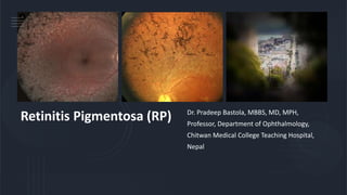
  • 1. Retinitis Pigmentosa (RP) Dr. Pradeep Bastola, MBBS, MD, MPH, Professor, Department of Ophthalmology, Chitwan Medical College Teaching Hospital, Nepal
  • 2. References • American Academy of Ophthalmology • Wolff’s Ocular Anatomy • Myrin Yanoff • Ryan’s Retina 5th Edition • Clinical Ophthalmology by Jack J. Kanski • The Massachusetts Eye and Ear Infirmary Illustrated Manual of Ophthalmology • AIOS Ready Reckoner in Ophthalmology • Duke Elder Book of Ophthalmology • Various articles from Internet • Presentations of Dr. Pradeep Bastola
  • 3. Differential diagnosis of Night Blindness (Nyctalopia) • Xerophthalmia • Retinitis pigmentosa • Primary open angle glaucoma/Advanced glaucoma • High myopia • Post pan-retinal photocoagulation • Congenital rod dystrophy • Congenital stationary night blindness • Familial night blindness • Pharmacologically constricted pupil • Pontine hemorrhage • Opioid poisoning cases • Cuneiform type of cortical cataract
  • 4. Retinitis pigmentosa (RP) is a clinically and genetically heterogeneous group of inherited retinal disorders characterized by diffuse progressive dysfunction of predominantly rod photoreceptors with subsequent degeneration of cone photoreceptors and the retinal pigment epithelium (RPE). Visual impairment usually manifests as night blindness and progressive visual field loss. Its prevalence is 1:3000 to 1:5000 RP may be seen in isolation (Typical RP), in association with systemic disease or with atypical presentation (Atypical RP).
  • 5. Inheritance: Sporadic (23%) Autosomal recessive (20%) Autosomal dominant (43%) X Linked (8%) Undetermined Inheritance (6%) Prevalence: 1 in 5000, appears in childhood and slowly progresses No racial predilection, M:F::3:2, usually always bilateral Primary retinal dystrophy affecting the rods more than cones and is usually hereditary ICD-10 H35.5
  • 6. Risk Factors • There are no known risk factors for RP other than genetic predisposition. • RP may occur as an isolated sporadic disorder or be inherited as autosomal dominant, autosomal recessive, or X-linked. • Mutation of the rhodopsin gene has been reported in many RP cases. • RP is also associated with certain systemic disorders which are usually autosomal recessive. • Many genes associated with RP have been identified and patients can undergo genetic testing. • Association with RPE65 is important as there is now effective gene therapy available for these patients.
  • 7. Molecular genetics of RP • More than 100 gene loci that cause RP have been mapped or identified. • Genes that cause RP can be categorized into those that affect the phototransduction cascade, the retinoid cycle, photoreceptor structure, or other biological function of photoreceptor and retinal pigment epithelium. • The most frequent known causes are mutations in the rhodopsin (phototransduction cascade), USH2A (photoreceptor structure), or RPGR (maintenance of cilia or ciliated cells with a possible role in trafficking) genes. • Patients with the same gene defect can have variable severity of disease at a given age. • Despite recent advances, about 50% of cases still have an unknown molecular genetic basis. There is a genetic treatment for RPE65 defects.
  • 8. Clinical features Symptoms/Signs  Night blindness (Nyctalopia): Diminution of vision in dim light  Diminution of vision (Painless gradual, progression depends upon the type of inheritance, AD with best prognosis and AR with worst prognosis)  Dark adaption (affected lately)  Peripheral visual field loss leading to tubular vision in advanced cases
  • 9. Fundus changes (Important) – Triad of pigments, attenuated arterioles and waxy pale optic disc • Retinal pigmentary changes: Typically, perivascular resembling bone corpuscles in shape found in equatorial region but later spread both anteriorly and posteriorly. • Retinal arterioles: Attenuated and later become thread like • Optic disc: Waxy pale but consecutive type of optic atrophy • Presence of Colloid bodies, choroidal sclerosis, cystoid macular edema, atrophic or cellophane maculopathy are also been seen in retina
  • 10. Visual field changes • Ring shaped scotoma later the scotoma increase anteriorly and posteriorly and finally a central tubular vision is only left. • Eventually it is also lost, and the patient becomes blind. ERG/EOG: Sub normal eventually extinguished
  • 11. Progression of retinitis pigmentosa Fine dust like pigmentation Perivascular ‘bone-spicule’ pigmentation Initially mid-peripheral Unmasking of large choroidal vessels Optic disc pallor Maculopathy Arteriolar attenuation Anterior and peripheral spread
  • 12. Associations of retinitis pigmentosa (RP) Ocular: Myopia, primary open angle glaucoma, microphthalmos, conical cornea and posterior sub capsular cataract Systemic: 25% patients have systemic association Laurence Moon Bardet Biedl syndrome: RP, obesity, hypogonadism, polydactyly, mental deficiency Bassen-Kornzweig syndrome: AR, abetalipoproteinemia, RP, diarrhea, Microsomal TG Protein (MTP) Deficiency disease.
  • 13. Ocular associations of retinitis pigmentosa Cataract (very common) Keratoconus (uncommon) Optic disc drusen (uncommon) Vitreous degeneration (common) Open-angle glaucoma (uncommon Myopia (common)
  • 14. Cockayne’s syndrome: RP, Infantile deafness, dwarfism, mental retardation, nystagmus, ataxia Refsum’s disease: RP, Peripheral neuropathy, cerebellar ataxia Usher’s syndrome: RP and labyrinthine deafness Hallgren’s syndrome: RP, Vestibulo cerebellar ataxia, congenital deafness, mental deficiency Kearns- Sayre Syndrome: RP, AV conduction defects, Mitochondrial transmission, progressive external Ophthalmoplegia, premature death
  • 15. Atypical RP • Retinitis pigmetosa sine pigmento: All features of RP but no visible pigments • Sectoral RP: Only a sector of retina involved • Pericentric RP: Pigments around the macula, rest all features present • Retinitis Punctata albescens: Presence of innumerable while dots scattered all over the retina other features include attenuated vessels, night blindness and visual field changes. • Pigmented paravenous chorio-retinal atrophy: Retinal pigments along the veins, usually sporadic, etiology unknown
  • 16. Retinitis punctata albescens Presents - usually under 30 years of ages • Scattered white dots extending from posterior pole to periphery • Subsequent development of ‘bone-spicule’ pigmentation Probably variant of retinitis pigmentosa Prognosis - poor ERG - reduced
  • 17. Fundus albipunctatus Inheritance - recessive Multitude of tiny yellow-white spots Extend from posterior pole to mid-periphery Congenital stationary night blindness Prognosis - excellent ERG - reduced Fovea spared
  • 19. Diagnosis The diagnosis of retinitis pigmentosa is established when the following findings are present. • Bilateral involvement (can be asymmetric) • Impairment of night vision and loss of peripheral vision • Rod dysfunction evidenced by elevated rod final threshold on dark adaptation and/or rod responses on ERG testing that are either reduced in b-wave amplitude and prolonged in implicit time or are essentially non-detectable (extinguished ERG) • Progressive loss in photoreceptor function
  • 20. Investigations • Visual fields: Defects leading to tubular vision in advanced RP • Electroretinogram (ERG): Reduction in a and b wave amplitudes • Dark adaptometry: Prolonged in RP • Electrooculogram (EOG): Abnormal in RP • Optical coherence tomography (OCT): Macular edema detection • Fundus fluorescein angiography (FFA): Detects early deterioration of RPE
  • 21. Treatment • Vitamin A, Vit E, and Placental extracts to stop progression • Correct refractive error • Acetazolamide, steroids, anti VEGF for cystoid macular edema (CME) • Low vision aid • Rehabilitation • Genetic counselling, and gene therapy • FDA approved Humanitarian Device, called the ARGUS II implant, which may help patients with end-stage RP, only indicated in RP patients with a visual acuity (VA) of perception of light to NPL. CurrentlythereisnocureforRetinitisPigmentosa
  • 22. Prognosis • Autosomal dominant RP has the best prognosis, with most patients under 30 years having a visual acuity of 20/30 or better. • X-linked is the most severe form with appreciable impairment of central visual acuity to 20/200 or less by the fifth decade of life. • Autosomal recessive and sporadic cases were intermediate in severity.
  • 23. Further reading Boyd K, Vemulakonda GA. Retinitis Pigmentosa. American Academy of Ophthalmology. EyeSmart® Eye health. https://www.aao.org/eye- health/diseases/retinitis-pigmentosa-list. Accessed March 25, 2019.
  • 25. Definition (World Health Organization, 1976) • Xerophthalmia refers to the spectrum of ocular disease caused by severe Vitamin A deficiency (VAD). • Vitamin A serves several essential functions in the eye, and deficiency can lead to a constellation of ocular signs and symptoms that affect the conjunctiva, cornea, and retina. • Xerophthalmia continues to be a leading cause of preventable blindness in developing countries. • Xerophthalmia signifies a severity of VAD that causes significant morbidity and mortality from malnutrition and increased susceptibility to mucosal infections.
  • 26. Epidemiology • VAD is among the leading causes of blindness worldwide, estimated to blind half a million children each year • The World Health Organization (WHO) estimated that 228 million children have VAD, causing 1-3 million childhood deaths and 5-10 million cases of eye disease. • VAD is especially prevalent in Africa and South-East Asia, where young children and pregnant women are disproportionally affected
  • 27. VAD usually involves a malabsorptive process, such as inflammatory bowel disease or post- gastric bypass surgery, or a severely restrictive diet. VAD is a public Health problem in more than half of all the countries. Vitamin A deficiency affects approximately 21 percent of the developing world's preschool- aged children and leads to the deaths of over 800,000 women and children each year. Prevalence of VAD very low in developed world, in the US (2013), 0.3% According to Nepal Blindness Survey (NBS, 1981), nutritional cause (0.9%) was also a leading cause of blindness, Xerophthalmia was the major nutritional deficiency noted then. Prevalence of VAD in Nepal before Nepal National Vitamin A program was 2-8%.
  • 28. Nepal National Vitamin A Program • Nepal Government has initiated National Vitamin A Program (NVAP) in 1993 to improve Vitamin A status of the children aged 6‐59 months and reduce child mortality. • This program has been recognized as a global success story of public health interventions with consistent coverage over 85%. • It initially covered 8 districts and was scaled up nationwide by 2002. • FCHVs distribute the capsules to the targeted children twice a year through a campaign style activity.
  • 29. • Biannual deworming tablets distribution: Family Welfare Division, DoHS has been implemented biannual deworming tablets distribution to the children aged 12-59 months aiming to reduce childhood anemia with control or parasitic infestation through public health measures. • This activity is integrated with biannual Vitamin A supplementation to the children aged 6-59 months, which takes place nationally in every ward on first week of Baisakh and Kartik each year.
  • 30. Government’s initiations to control of vitamin A deficiency in Nepal • The biannual supplementation of high dose vitamin A capsules to 6–59 months children. • Postpartum vitamin A supplementation for mothers within 42 days of delivery. • Strengthen implementation of vitamin A treatment protocol for severe malnutrition, persistent diarrhoea, measles and xerophthalmia. • Nutrition education to promote dietary diversification and consumption of vitamin A rich foods. • Ensuring the availability of vitamin, A capsules at health facilities. • Increase awareness of importance of vitamin A supplementation. • The biannual distribution of vitamin A capsules to 6- to 59 months children through FCHVs. • Advocate for increased home production, consumption and preservation of vitamin A rich foods. • Strengthen the use of the vitamin A Treatment protocol.
  • 31. Etiology of Xerophthalmia (VAD) Reduced intake of Vitamin A Impaired absorption of Vitamin A Reduced Storage of Vitamin A • Inadequate food supply • Chronic alcoholism • Highly selective dieting • Dysphagia • Mental illness • Crohn's disease • Celiac disease • Pancreatic insufficiency • Short bowel syndrome • Chronic diarrhea • Inflammatory bowel disease • Upper gastrointestinal surgery • Giardiasis • Abetalipoproteinemia • Liver disease • Cystic fibrosis
  • 32. Recommended daily intake of Vitamin A • The recommended dietary allowance of vitamin A is 700ug/day in females and 900ug/day in the males. • For children and pregnant or lactating women, the recommended amount is 300-900, 770, and 1300ug/day, respectively. • Children aged 1-5 years old require a minimum of 200ug/day to prevent symptomatic VAD. • Fat soluble Vitamin, 80-90% stored in the Liver Dietary sources of preformed vitamin A include dark leafy greens, orange-colored vegetables, fish liver oils, liver, egg yolks, butter, and vitamin A-fortified dairy products.
  • 33. Risk factors • Living in an endemic area • Low socioeconomic status • Malnutrition • Maternal malnourishment (affects Vitamin A concentration in breastmilk) • Zinc deficiency: inadequate zinc can depress the hepatic synthesis of retinol-binding protein (RBP), which is required for metabolism of retinol from the liver. Zinc may also play a role in the conversion of beta-carotene to retinol via the enzyme 15-15’-dioxygenase. • Co-existing measles or other respiratory/diarrheal illness • Vitamin A has several essential functions in the body, including cell development, metabolism, immune function, vision, and reproductive function.
  • 34. Diagnosis (WHO Classification of Xerophthalmia, 1976 based on clinical signs) Grade of xerophthalmia Peak age group (years) Type of deficiency XN: Night blindness 2-6; adult women Longstanding. Not blinding X1A: Conjunctival xerosis 3-6 Longstanding. Not blinding X1B: Bitot's spots 3-6 Longstanding. Not blinding X2: Corneal xerosis 1-4 Acute deficiency. Can be blinding X3A: Corneal ulcer <1/3 cornea 1-4 Severe acute deficiency. Blinding X3B: Corneal ulcer/keratomalacia1/3 cornea or greater 1-4 Severe acute deficiency. Blinding XS: Corneal scarring (from X3) >2 Consequence of corneal ulceration XF: Xerophthalmos fundus Adults Longstanding. Not blinding. Rare
  • 35. Night blindness (Grade XN): Defective vision in dim light or night blindness is one of the most common manifestations of VAD, especially in children aged 2-6 years or pregnant or lactating women. Although it is considered one of the earliest manifestations, children with VAD may develop one of the more severe signs, such as corneal ulcers, after infection or diarrhoea without any of the classically early signs. Children may not be able to verbalize their symptoms, and parents need to be asked if they have noticed their children behaving differently in the dark, e.g., becoming less active or fearful. Night blindness is both a sensitive and specific indicator for serum retinol levels. Conjunctival xerosis (Grade X1A): Conjunctival xerosis is characterized by a dull and dry appearance of the conjunctiva with slight wrinkling. It is caused by the loss of goblet cells and insufficient mucin secretion, and it can be subtle and difficult to detect clinically.
  • 36. Bitot spots (Grade X1B): Bitot spots are collections of desquamated, keratinized epithelial cells mixed with the gas-forming bacteria Corynebacterium xerosis. • They appear as triangular patches of foamy, whitish, opaque deposits, typically located on the bulbar conjunctiva near the limbus at the 3 and 9 o’clock positions. They are more common temporally. Corneal xerosis (Grade X2): Conjunctival xerosis is characterized by a dull and hazy appearance of the cornea that is caused by drying of the cornea secondary to conjunctival gland dysfunction. • It may initially present with bilateral punctate corneal epithelial erosions, and it can quickly progress to the stage of corneal ulceration. • Up to this stage, high-dose Vitamin A supplementation can result in the full preservation of vision.
  • 37. Corneal ulceration (Grade X3A and B): Corneal xerosis can lead corneal ulceration and melting if not treated urgently. Keratomalacia, the melting away of the cornea by liquefactive necrosis, is the most severe form of xerophthalmia, which can perforate and destroy the cornea in just a matter of days. A child who appears relatively healthy but develops keratomalacia should be questioned about a recent history of measles or diarrhea, which could rapidly deplete already deficient vitamin A stores. Corneal scar (Grade XS): Corneal scarring due to VAD is often symmetric and bilateral. Other causes of corneal scarring must be ruled out.
  • 38. Xerophthalmic fundus (Grade XF): Prolonged VAD can lead to structural changes in the retina. Fundus changes appear as small, white, deep retinal lesions scattered throughout the posterior pole.
  • 39. Work up/Investigations • History, and physical examination • Clinical signs • Serum vitamin A/retinol (reference range: 20-60 mcg/dL). (VAD - related ocular symptoms have been shown to develop at concentrations <10mcg/dL) • Serum retinol binding protein (reference range: 30-75 ug/ml). • Serum zinc (reference range: 75-120 mcg/dL)
  • 40. • Dark adaptometry and night vision threshold tests - Prolonged • Electroretinogram (ERG): Retinopathy from VAD is associated with decreased amplitude • Impression cytology: Conjunctival specimens can be viewed for the presence of goblet cells, a decrease in normal amount is considered an effective measurement of VAD • Liver biopsy: Considered the gold standard for evaluating total body vitamin A, not practiced except for research
  • 41. Differential Diagnosis • Parasitic eye disease, e.g., Acanthamoeba keratitis or Onchocerciasis • Trachoma • Allergic conjunctivitis • Viral conjunctivitis • Dry eye syndrome • Retinitis pigmentosa and other retinal dystrophies that can present with night blindness • Pinguecula/pterygium (may resemble Bitot spots)
  • 42. Treatment (Oral or Intramuscular Vitamin A supplement) Keratomalacia should be treated as a medical emergency, as it is an indicator of very severe VAD Vitamin A Dosage International Units (IU), (IM dose half of oral dose) Young infants 0-5 months1 50,000 Older infants 6-11 months (less than 8kgs)1 100,000 Children (males: 12 months or more; females 12 months to 12 year and 50 y or more)1 200,000 Women (13-49 y) with night blindness and/or Bitot's spots 10,000 every day or 25,000 every week for at least 3 mo Women (13-49 y) with active corneal lesions 200,000 on days 1, 2, and 14 Vitamin A treatment regimens. 1 Schedule: severe malnutrition, day 1; measles, days 1 and 2; xerophthalmia, days 1, 2, and 14.
  • 43. Patients with concomitant zinc deficiency should also undergo zinc supplementation. Keratomalacia in Xerophthalmia is treated in line with the Bacterial Corneal Ulcer. VAD associated with malabsorptive, or other processes is treated based on the severity of deficiency and provider discretion, usually involving daily treatment. Localized ocular treatment includes intense lubrication, topical retinoid acid, and management of perforation. Patient education is a large component of management, and patients and their families should be educated on dietary sources of vitamin A and well-balanced, nutrient-rich diets. Alcohol abuse should be addressed if applicable. In resource poor countries, large-scale vitamin A supplementation programs are in place eg, Nepal, India, Sub Saharan Africa
  • 44. Prognosis Nearly 2/3 of children with keratomalacia die within months. Mortality in children with night blindness is triple the mortality found in children with subclinical VAD, and mortality in children with both Bitot spots and night blindness is reported to be nine times that of children with subclinical VAD. With prompt treatment of high-dose vitamin A, the early Ophthalmologic signs, such as a rod function, conjunctival xerosis, and night blindness, can resolve completely within about 2 months of supplementation, without long-term sequelae. Corneal xerosis and ulceration may also improve; however, they can lead to scarring and permanent vision loss.