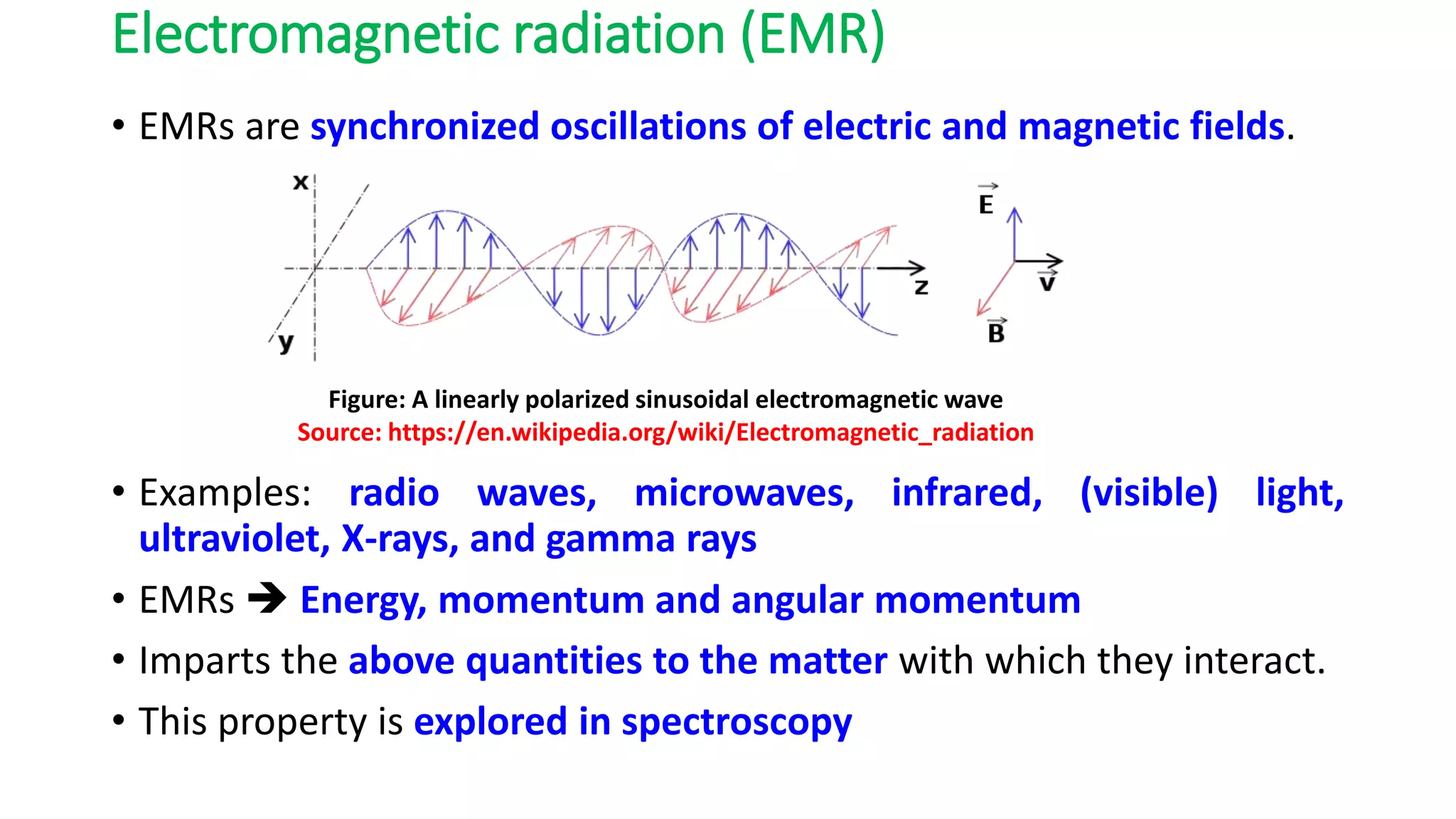
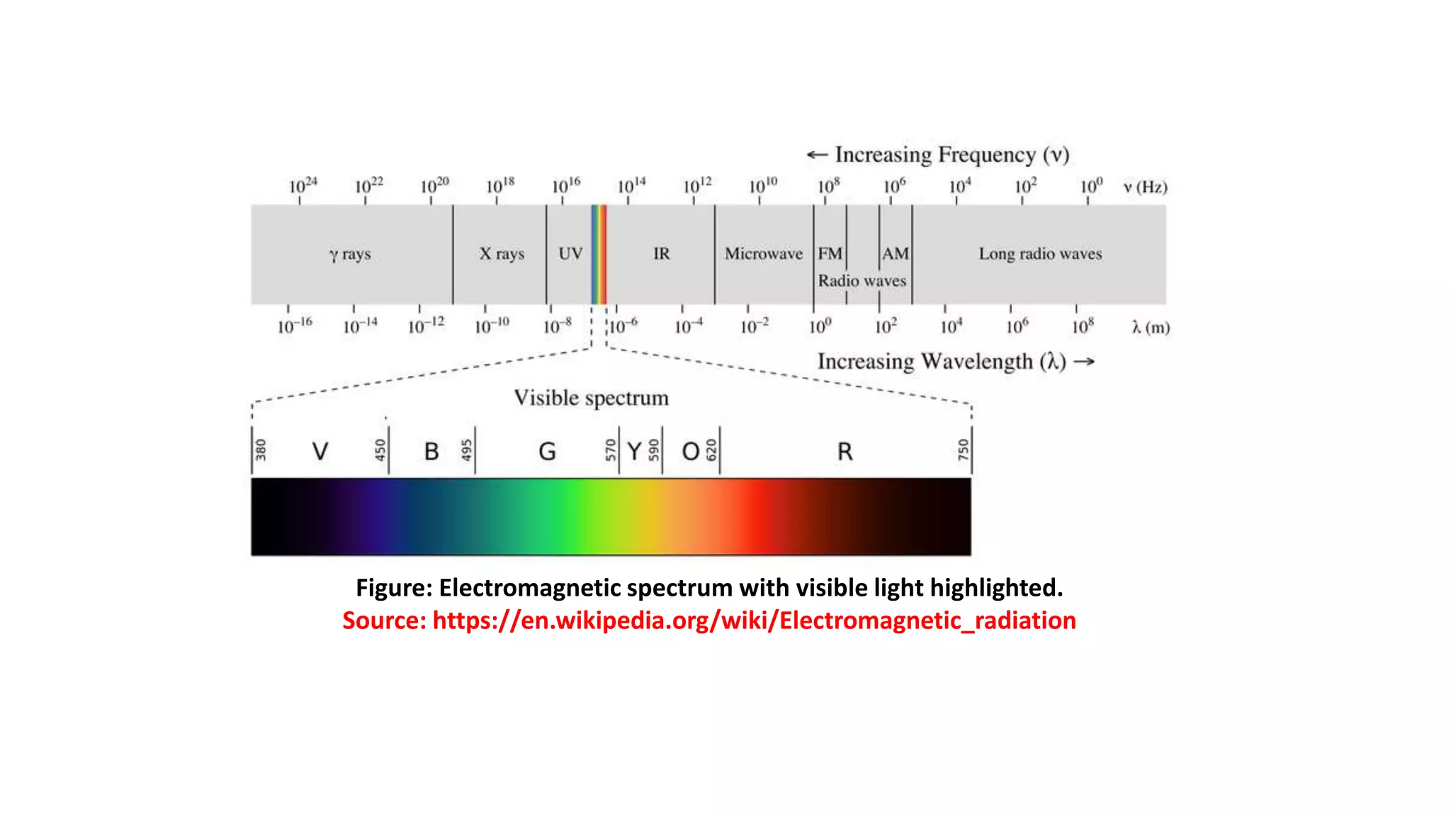
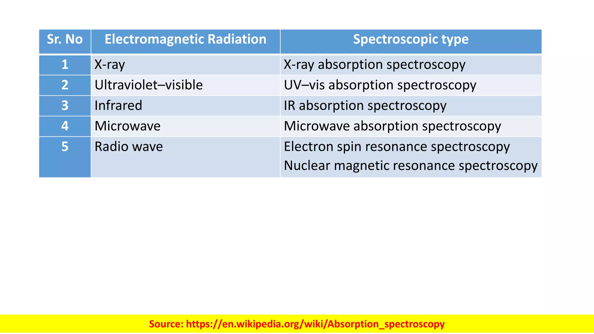
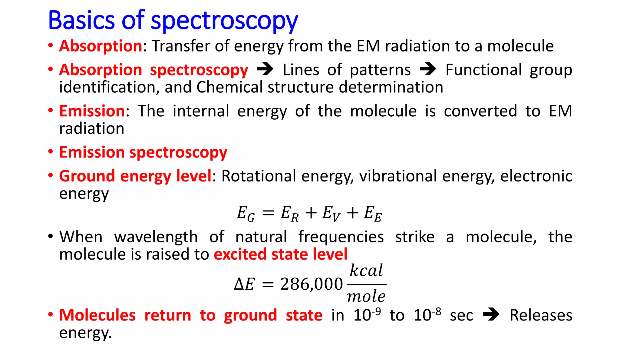
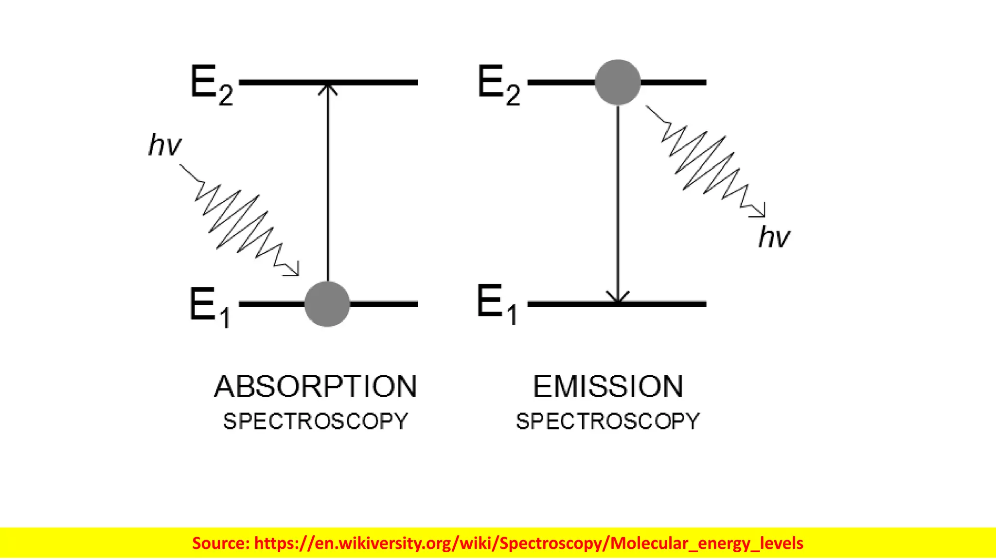
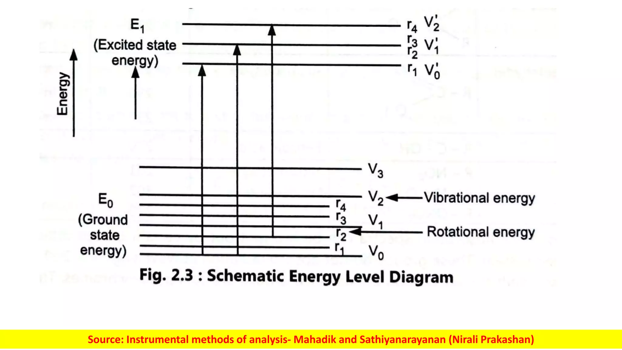
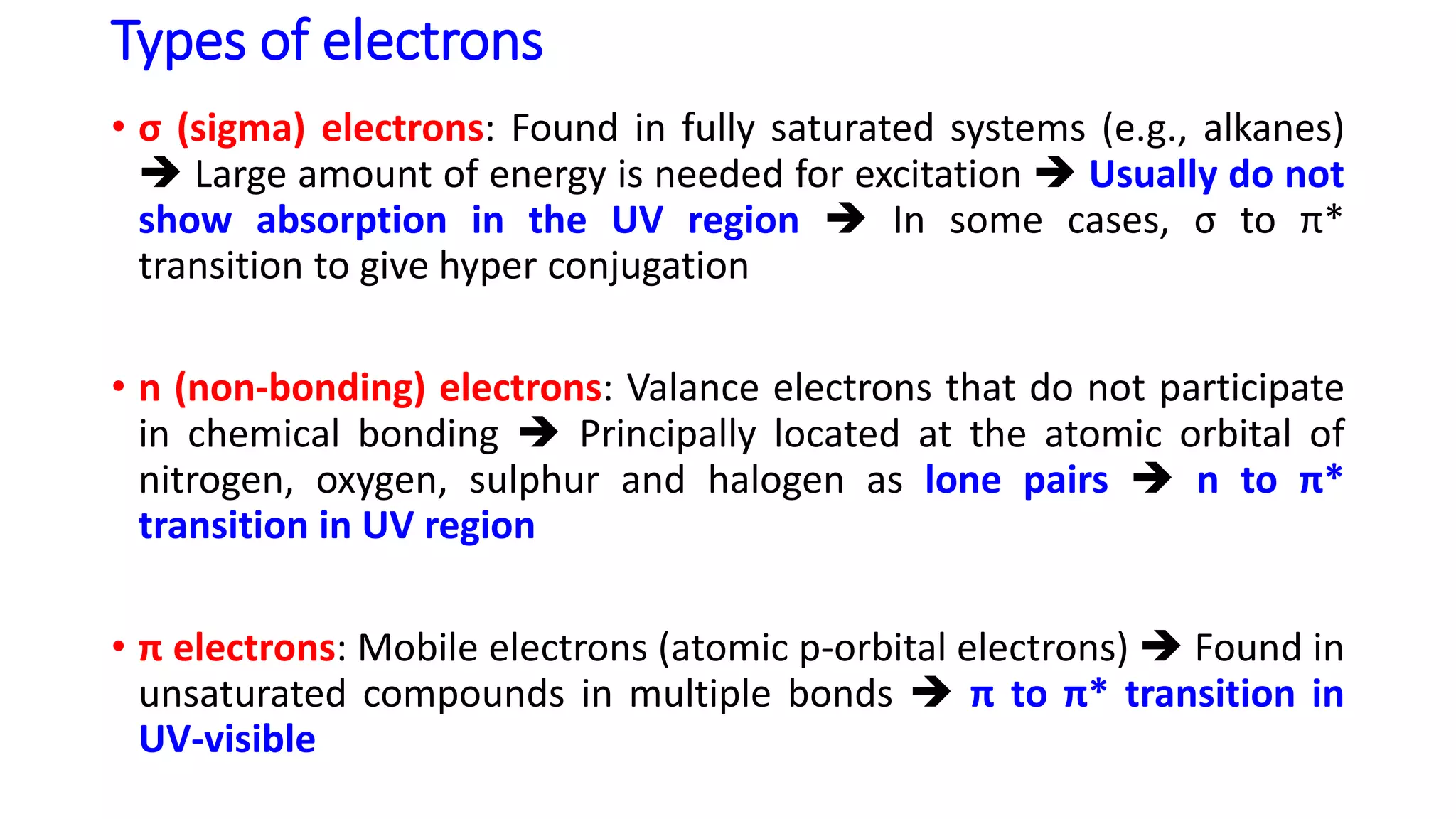
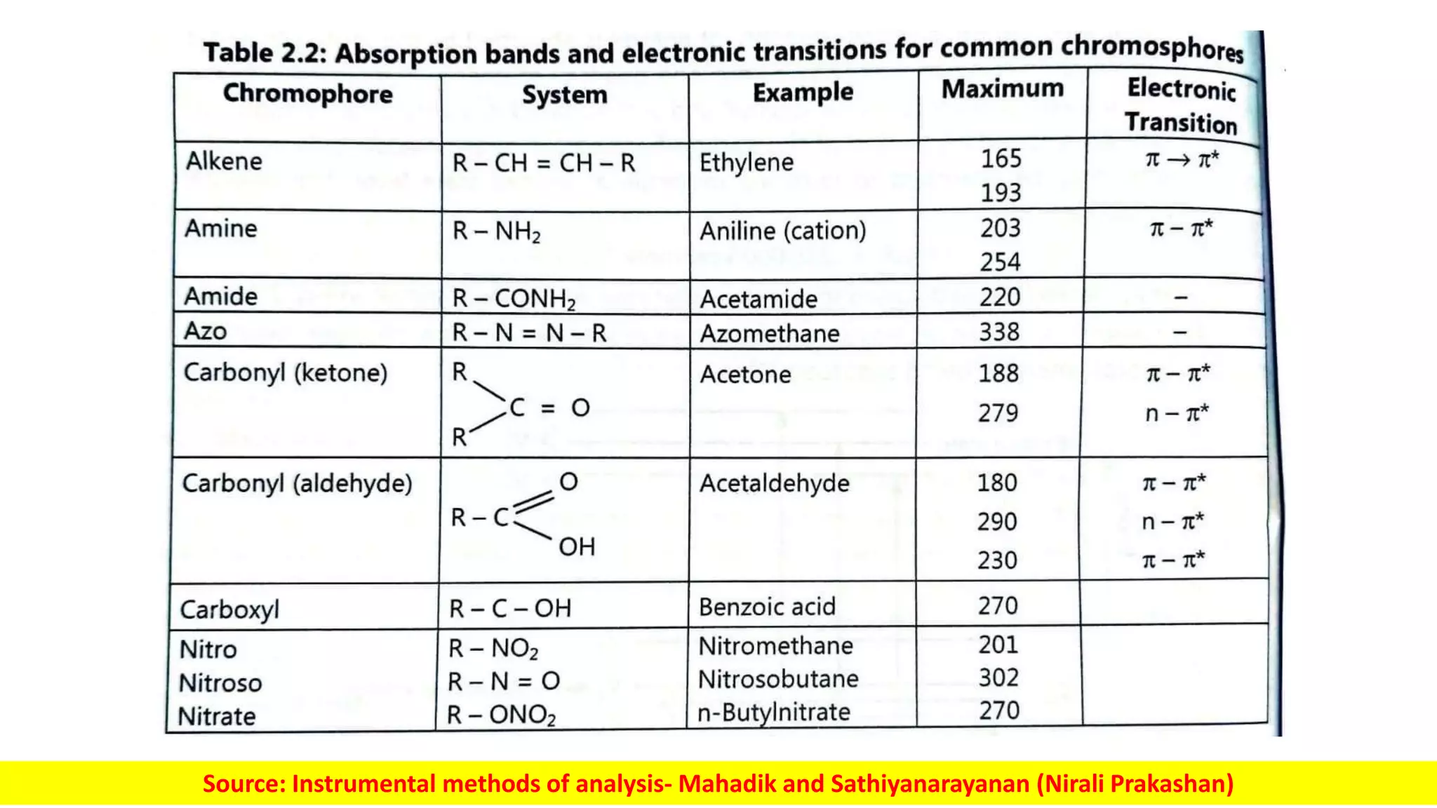
2. Electromagnetic radiation (EMR)
• EMRs are synchronized oscillations of electric and magnetic fields.
• Examples: radio waves, microwaves, infrared, (visible) light,
ultraviolet, X-rays, and gamma rays
• EMRs Energy, momentum and angular momentum
• Imparts the above quantities to the matter with which they interact.
• This property is explored in spectroscopy
Figure: A linearly polarized sinusoidal electromagnetic wave
Source: https://en.wikipedia.org/wiki/Electromagnetic_radiation
4. Class Frequency Wavelength Energy per photon
Ionizing radiation γ Gamma rays 300 EHz 1 pm 1.24 MeV
30 EHz 10 pm 124 keV
HX Hard X-rays
3 EHz 100 pm 12.4 keV
SX Soft X-rays
300 PHz 1 nm 1.24 keV
30 PHz 10 nm 124 eV
EUV Extreme ultraviolet
3 PHz 100 nm 12.4 eV
NUV Near ultraviolet
300 THz 1 μm 1.24 eV
NIR Near infrared
30 THz 10 μm 124 meV
MIR Mid infrared
3 THz 100 μm 12.4 meV
FIR Far infrared
300 GHz 1 mm 1.24 meV
Microwaves and radio
waves
EHF Extremely high frequency
30 GHz 1 cm 124 μeV
SHF Super high frequency 3 GHz 1 dm 12.4 μeV
UHF Ultra high frequency 300 MHz 1 m 1.24 μeV
VHF Very high frequency 30 MHz 10 m 124 neV
HF High frequency 3 MHz 100 m 12.4 neV
MF Medium frequency 300 kHz 1 km 1.24 neV
LF Low frequency 30 kHz 10 km 124 peV
VLF Very low frequency 3 kHz 100 km 12.4 peV
ULF Ultra low frequency 300 Hz 1000 km 1.24 peV
SLF Super low frequency 30 Hz 10000 km 124 feV
ELF Extremely low frequency 3 Hz 100000 km 12.4 feV
Table: The range of frequencies (the spectrum) of electromagnetic radiation and their respective wavelengths and photon energies
Source: https://en.wikipedia.org/wiki/Electromagnetic_spectrum
5. Sr. No Electromagnetic Radiation Spectroscopic type
1 X-ray X-ray absorption spectroscopy
2 Ultraviolet–visible UV–vis absorption spectroscopy
3 Infrared IR absorption spectroscopy
4 Microwave Microwave absorption spectroscopy
5 Radio wave Electron spin resonance spectroscopy
Nuclear magnetic resonance spectroscopy
Source: https://en.wikipedia.org/wiki/Absorption_spectroscopy
6. Basics of spectroscopy
• Absorption: Transfer of energy from the EM radiation to a molecule
• Absorption spectroscopy Lines of patterns Functional group
identification, and Chemical structure determination
• Emission: The internal energy of the molecule is converted to EM
radiation
• Emission spectroscopy
• Ground energy level: Rotational energy, vibrational energy, electronic
energy
𝐸𝐺 = 𝐸𝑅 + 𝐸𝑉 + 𝐸𝐸
• When wavelength of natural frequencies strike a molecule, the
molecule is raised to excited state level
∆𝐸 = 286,000
𝑘𝑐𝑎𝑙
𝑚𝑜𝑙𝑒
• Molecules return to ground state in 10-9 to 10-8 sec Releases
energy.
10. Types of electrons
• σ (sigma) electrons: Found in fully saturated systems (e.g., alkanes)
Large amount of energy is needed for excitation Usually do not
show absorption in the UV region In some cases, σ to π*
transition to give hyper conjugation
• n (non-bonding) electrons: Valance electrons that do not participate
in chemical bonding Principally located at the atomic orbital of
nitrogen, oxygen, sulphur and halogen as lone pairs n to π*
transition in UV region
• π electrons: Mobile electrons (atomic p-orbital electrons) Found in
unsaturated compounds in multiple bonds π to π* transition in
UV-visible
11. Absorption spectra
• Due to electronic level transitions in the groups present in a
molecule.
• Chromophore: The group that absorbs in visible and UV wavelengths
• Auxochrome: Groups that have non-bonding valence electrons, and
do not absorb radiation at wavelengths >200 nm Modify and shift
the absorption bands of the chromophores
• Spectral shifts
Source: Instrumental methods of analysis- Mahadik and Sathiyanarayanan (Nirali Prakashan)