
Recommended
More Related Content
What's hot
What's hot (20)
Similar to Causes, Risk Factors and Prevention of Peri-implantitis
Similar to Causes, Risk Factors and Prevention of Peri-implantitis (20)
More from PiyaliBhattacharya10
More from PiyaliBhattacharya10 (13)
Recently uploaded
Recently uploaded (20)
Causes, Risk Factors and Prevention of Peri-implantitis
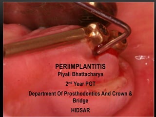
- 1. Piyali Bhattacharya 2nd Year PGT Department Of Prosthodontics And Crown & Bridge HIDSAR PERIIMPLANTITIS
- 2. Peri-implantitis is a progressive and irreversible disease of implant-surrounding hard and soft tissues and is accompanied with bone resorption, decreased osseointegration , increased pocket formation and purulence.* *Khammissa RAG, Feller L, Meyerov R, Lemmer J: Peri-implant mucositis and peri-implantitis: clinical and histopathological characteristics and treatment. SADJ 2012, 67(122):124–126 Definition
- 4. Peri-implant Mucosa Physiological Periodontium 1. Desmosomes and hemidesmosomes of epithelium and junctional epithelium (biological width) are linked with the contact surface 2. Direct bone-to-implant contact 3. Subepithelially more collagen fibers and less fibroblasts/vessels 4. Parallel collagen fibers in relation to implant surface 5. Biologic width 3.08 mm (includes sulcus) 6. Probing depth 2.5–5.0 mm (depending on previous soft tissue depth 7. Vascularity is greater 8. Connective tissue consist of Only two groups: parallel and circular fibers; no attachment to the implant surface ↑ collagen, ↓fibroblasts 1. Desmosomes and hemidesmosomes of epithelium and junctional epithelium (biological width) are linked with the contact surface 2. Anchoring system of root cementum, alveolar bone and desmodontic fibers 3. Subepithelially more fibroblasts and vessels 4. Dentogingival, dentoperiostal, circular and transseptal fiber orientation 5. Biologic width 2.04–2.91 mm 6. Probing depth 3 mm in health 7. Vascularity is greater 8. Connective tissue consist of 12 groups: six insert perpendicular to tooth surfaces↓collagen, ↑ fibroblasts *An Implant Is Not a Tooth: A Comparison of Periodontal Indices, Dental Implant Prosthetics, 2nd Edition, Carl.E.Misch
- 5. IMPLANT QUALITY SCALE: ICOI CONSENSUS PISA CONFERENCE, ITALY, 2008. Group Management Clinical consideration I.Success(optimal health) Normal maintenance a. No pain or tenderness upon function b. 0 mobility c. <2 mm radiographic bone loss from initial surgery d. Probing depth <5 mm e. No exudate history II. Survival (satisfactory health) Reduction of stresses, Shorter intervals between hygiene appointments, Gingivoplasty, Yearly radiographs No pain 0 mobility 2–4 mm radiographic bone loss Probing depth 5–7 mm No exudate history
- 6. III. Survival (compromised health) Reduction of stresses, Drug therapy (antibiotics, chlorhexidine) Surgical reentry and revision, Change in prosthesis or implants a. No pain upon function b. 0 mobility c. Radiographic bone loss >4 mm d. Probing depth >7 mm e. May have exudate history IV. Failure (clinical or absolute failure) Removal of implant Pain upon function Mobility Radiographic bone loss > 1 2 length of implant Uncontrolled exudate No longer in mouth *An Implant Is Not a Tooth: A Comparison of Periodontal Indices, Dental Implant Prosthetics, 2nd Edition, Carl.E.Misch
- 7. ETIOPATHOGENESIS Zitzmann et al. quantified the incidence of the development of peri-implantitis in patients with a history of periodontitis almost six times higher than in patients with no history of periodontal inflammation. *Zitzmann NU, Walter C, Berglundh T: Ätiologie, Diagnostik und Therapie der Periimplantitis – eine Übersicht. Deutsche Zahnärztliche Zeitschrift 2006, 61:642–649
- 8. • Based on the Consensus Report of the Sixth European Workshop in Periodontology, Lindhe & Meyle reported an incidence of mucositis of up to 80% and of peri-implantitis between 28% and 56% • The fact that bone remodeling processes often result in marginal bone loss during the first weeks after abutment connection which cannot be regarded as peri-implantitis. • This led to the recommendation to take a radiograph after insertion of the suprastructure and to consider it as a basis for any future assessment of peri-implant bone loss. *Lindhe J, Meyle J: Peri-implant diseases: consensus report of the sixth european workshop on periodontology. J Clin Periodontol 2008, 35:282–285.
- 9. • In contrast to periodontitis, peri-implantitis lesions harbor bacteria that are not part of the typical periodontopathic microbiota. In particular, Staphylococcus aureus appears to play a predominant role for the development of a peri-implantitis. This bacterium shows an high affinity to titanium and has according to the results of Salvi et al. * Salvi GE, Fürst MM, Lang NP, Persson GR: One-year bacterial colonization patterns of Staphylococcus aureus and other bacteria at implants and adjacent teeth. Clin Oral Implants Res 2008, 19:242–248.
- 10. RISK FACTORS AND PREVENTION • Implant loss may occur as “early implant loss” up to one year after implant insertion and “delayed implant loss” with a time period of more than one year after implant insertion - • Smoking with additional significantly higher risk of complications in the presence of an positive combined IL-1 genotype polymorphism.** • History of periodontitis. • Lack of compliance and limited oral hygiene (including missing checkups). • Charyeva O, Altynbekov K, Zhartybaev R, Sabdanaliev A: Long-term dental implant success and survival–a clinical study after an observation period up to 6 years. Swed Dent J 2012, 36:1–6. ** Gruica B, Wang H-Y, Lang NP, Buser D: Impact of IL-1 genotype and smoking status on the prognosis of osseointegrated implants. Clin Oral Implants Res 2004, 15:393–400.
- 11. • Systemic diseases (e.g. mal-adjusted diabetes mellitus, cardiovascular disease, immuno- suppression). • Soft tissue defects or poor-quality soft tissue at the area of implantation (e.g. lack of keratinized gingiva).** • History of one or more failures of implants. • Heitz-Mayfield LJA: Peri-implant diseases: diagnosis and risk indicators. J Clin Periodontol 2008, 35:292–304. ** Lagervall M, Jansson LE: Treatment outcome in patients with peri-implantitis in a periodontal clinic- a retrospective study. J Periodontol 2012, 84:1365–1373.
- 12. ETIOLOGY OF RETROGRADE PERI- IMPLANTITIS* • It is defined as clinically symptomatic peri-apical lesion that develops within the first few months after implant insertion while the coronal portion of the implant sustains a normal bone to implant interface. It can be an active or an inactive lesion. • Overpreparation or overheating of osteotomy sites • Presence of a foreign body Iatrogenic causes (e.g. “cementitis”). • Persistent or incompletely debrided periapical lesion. *Quirynen M, Vogels R, Alsaadi G, Naert I, Jacobs R, van Steenberghe D,et al. Predisposing conditions for retrograde peri-implantitis, and treatment suggestions. Clin Oral Implants Res 2005;16:599-608.
- 13. • Moreover, smoking has been shown to be a predictor for implant failure • The extent of osseointegration as well as the oral hygiene around dental implants was found to be reduced among smokers SMOKING • The presence of periodontitis or cigarette smoking increased the risk for peri-implantitis up to 4.7-fold as reported by Wallowy et al. *Clementini M, Rossetti PH, Penarrocha D, Micarelli C, Bonachela WC, Canullo L: Systemic risk factors for peri-implant bone loss: a systematic review and meta-analysis. Int J Oral Maxillofac Surg 2014, 43:323–334. **Kasat V, Ladda R: Smoking and dental implants. J Int Soc Prev Commun Dent 2012, 2:38–41
- 14. GENDER, AGE, MAXILLA vs MANDIBLE • Evidence of predictors for implant success such as gender or age could not be found but for the jaw of treatment (maxillary versus mandibular implants). • In a study by Vervaeke et al. maxillary implants were at a significantly higher risk for peri-implant bone loss compared to mandibular implants
- 15. HISTORY OF PERIODONTITIS • The type of dentition (edentulous versus partially edentulous) may influence the colonization of peri-implant tissues with periodontal pathogens.
- 16. • Across an observation period of 10 years in a group of patients with periodontitis, the previously eliminated bacterial strains of Aggregatibacter actinomycetemcomitans and Porphyromonas gingivalis could again be detected in the oral mucosa. * Malo P, Rigolizzo M, Nobre M, Lopes A, Agliardi E: Clinical outcomes in the presence and absence of keratinized mucosa in mandibular guided implant surgeries: a pilot study with a proposal for the modification of the technique. Quintessence Int 2013, 44:149–157.
- 17. CEMENTITIS • “cementitis” may be regarded as the most important identifiable iatrogenic risk factor since its first description by Wilson et al. in 2009 . Wilson TG Jr: The positive relationship between excess cement and peri-implant disease: a prospective clinical endoscopic study. J Periodontol 2009, 80:1388–1392
- 18. • The latter group revealed that residual dental cement in a group of patients with clinical or radiographic signs of peri-implant disease was present in 81% of the sites. After its removal, clinical signs were absent in 74% of the affected sites. • Korsch et al. found that the removal of cement remnants led to a decrease of the inflammatory response by almost 60%* * Korsch M, Obst U, Walther W: Cement-associated peri-implantitis: a retrospective clinical observational study of fixed implant-supported restorations using a methacrylate cement. Clin Oral Implants Res 2014, 25:797–802.
- 19. LACK OF KERATINIZED GINGIVA • Lang and Loe advocate a minimum 2 mm of keratinized gingiva and 1 mm of attached gingiva to maintain gingival health * Lang NP, Loe H: The relationship between the width of keratinized gingiva and gingival health, J Periodontol 43:623–627, 1972.
- 20. a number of benefits are present wit keratinized mucosa- a) The color, contour, and texture of the soft tissue drape should be similar around implants and teeth when in the esthetic zone. b) The interdental papillae should ideally fill the interproximal spaces. c) A high smile line often exposes the free gingival margin and interdental papillae zones.
- 21. d) The keratinized tissue is more resistant to abrasion. As a result, hygiene aids are more comfortable to use, and mastication is less likely to cause discomfort. e) In a two-stage protocol, the implant is less likely to become exposed during the healing process. The formation of an interdental/implant papillae is completely unpredictable with mobile un-keratinized tissues.
- 22. • The presence of keratinized tissue next to an oral implant presents some unique benefits compared to natural teeth. Keratinized gingiva has more hemidesmosomes; thus, the JEA zone may be of benefit when in keratinized tissue. Whereas the orientation of collagen fibers in the connective tissue zone of an implant may appear perpendicular to the implant surface, these fibers in mobile, nonkeratinized tissue run parallel to the surface of the implant.
- 23. PLATFORM SWITCH • Abutment is located horizontally between implant and crown) can complicate probing and, thus, hide the true extension of peri-implantitis. • Nevertheless, studies have indicated that platform switch might be an important protective factor against peri-implant disease.* * Vandeweghe S, De Bruyn H: A within-implant comparison to evaluate the concept of platform switching: a randomised controlled trial. Eur J Oral Implantol 2012, 5:253–262
- 24. Internal connections with inward located microgap, should be preferred
- 25. IMPLANT LOSS / IMPLANT FAILURES Implant loss can be differentiated on the basis of the following additional factors- • Overloading of the implant, • Faults in material and techniques, • Poor bone quality at the implant area, • Systemic diseases and drug therapies, which inhibit bone modulations according to “Wolff ’s law” (bone density and strength increase with stress - and vice versa). Steigenga JT, al-Shammari KF, Nociti FH, Misch CE, Wang H-L: Dental implant design and its relationship to long-term implant success. Implant Dent 2003, 12:306–317.
- 26. DIAGNOSIS OF PERI-IMPLANTITIS • According to Mombelli and Lang, the parameters to be assessed include: a) Peri-implant probing: A rigid plastic probe is ideal.probing depth around the connective tissue attachment of 1–1.5 mm. * Pulluri, et al.: Treatment for peri-implantitis, International Journal of Oral Health Sciences | Volume 7 | Issue 2 | July-December 2017
- 27. b) BOP: It has been shown that it is not a reliable predictor for progression of periodontal disease, instead its absence is a much better predictor for stability. * Pulluri, et al.: Treatment for peri-implantitis, International Journal of Oral Health Sciences | Volume 7 | Issue 2 | July-December 2017 57
- 28. c) Mobility: Implant mobility is an indication of lack of osseointegration, but it is of no use in diagnosing early implant disease, rather it shows the final stages of de-integration. Periotest or RFA can be used to assess the stability of an implant. * Pulluri, et al.: Treatment for peri-implantitis, International Journal of Oral Health Sciences | Volume 7 | Issue 2 | July-December 2017
- 29. d) Radiography: Bone loss is a definite indicator for peri-implantitis. The distance from implant shoulder to the alveolar bone crest is a reliable parameter providing the radiographs are properly standardized. * Pulluri, et al.: Treatment for peri-implantitis, International Journal of Oral Health Sciences | Volume 7 | Issue 2 | July-December 2017
- 30. SURGICAL TREATMENT OF PERI-IMPLANTITIS A. Surface polishing/implantoplasty The main objective of this therapy is to arrest the progression of the disease and to achieve a maintainable site by the patient. * Pulluri, et al.: Treatment for peri-implantitis, International Journal of Oral Health Sciences | Volume 7 | Issue 2 | July-December 2017
- 31. • Implant topography should be altered with high-speed diamond burs and polishers to produce smooth continuous surfaces. Performed before any osseous resective therapy is initiated and with profuse irrigation Pulluri, et al.: Treatment for peri-implantitis, International Journal of Oral Health Sciences | Volume 7 | Issue 2 | July-December 2017 57
- 32. GUIDED BONE REGENERATION Pros • In 2007, Roos-Jansaker et al. were able to show a good response to therapy (implants pocket reduction of 2.9– 3.4 mm and new bone fill of 1.4–1.5 mm) for peri-implantitis sites treated with either bone grafts alone or bone grafts in conjunction with a resorbable collagen membrane. Cons • However, in a recent systematic review by Sahrmann et al. in 2011 have concluded that complete fill of the bony defect using guided bone regeneration (GBR) seems not to be a predictable outcome. * Smeets et al. , Definition, etiology, prevention and treatment of peri-implantitis – a review Head & Face Medicine 2014, 10:34
- 33. • The mucosal health status is not considered in most studies. Well-controlled trials are further advocated to determine predictable treatment protocols for the successful regenerative treatment of peri-implantitis using GBR technique. * Smeets et al. , Definition, etiology, prevention and treatment of peri-implantitis – a review Head & Face Medicine 2014, 10:34
- 34. • Resective surgery can be carried out for the elimination of peri-implant lesions, whereas regenerative therapies may be applicable for defect filling. • Ostectomy and Osteoplasty: It is indicated in moderate to severe horizontal bone loss, moderate (<3 mm) vertical bone defects (1 and 2 wall bone defects), reduce the overall pocket depth and implant position in the un-esthetic area. Peri-implant Resective Therapy
- 35. • Resective surgery has been shown to be effective in reduction of BOP, PDs and clinical signs of inflammation. • Implantoplasty can be considered a useful adjunct along with resective surgery to smooth and decontaminate portions of an implant left exposed by peri-implantitis lesions.
- 36. TREATMENT OF RETROGRADE PERI-IMPLANTITIS • Implant extraction, • peri-apical surgery, • debridement, • regenerative procedure, • local decontamination (antimicrobials/lasers), and • use of antibiotics. Pulluri, et al.: Treatment for peri-implantitis, International Journal of Oral Health Sciences | Volume 7 | Issue 2 | July-December 2017 57
- 37. CONSENSUS STATEMENT: NONSURGICAL INTERVENTION THIRD EAO CONSENSUS CONFERENCE 2012: • Peri-implant mucositis can be treated successfully. • Peri-implantitis has limited success with mechanical debridement • Clinical recommendations • Patients should be monitored regularly for: • Plaque control • Signs of peri-implant inflammation • BOP • A regular maintenance program for the long-term management of peri-implantitis lesions Pulluri, et al.: Treatment for peri-implantitis, International Journal of Oral Health Sciences | Volume 7 | Issue 2 | July-December 2017 57
- 38. CUMULATIVE INTERCEPTIVE SUPPORTIVE THERAPY • In 2004, it was modified and called AKUT-concept by Lang et al. Probing Depth <3 Plaque index <1 and BOP -ve Plaque index > 1 and BOP +ve Mechanical debriding and scaling and polishing A Probing Depth 4-5 mm Antiseptic Cleaning 0.1% chlorohexidine gel 2X daily for 3-4 weeks B C Probing Depth >5mm BOP +ve , no cratering BOP +ve , cratering < 2mm BOP +ve , bone loss >2 mm Local or systemic antibiotic therapy Resective or Regenerative surgery D + + + + + +
- 39. DECISION MAKING TREE FOR PERI-IMPLANTITIS TREATMENT MOBILITY YES NO Failed Implant Remove Implant Bone Loss Yes: Failing Implant No: Ailing Implant Extent of Bone Loss Periimplant Mucositis a. Supra and sub gingival biofilm removal. b. Adjunctive antimicrobials (local and systemic) Surgical treatment: a. Apically positioned flap with osseous resection b. Guided Bone Regeneration c. Flap Surgery >1/2 fixture <1/2 fixture Remove implant Re-develop site Check amount of bone loss >2mm <2mm a. Non surgical treatment: Scaling root, planing b. Surface Decontamination: Antibiotic , laser
- 40. CONCLUSION • A review by Heitz-Mayfield and Andrea Mombelli in 2014 showed that successful treatment outcomes 12 months following therapy of peri-implantitis could be achieved in a majority of patients. Their recommendations include: a) Pretreatment phase should include oral hygiene assessment, smoking cessation, and prosthetic evaluation b) Surgical access when the resolution of peri-implantitis is not achieved with nonsurgical treatment
- 41. c)Postoperative systemic antibiotics and mouth washes during the healing period must be advocated d)3 to 6 months maintenance, including oral hygiene instruction and supramucosal biofilm removal
- 42. REFERENCES 1. Khammissa RAG, Feller L, Meyerov R, Lemmer J: Peri-implant mucositis and peri-implantitis: clinical and histopathological characteristics and treatment. SADJ 2012, 67(122):124–126 2. An Implant Is Not a Tooth: A Comparison of Periodontal Indices, Dental Implant Prosthetics, 2nd Edition, Carl.E.Misch 3. Zitzmann NU, Walter C, Berglundh T: Ätiologie, Diagnostik und Therapie der Periimplantitis – eine Übersicht. Deutsche Zahnärztliche Zeitschrift 2006, 61:642–649 4. Lindhe J, Meyle J: Peri-implant diseases: consensus report of the sixth european workshop on periodontology. J Clin Periodontol 2008, 35:282–285. 5. Salvi GE, Fürst MM, Lang NP, Persson GR: One-year bacterial colonization patterns of Staphylococcus aureus and other bacteria at implants and adjacent teeth. Clin Oral Implants Res 2008, 19:242–248. 6. Charyeva O, Altynbekov K, Zhartybaev R, Sabdanaliev A: Long-term dental implant success and survival–a clinical study after an observation period up to 6 years. Swed Dent J 2012, 36:1–6. 7. Gruica B, Wang H-Y, Lang NP, Buser D: Impact of IL-1 genotype and smoking status on the prognosis of osseointegrated implants. Clin Oral Implants Res 2004, 15:393–400.
- 43. 8. Heitz-Mayfield LJA: Peri-implant diseases: diagnosis and risk indicators. J Clin Periodontol 2008, 35:292–304. 9. Lagervall M, Jansson LE: Treatment outcome in patients with peri-implantitis in a periodontal clinic- a retrospective study. J Periodontol 2012, 84:1365–1373. 10. Quirynen M, Vogels R, Alsaadi G, Naert I, Jacobs R, van Steenberghe D,et al. Predisposing conditions for retrograde peri-implantitis, and treatment suggestions. Clin Oral Implants Res 2005;16:599-608. 11. Clementini M, Rossetti PH, Penarrocha D, Micarelli C, Bonachela WC, Canullo L: Systemic risk factors for peri-implant bone loss: a systematic review and meta- analysis. Int J Oral Maxillofac Surg 2014, 43:323–334. 12. Kasat V, Ladda R: Smoking and dental implants. J Int Soc Prev Commun Dent 2012, 2:38–41 13. Malo P, Rigolizzo M, Nobre M, Lopes A, Agliardi E: Clinical outcomes in the presence and absence of keratinized mucosa in mandibular guided implant surgeries: a pilot study with a proposal for the modification of the technique. Quintessence Int 2013, 44:149–157. 14. Wilson TG Jr: The positive relationship between excess cement and peri-implant disease: a prospective clinical endoscopic study. J Periodontol 2009, 80:1388–1392
- 44. 15. Korsch M, Obst U, Walther W: Cement-associated peri-implantitis: a retrospective clinical observational study of fixed implant-supported restorations using a methacrylate cement. Clin Oral Implants Res 2014, 25:797–802. 16. Lang NP, Loe H: The relationship between the width of keratinized gingiva and gingival health, J Periodontol 43:623–627, 1972. 17. Vandeweghe S, De Bruyn H: A within-implant comparison to evaluate the concept of platform switching: a randomised controlled trial. Eur J Oral Implantol 2012, 5:253– 262 18. Steigenga JT, al-Shammari KF, Nociti FH, Misch CE, Wang H-L: Dental implant design and its relationship to long-term implant success. Implant Dent 2003, 12:306–317 19. Pulluri, et al.: Treatment for peri-implantitis, International Journal of Oral Health Sciences | Volume 7 | Issue 2 | July-December 2017 20. Smeets et al. , Definition, etiology, prevention and treatment of peri-implantitis – a review Head & Face Medicine 2014, 10:34