1. Patrick Guvele , Abhilash Sasidharan, Nancy A. Monteiro-Riviere
Nanotechnology Innovation Center of Kansas State University, Kansas State University, Manhattan, KS.
Nanomaterials (NMs) can be broadly defined as materials with at
least one dimension in the size range of 1–100 nm.
Silver nanoparticles (Ag NP) have found numerous applications in
the biomedical field owing to its anti-microbial properties, and are
currently used in various consumer products.
The health and safety of Ag NP are of a great concern due to their
increasing availability and use in consumer products and biomedical
applications.
This calls for a detailed study on the nano-bio interactions of Ag NP
in human primary endothelial cells.
Silver Nanoparticle Interaction with Human Cells
INTRODUCTION
MATERIALS and METHODS
RESULTS
CELLULAR UPTAKE
Nanoparticles: BiopureTM 40 & 80 nm BPEI and lipoic acid coated Ag
NPs (1 mg/ml) were obtained from nanoComposix (San Diego, CA).
Cell culture: HUVEC were procured from Lonza (Lonza, Walkersville,
MD) and cultured in EGM-2 medium. Cells were incubated in a
humidified atmosphere of 5% CO2 at 37°C.
Physicochemical characterization of nanomaterials: Transmission
electron microscopy (TEM) was used to measure the average diameter
and morphology of the both Au and AgNP. For high resolution TEM, 5µl
of each NP suspensions were drop onto carbon coated grids. Samples
were visualized with the Tecnai G2 Spirit BioTWIN with an acceleration
voltage of 120 kV. Dynamic light scattering (DLS) and zeta-potential
analysis (Malvern Zetasizer Nano ZS, UK) was performed to study the
size distribution and surface charge at a concentration of 50 µg/ml at
25°C
Cell viablity analysis: Alamar blue assay was used to evaluate the cell
viability. When cells reached 80% confluency, they were harvested and
1 × 10⁴ cells/ml were seeded in 96 well plates and incubated for 24 h at
37 °C. The cells were then treated with different concentrations of Ag
NP for 24 h at 37 °C and Alamar Blue assay was performed.
Fluorescence was recorded using a fluorescence microplate reader
using 560/590 nm ex/em filter settings.
OBJECTIVE
To study the role of physicochemical properties such as size, surface
charge and surface chemistry of Ag NP on interaction with human
primary cells.
CONCLUSIONS
TOXICITY ANALYSIS
A. OPTICAL MICROGRAPHS
B. TRANSMISSION ELECTRON MICROGRAPHS (TEM)
Table 1. Physicochemical characterization analysis
Figure 2. Transmission electron micrographs (TEM) and dynamic
light scattering (DLS) analysis of BPEI and Lipoic AgNP.
Figure 1. Transmission electron micrographs (TEM) and dynamic light
scattering (DLS) analysis of 40nm BPEI and Lipoic AgNP.
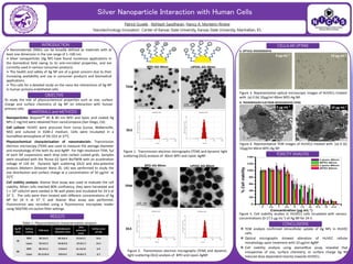
Figure 3. Representative optical microscopic images of HUVECs treated
with (a) 0 (b) 10µg/ml 40nm BPEI-Ag NP.
Figure 4. Representative TEM images of HUVECs treated with (a) 0 (b)
10µg/ml 40nm BPEI-Ag NP.
TEM analysis confirmed intracellular uptake of Ag NPs in HUVEC
cells.
Optical micrographs showed alteration of HUVEC cellular
morphology upon treatment with 10 µg/ml AgNP.
Cell viability analysis using alamarBlue assay revealed that
irrespective of size, surface chemistry, or surface charge Ag NP
induced dose dependent toxicity towards HUVECs.
Figure 5. Cell viability studies in HUVECs cells incubated with various
concentrations (0-17.5 µg mL-1) of Ag NP for 24 h.
0 µg mL-1
10 µg mL-1
0 µg mL-1
10 µg mL-1