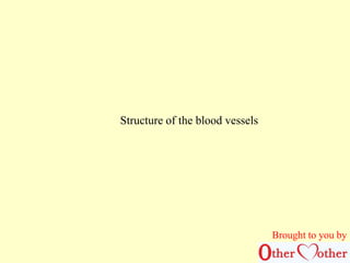
10. TS Artery
lumen
tunica intima
tunica media
tunica adventitia
endothelium lining
muscle layer
with elastic
fibres
connective tissue
Brought to you by
22. Words to use to complete the sheet describing the structure of arteries and veins
(some words may be used once, some more than once and some not at all)
High
Low
Present
Absent
Thin
Thick
Squeeze
Narrow
Backflow
Large
Small
Friction
Smooth
Elastic
Fibres
Muscles
Anchors the vessels in the tissues
And helps to maintain the blood flow
Also prevents over-expansion to maintain the pressure
By controlling the diameter
Brought to you by
23. Describe, and account for, the similarities and
differences in the structure of arteries and veins.
To be done in continuous prose
(no tables, no bullet points)
Brought to you by
31. Checkpoint 1.2. How the structure of an
artery, a vein and a capillary is related to the
function of the vessel.
• Refer to Fig 1.10 B
Brought to you by
32. Artery.
Outer layer of connective
tissue with fibres of collagen
(a strong fibrous protein)
makes the outer wall tough to
prevent over-stretching and to
protect against the pressure
exerted by other organs
rubbing against it.
Thick walls containing lots of elastic fibres (made from a protein called
elastin) and smooth muscle cells.
•Elastic fibres allow walls to stretch when blood pumped at high speed and
high pressure into arteries by contraction of ventricles; elastic recoil when the
pressure drops as the ventricles relax pushes the blood forward to maintain
the flow and the pressure.
•The smooth muscles contract to control how far the artery stretches and so
controls the diameter of the artery, which also maintains the pressure. (NB.
The muscles do not contract to pump the blood in the arteries!)
Brought to you by
33. The narrow lumen
helps maintain the blood
at higher pressure.
No valves
because forward blood flow is
maintained by the heart and
elastic recoil of the arteries.
Brought to you by
34. Vein
Outer layer of connective
tissue with fibres of collagen
(a strong fibrous protein)
makes the outer wall
tough to prevent over-stretching
and to protect
against the pressure
exerted by other organs
rubbing against it.
Thin walls with few elastic fibres and smooth muscle.
Blood flows slowly under low pressure; there is no
pulse so the walls do not need to stretch and recoil.
Wide lumen.
Brought to you by
35. Distribution of blood in the circulatory system
• Heart 3%
• Pulmonary circulation to lungs 10%
• Systemic circulation 87%
• Arteries 17%
• Capillaries 5%
• Veins 65%
Brought to you by
36. Has pocket valves that prevent the backflow of blood.
Blood in the vein is pushed forward by the increase in pressure
produced by the contraction of the nearby skeletal muscles which
the vein run through.
When the muscles relax and stop pressing the pressure
drops and the valves prevent the blood flowing
backwards. Brought to you by
37. Capillaries
Very narrow
Lie close to all cells in the body
Capillary endothelial cell Red blood cell
Capillary wall is one cell thick Very small lumen
Narrow diameter slows down blood flow
to allow time for exchange between blood and surrounding cells to take
place more efficiently
Thin walls only one cell thick
to ensure maximum rate of transfer between blood and
surrounding tissue fluid
Brought to you by
38. Atherosclerosis. Light micrograph of a cross section through an artery
with mild atheroma. The artery wall is pink. The formation of a fatty
plaque or atheroma (grey, centre) has greatly narrowed the size of the
artery lumen (white, centre). This causes a considerable reduction in
blood flow. When this occurs in the arteries leading to the heart
symptoms of angina pectoris (gripping pains in the chest) are
frequently experienced. In severe cases heart attacks or strokes may
occur. Atherosclerosis is principally caused by high fat diets, cigarette
smoking, obesity and inactivity
Brought to you by
39. Atheroma & thrombus.
Coloured light micrograph of a
section through an artery almost
completely blocked by
atherosclerosis and a thrombus.
The large red mass in the centre
is a thrombus, an abnormal blood
clot. This is attached to a part of
the arterial wall that has
thickened with atheroma (yellow-red),
a fatty deposit containing
fibrous tissue, dead cells &
cholesterol. Atherosclerosis is the
biggest cause of death in the UK.
It causes progressive narrowing
of the arteries by deposits of
atheroma, and encourages the
formation of abnormal clots that
can block arteries. Fatal
complications of atherosclerosis
include heart attack and Brought to you by
40. Atherosclerosis. Light
micrograph of a cross section
through an artery obstructed
with an atheroma plaque. The
artery (at upper left) has a
central lumen (black), where
blood flows. Bordering the
lumen is a fibrous and fatty
deposit of a plaque on the
arterial wall. This can be seen
as a dark grainy irregular
deposit on the inner wall.
Surrounding the plaque is the
dark artery wall muscle with an
inner layer of lighter
endothelium. Atherosclerosis,
the thickening of the artery
walls, is mainly due to a fatty
diet high in cholesterol. This can
result in clot formation or
severe artery blockage which
may lead to heart attack.
Brought to you by
41. Atheroma. Cutaway illustration of
the inside of a human artery showing
fatty plaques of atheroma. The artery
has three distinct layers. The tunica
adventitia (outer layer) is fibroelastic
and the tunica media (middle layer)
is muscular. The inner layer, the
tunica intima, is composed of a layer
of endothelial cells (large, orange)
supported by connective tissue.
Atheroma (green and yellow, centre
right) is a mixture of low- density
lipoproteins, decaying muscle cells,
fibrous tissue, blood platelets and
cholesterol. It has narrowed the
artery and caused thinning and
damage to the endothelial layer
(atherosclerosis).
Brought to you by
42. Heart disease. Coloured 3-D computed tomography (CT) scan of the heart of sixty
year old patient with heart disease. The left coronary artery (pink) is on the top of the
heart, and it supplies the heart with oxygenated blood. The left hand branch is the
anterior interventricular artery, which has become narrowed near the top (highlighted
area). Stenosis, or narrowing, of arteries leads to reduced blood flow to the areas
served by the artery. If the artery becomes completely blocked these areas die,
causing myocardial infarction, or heart attack. Risk factors for stenosis include
obesity, smoking, diabetes and a family history of the condition.
Brought to you by
43. Coloured angiogram taken
during a percutaneous
transluminal coronary
angioplasty (PTCA) to the
right coronary artery. It is
done to treat a severe
stenosis (narrowing,
upper centre left) caused
by plaques of atheroma
lining the inside of the
artery; the blood flow is
also impaired by a clot
seen in the same area
just below the stenosis
Brought to you by
44. Heart disease. Coloured 3-D computed tomography (CT) scan of the heart of sixty
year old patient with heart disease. The left coronary artery (pink) is on the top of the
heart, and it supplies the heart with oxygenated blood. The left hand branch is the
anterior interventricular artery, which has become narrowed near the top (highlighted
area). Stenosis, or narrowing, of arteries leads to reduced blood flow to the areas
served by the artery. If the artery becomes completely blocked these areas die,
causing myocardial infarction, or heart attack. Risk factors for stenosis include obesity,
smoking, diabetes and a family history of the condition.
Brought to you by
45. Heart disease. Coloured angiogram (X-ray) of the coronary (heart) arteries of a
patient with heart disease. Coronary arteries (orange) supply the heart muscle with
oxygenated blood. Stenosis (narrowing) of the blood vessels is seen at left. Stenosis is
usually due to atherosclerosis, where fatty deposits of atheroma form on the inner
walls of arteries. It may also be due to abnormal blood clots (thrombi) blocking part of
an artery. Lack of blood to the heart muscle causes angina (severe chest pain) and can
lead to a heart attack (death of part of the heart muscle). Atherosclerosis is usually
caused by a high-cholesterol diet, but smoking and inactivity are also risk factors.
Brought to you by
46. Heart bypass grafts. Artwork of a heart that has had a blockage of the coronary
arteries treated by coronary artery bypass graft (CABG) surgery. The coronary arteries
are the small blood vessels seen running over the outer surface of the heart. They
supply oxygenated blood to keep the heart muscle pumping, and a blockage can cause
a fatal heart attack. The solution is to harvest arteries from elsewhere in the body and
use them to bypass the blockage. Three grafts are seen running from the aorta, the
main body artery, back to the coronary arteries, secured by sutures (black). Three
grafts makes this a triple bypass operation, indicating an advanced state of heart
disease. Brought to you by
47. This platform has been started by Parveen
Kumar Chadha with the vision that nobody
should suffer the way he has suffered because of
lack and improper healthcare facilities in India.
We need lots of funds manpower etc. to make
this vision a reality please contact us. Join us as
a member for a noble cause..
Brought to you by
48. Our views have increased the
mark of the 30,000
Thank you viewers
Looking forward for franchise,
collaboration, partners.
Brought to you by
49. Contact Us:-
011-25464531, 011-41425180, 011-
+91-9818308353,+69612-17387
Brought to you by
othermotherindia@g9m8a1i8l.5c6o9m476
www.other-mother.in
Saxbee Consultants Details :-www.parveenchadha.com
https://cparveen.wix.com/other-mother
https://twitter.com/othermotherindi
http://www.linkedin.com/profile/view?id=326103341&trk=nav_responsive_tab_profile
https://www.facebook.com/pages/Other-Mother-Nursing-Crusade/224235031114989?ref=hl
A WORLDWIDE MISSITION
JOIN US