
Recommended
More Related Content
Similar to Axial skeleton bones
Similar to Axial skeleton bones (20)
More from NwaOsman
More from NwaOsman (11)
Recently uploaded
Recently uploaded (20)
Axial skeleton bones
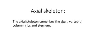
- 1. Axial skeleton: The axial skeleton comprises the skull, vertebral column, ribs and sternum.
- 3. Skull: The skull includes all of the bones of the head. The head consists of the cranium and face and accordingly the bones are divided in to cranial bones (enclose the brain), and facial bone (forming the skeleton of oral and nasal cavities).
- 4. The cranial bones (encloses the brain) include: 1- Occipital bone. 2- Parietal bone. 3- Interparietal bone. 4- Temporal bone. 5- Frontal bone. 6- Sphenoid bone. 7- Ethmoid bone.
- 6. 1- The occipital bone: a- It is situated at the caudal part of the cranium. b- Its lower part is perforated centrally by the foramen magnum. c- The lateral parts bear the occipital condyles, which articulate with the atlas. d- Lateral to the condyle is the jugular process. e- Between the root of this process and the occipital condyle is the condyloid fossa, in which there is the hypoglossal foramen.
- 7. f- The basilar part is strong, which extends forward from the ventral margin of the foramen magnum and fused with the body of the sphenoid bone at basilar tubercles. g- The external surface of the occipital bone is crossed by the nuchal crest which joins the temporal crest laterally. h- The external occipital protuberance is present on which the ligamentum nuchae is attached.
- 8. 2- The parietal bone: a- The two parietal bones form the greater part of the roof of the cranium; they unite in the middle line called external parietal crest (external sagittal crest). b- Each parietal bone is quadrilateral in outline and has two surfaces and four borders. c- The external (parietal surface) is convex. d- The internal (cerebral surface) is concave.
- 9. 3- The interparietal bone: a- This bone is centrally placed between the two parietal bones. b- It is usually described as a single bone. c- The caudal border joins the occipital bone. d- The cranial and lateral borders are united with the parietal bones.
- 10. 4- The temporal bone: a- The temporal bone forms the greater part of the lateral wall of the cranium. b- It is situated between the occipital behind, the parietal dorsally, the frontal in front and sphenoid ventrally. c- It consists of: squamous temporal bone, tympanic temporal bone and petrous temporal bone. •
- 12. (The squamous temporal bone includes the following): 1- zygomatic process: it is connects with the zygomatic bone to make the zygomatic arch. 2- mandibular fossa: this is form when the mandible attaches to form the temporomandibular joint
- 13. (The tympanic temporal bone includes the following): 1- Tympanic bulla: it is large bulbus part on the ventral side of the skull. 2- External acoustic meatus: it is bony canal for the ear.
- 14. (The petrous temporal bone): 1- Is placed between the occipital bone behind and the parietal bone in front, it consists of mastoid process. 2- The muscular process is a sharp spine which project from the base of petrous temporal bone.
- 15. 5- The frontal bone: a- the frontal bones are situated between the parietal behind and the nasal bones in front. b- Each is irregularly quadrilateral, and consists of nasofrontal and orbital parts.
- 16. 6- The sphenoid bone: a- The sphenoid bone is situated at the base of the cranium. b- It consists of body, pair of wings and two pterygoid processes. Wings of sphenoid bone: 1- They are irregularly quadrilateral in outline. 2- At the junction of wing with the body there is a small groove which leads forward to the pterygoid canal. Two Pterygoid processes: 1- Arise from the wings of the sphenoid bone. 2- Its root is perforated by the alar foramen.
- 18. 7- The ethmoid bone: It lies in front of the body and the wings of the sphenoid bone, or located between the eyes on the rostral side of the brain. This includes the following: a- Cribriform plate: 1- Is a shelf like partition between the cranial and nasal cavities. 2- Each half of the cribriform plate forms a deep oval cavity (ethmoidal fossa) which lodges the olfactory bulb. 3- The plate is perforated by numerous small foramina for the passage of the olfactory nerve filaments.--
- 19. a- Cribriform plate: 1- Is a shelf like partition between the cranial and nasal cavities. 2- Each half of the cribriform plate forms a deep oval cavity (ethmoidal fossa) which lodges the olfactory bulb. 3- The plate is perforated by numerous small foramina for the passage of the olfactory nerve filaments.--
- 20. b- Perpendicular plate: 1- Is a median plate and forms the caudal part of the nasal septum. 2- Its cranial border is continuous with the septal cartilage. 3- Its caudal border projects in to cranial cavity. 4- Its dorsal border joins the frontal bone. 5- And its ventral border is received in to the groove of the vomar. c- Ethmoid labyrinth: it is the scroll like located on the plate.