
Recommended
More Related Content
What's hot
What's hot (20)
Similar to Lung cancer
Similar to Lung cancer (20)
More from NK
More from NK (12)
Recently uploaded
Recently uploaded (20)
Lung cancer
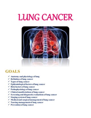
- 1. LUNG CANCER GOALS Anatomy and physiology of lung Definition of lung cancer Types of lung cancer Epidemiologicalfactors oflung cancer Risk factors of lung cancer Pathophysiologyof lung cancer Clinical manifestations of lung cancer Screening and diagnostic evaluations of lung cancer Staging systemof lung cancer Medicaland surgicalmanagementof lung cancer Nursing managementof lung cancer Prevention of lung cancer
- 2. Anatomy and physiology LUNGS are a pair of sponge-like cone shaped organs in the chest, part of our respiratory system. They are covered by a thin covering called pleura which protects and helps lungs move back and forth as they expand and contract during breathing. A thin dome shaped muscle below the lungs called diaphragm separates the chest from abdomen, moves up and down during breathing forcing air in and out of the lungs. Left lung is smaller because the heart occupies spaceon left side. Right lung has three lobes whereas the left one has two lobes. Function Main function of the lungs is to exchange gases between the air we breath and the blood. When we breathe in, oxygen enters into the bodythrough the lungs and when we breathe out, carbon dioxide is sent out of the body. Passageofair :- Air enters the lungs through nose or mouth via windpipe which divides into two airways going into the right and left lung each. These airways are called bronchi. Inside each lung the bronchus divides further into smaller tubes, the secondarybronchi which again sub divide into smaller branches called bronchioles. At the end of the bronchioles are tiny sacs k/a alveoli. Many tiny blood vessels that run through these alveoli perform the function of exchange of gases.
- 3. Definition Lung canceroriginates from the tissues of the lung, usually from cells lining the air passages. It can spread to distant parts of the bodythrough blood and lymphatic vessels, called metastasis. Nearly 40% of those people newly diagnosed with lung cancer already have metastasis to other parts of the bodye.g. lymph nodes, liver, bone, brain, adrenal gland etc. Types These types are diagnosed based on how the cells look under a microscope. The two main types are:- 1. Small cell lung cancer(SCLC) – accounts for 15% of cases and found mostly in heavy smokers. Generally starts in one of the larger breathing tubes, grows fairly rapidly and is likely to be large by the time of diagnosis. Spread more quickly and aggressively. 2. Non small cell lung cancer(NSCLC) – more than 80% of all lung cancers. Grows more slowly. Three major sub-types :- Squamous cell carcinoma Adenocarcinoma Large cell carcinoma Occurrence 30-35% 25-30% 10-20% Location(arise from:-) Bronchial epithelium Bronchiole mucus gland May be located centrally, mid lung or peripherally Growth Slow, metastasis not common Slow Slow, metastasis may occurto kidney, liver & adrenals Cavity May occur Rarely Common Majorrisk factor - Strongly linked to cigarette smoking -
- 4. Epidemiologic factors o Second most common cancer in men & fifth, in both men & women together. o Lung cancer statistics in India (on the basis of Globocan2018 records):- o New cases in men – 48,698 cases per year & in women – 19,097 cases per year. o Deaths in men – 45,363 in a year & women – 18,112 in a year. o The mean age for getting lung cancer – 54.6 years. The majority of lung cancer patients are more than 65 years of age. o Males predominates with a male/female ratio of 4.5:1& this ratio varies with age and smoking status. The ratio increases progressively up to 51-60 years and then remains the same. o The smoker to non-smoker ratio is high up to 201 in various studies. Risk factors A) Modifiable:- Tobaccosmoke – 80 % deaths of lung cancer, leading one. Cigarette smoking increases a person’s chance of getting lung cancer by 15 to 39 times. In India, 87% of male & 85% of female patients with lung cancer have history of active tobacco smoking & 3% patients found having history of passive tobacco exposure. Beedi found more carcinogenic than cigarette smoke. Exposure to other cancercausing agents in workplace – radioactive such as uranium, inhaled chemicals such as beryllium, silica, coalproducts, etc Exposure to asbestosin mines, textile plants – microscopic in nature & when inhaled, get lodged in the soft internal tissue of respiratory system & can irritate the lungs, Talc and talcum powder – may contain asbestos in natural form. Certain dietary supplements - smokers taking beta carotene supplements, increased risk of lung cancer. Vitamin A deficiency increases the chance of developing squamous cell carcinoma of lung in smokers. B) Non-modifiable:- Family history of lung cancer in first degree relatives increases the risk. Personalhistory of lung cancer in one lung can develop lung cancer in other lung. The cancer survivors who have been given radiation therapy to chest area are also at a higher risk.
- 5. Pathophysiology Clinical manifestations o Persistent cough that gets worse despite usual treatment o Chest pain, worse with deep breathing, coughing or laughing o Coughing up flank blood o Coughing up phlegm with traces of blood in it o Hoarseness o Unexplained weight loss and loss of appetite o Shortness of breath o Feeling tired or weak infections such as bronchitis and pneumonia o Jaundice if cancer spread to the liver Following symptoms are usually associated with more advanced lung cancer stage:- o Difficulty in swallowing o Hoarseness in voice o Swelling in the neck caused by enlarged lymph nodes.
- 6. Screening 1. Chest X-ray 2. Sputum cytology 3. Low dosespiral/helical CT scan No guidelines for lung cancer screening in India. As per the guidelines of The American Cancer Society, if a person meet all the following criteria, he/she should go for lung cancer screening :- o Age between 55 & 74 years o 30-pack-years of smoking history (calculated as number of packs of cigarettes multiplied by the number of years personhave been smoking) o Still smoking or have quit in the last 15 years. o Fairly good health (no symptoms of lung cancer or serious medical problems or metal implants or prior history of lung cancer treated) Screening should be done every year till the age of 74 years or till any symptoms appear. Diagnostic evaluation :- Medical history and physical examination Blood tests – CBC, Blood chemistry tests Imaging tests:- o ChestX-ray – to look for a mass in the lungs. o CT scan– more likely to show masses than X-ray and can also provide information about the size, shape & position of lung tumors and help find spread of the cancer to lymph nodes or other organs. o CT guided needle biopsy – to take some tissue for diagnosis. o MRI – to look for spread of lung cancer to brain and spinal cord to help in staging of the cancer.
- 7. o PET scan– to see if the cancer has spread to lymph nodes or other areas. Also helpful if dr thinks that lung cancer has spread but does not know where. Also determines whether surgery can be done or not. Laboratory tests:- o Needle biopsy – it can be done by fine needle aspirationbiopsy(FNAB) where a very fine needle with syringe used to withdraw or aspirate cells & fragments from the mass or core biopsyin which a larger needle is used to remove small cylinders or core of tissue. o Sputum cytology– best method is to submit early morning sputum samples obtained after a deep cough for 3 days in a row, to see it under a microscopeto look for cancer cells. o Bronchoscopy– to see for tumors or blockages in the major airways or bronchi through bronchoscope. o Thoracocentesis – performed if there is a buildup of fluid around the lungs which can cause difficulty in breathing. A hollow needle is inserted between the ribs to drain the fluid and check for tumor cells in the fluid (effusion cytology) Stages TNM staging • T0 – no primary tumor • T1 – tumor < 3cm and not reached Pleura • T2 – 1 or more, 3 – 7 cm, reached bronchus • T3 – 1 or more, > 7cm, chest wall • T4 – 1 or more, any size into space between the lungs • N0 – no lymph nodes • N1 – within lung, bronchus • N2 – around carina, mediastinum • N3 – collarbone on either side • M0 – not spread to distant organs or areas, other lung or lymph nodes • M1 – other lung, cancer cells in fluid around the lung • M2 – spread to distant lymph nodes or to other organs
- 9. Treatment Modalities A) Medical management :- Photodynamic therapy – treat early stage, cancer in the outer layers of the lung airways only Thoracentesis:- to drain fluid Lasertherapy :- treat very small tumors in the linings of airways, open up airways blocked. B) Pharmacological therapy Chemotherapy – either used before or after the surgery. Can be used as palliation to relieve pain or other symptoms of advanced cancer.
- 10. C) Radiation therapy – can be external beam radiation or brachytherapy. o can be either used after surgery to kill any cancer cells that may remain or can be used as the palliative therapy to relieve pain and other symptoms in advanced stage of lung cancer. D) Surgical management :- Lobectomy – entire lobe is removed Segmentectomyorwedge resection – only a part of the lobe is removed Pneumonectomy– removes entire lung Video assistedthoracic surgery(VATS) – treat early stage lung cancer in the outer parts of the lung with small incisions
- 11. E) Radiofrequency ablation – uses high energy radio waves to heat the tumor, a needle like probeis put through the skin to the tumor and electric current is passed through the probe which heats the tumor and destroys the cancer cells. F) Targeted drug therapy – drugs that target specific molecular targets in the tumor cells. Specifically kill cancer cells only, block their growth, prevent cancer from spreading and can stop tumors from growing by blocking signals inside the cancer cells. Their side effects from targeted therapy are minimal since they do not have any toxic effects on normal cells. Types :- ▪ Inhibitors of epidermal growth factorreceptor (EGFR) – Erlotinib, Gefitinib ▪ Monoclonalantibody againstEGFR – Cetuximab ▪ Inhibitors of vascularendothelial growth factor(VEGF) – Bevacizumab ▪ Inhibitors of EML4-ALK – Crizotinib G) Palliative care – aimed at relieving symptoms and improving a person’s quality of life. Issues addressed :- physical, emotional and coping, spiritual Nursing management Assessment:- • Monitor s/s of respiratory failure • Administer chemotherapy and other desired medications • Educate pt with their disease and its progression • Respiratory assessment and Lab investigations. • Pt’s knowledge and understanding to diagnosis and treatment • Pt’s anxiety level and supportsystem. Exposure to carcinogen Nursing diagnosis:- • Ineffective airway clearance related to increased tracheobronchial secretion • Ineffective breathing pattern related to decreased lung capacity • Altered nutrition less than bodyrequirement related increased metabolic demand and decreased food intake • Anxiety related to lack of knowledge • Pain related to the pressure of the tumor
- 12. Prevention No proven way to completely prevent lung cancer but there are steps to lower the risk of getting lung cancer :- i. No smoking – bestway of prevention. People who have never smoked have the lower risk of lung cancer. ii. Quitting smoking – if someone stop smoking before the age of 50 years, there is reduced risk of getting lung cancer by half in the next 10-15%. There are stop-smoking aids which can help to quit effectively. iii. Avoid second hand smoke – avoid smoking zones in public places or avoid people smoking around you. iv. Lower exposure to radon – Radonlevels should be checked in the area where radon is a known problem and people should take measures to reduce exposure. v. Lower exposure to workplace risk factors – people should follow employer’s precautions. vi. Dietary and lifestyle modifications – diet rich in fruits and vegetables along with regular physical activity. Bibliography Cancerindia.org Cancer.org