
Recommended
More Related Content
What's hot
What's hot (20)
Similar to Dermoid cyst
Similar to Dermoid cyst (20)
Recently uploaded
Recently uploaded (20)
Dermoid cyst
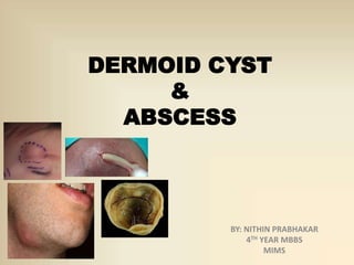
- 1. DERMOID CYST & ABSCESS BY: NITHIN PRABHAKAR 4TH YEAR MBBS MIMS
- 2. A cavity lined by Epithelium containing desquamated cells CONTENTS: mixture of sweat, sebum, desquamated epithelial cells, hair follicles SITES: along the lines of embryonic fusion(midline of the body or face)
- 3. CLINICAL TYPES: A. Congenital/sequestration dermoid:- External and internal angular dermoid (along the fusion of front-nasal and maxillary processes) Sublingual dermoid Pre- auricular & post-auricular dermoids B. Tubulo-dermoids C. Implantation dermoids D. Teratomatous dermoids
- 4. A. SEQUESTRATION DERMOID: PATHOGENESIS: 1 • DURING EMBRYONIC FUSION DERMAL CELLS GET SEQUESTRATED IN THE SUBCUTANEOUS PLANE 2 • THESE CELLS UNDERGO PROLIFERATION AND LATER UNDERGOES LIQUEFACTIVE NECROSIS RESULTING IN THE FORMATION OF THE CYST 3 • THE CYST SLOWLY GROWS N INDENTS THE MESODERM -BONY DEFECTS
- 5. CLINICAL FEATURES: AGE OF ONSET: presents in the 2nd or 3rd decade of life Swelling is insidious in onset & gradually progressive Smooth, soft ,non-tender swelling Skin over the swelling is pinchable Paget’s test: +ve Method: fix the swelling with two fingers(watching fingers) summit is indented with the index finger of other hand(displacing finger)yeilding sensation over the watching fingers
- 6. Swelling is fluctuant in nature Transillumination test: -ve Cough impulse : may be present if the swelling has intracranial extension Resorption & indentation of bone beneath the swelling DIFFERENTIAL DIAGNOSIS: LIPOMA SEBACEOUS CYST BURSITIS
- 7. INVESTIGATIONS: Blood: Total count, differential count ,Hb% , ESR Fine needle aspiration cytology (FNAC) X-RAY: reveals resorption and indentation of the underlying bone CT-SCAN: to indentify the size,shape& local spread of the swelling
- 8. TREATMENT: Excision under general anesthesia . Often neurosurgical approach is required by raising the osteocutaneous flaps of the cranium
- 9. B. TUBULODERMOIDS: Arises from the embryonic tubular structures Pathogenesis : Increased secretions from the lining epithelial cells of the ectodermal tube accumulation of the secretions foramtion of swelling Examples: thyroglossal cyst, ependymal cyst, postanal dermoid
- 10. C. IMPLANTATION / ACCQUIRED DERMOIDS: Seen in individuals like tailors,gardeners who sustain repeated minor injuries Sites: fingers,toes,feet Minor trauma epidermal cells gets buried in the subcutaneous space cyst formation Signs: painless,soft ,smooth,adherent to skin, tensely cystic,mobile swelling Managed by surgical excision under local anaesthesia
- 11. D. TERATOMATOUS DERMOID: A benign or malignant tumor arising from all the germ layers consisting of hair,teeth,cartilage, sebum and muscle Occurs in the testis, ovaries,mediastinium or retro peritoneum COMPLICATIONS OF DERMOIDS: INFECTIONS RUPTURE AND PRESSURE EFFECTS CALCIFICATION SURFACE ULCERATION
- 13. It is the collection of pus within the body Mainly of four types: i. Pyogenic abscess ii. Pyaemic abscess iii. Metastatic abscess iv. Cold abscess
- 14. PYOGENIC ABSCESS: It is the most commonest variety of abscess usually resulting from cellulitis or lymphadenitis Etiopathogenesis: Pathogens: staphylococcus aureus streptococcus pyogenes ,anaerobes Mode of infection may be direct, haematogenous,lymphatics or by extension from the adjacent tissue Organisms enter the tissue activation of immune cells release of mediators and lysozyme destruction of cells and release of proteinsfibrin deposition formation of pus and pyogenic membrane
- 15. CLINICAL FEATURES: Patient presents with fever associated with chills and rigors, throbbing type of pain over the swelling( Localised,red,tender,warm,soft,smooth swelling Visible pus & brawny indurations around the swelling Fluctuation: may or may not be present DIFFERENTIAL DIAGNOSIS: Haematoma Sarcoma Aneurysm? cold abscess
- 16. PYAEMIC ABSCESS: These are generally multiple in number This condition is caused when an Infective emboli circulating in the blood gets lodged in multiple places in the body n thus leading to abscess formation These emboli contain bulk of organisms derived from an infective focus such as vegetations of the valves , thrombus, skin bones The peculiarity of these abscess is that they lie in the subfascial plane(deeper lesions)& do not present the features of common abscess They are non reacting but the constitutional symptoms like weakness, weight loss ,fatiguability are severe Managed by evaluating the focus of infection, antibiotic therapy & drainage of surface abscess if any.
- 17. NOTE THE MULTIPLE LESIONS.
- 18. METASTATIC ABSCESS: These occurs as a spread from other abscess Ex: lung abscess spreading to the brain COLD ABSCESS: These are non reacting abscess caused due to chronic inflammation usually secondary to tubercular infection Caeseation of the lymph nodes, bones abscess formation Sites: commonly around the neck,axilla. also seen around the loin , back(pot’s spine),chest wall
- 20. INVESTIGATION • Blood: Total count will be elevated Differential count (lymphocytic elevation suspect TB) • Urine examination for presence of Glucose • Chest X-RAY in cases of lung abscess • USG Abdomen done as and when required • CT SCAN to identify the number size and shape of the abscess • And other investigation relevant to the organ is done(eg.LFT for liver abscess)
- 21. MANAGEMENT: • Medical management: Broad spectrum Antibiotic therapy is started • Surgical management:- . Incision and drainage of the abscess by HILTONS METHOD PROCEDURE: 1) The abscess is draped, cleaned using a sterile swab, presence of pus confirmed by aspirating the abscess 2)Under general or regional block anaesthesia,an incision is made parallel to the nuerovascular bundle using a No.11 blade
- 22. 3) The pyogenic membrane of the abscess is ruptured using a sinus forceps and the pus is collected in kidney tray. 4) With the little finger the loculi is inspected for remant purulent material 5) The abscess cavity is injected with normal saline and a drain like roller gauge is placed. 6) The wound is left open, which later heals by formation of granulation tissue
- 24. COMPLICATIONS: • Bacteremia,Septicemia and pyemia • Multiple abscess formation • Sinus and fistula formation • Pressure effects over the underlying structures • Antibioma formation: This is typically seen in case of breast abscess. • Metastasis to other organs