
Recommended
More Related Content
What's hot
What's hot (20)
Similar to #Ventilators
Similar to #Ventilators (20)
More from Nisar Arain
More from Nisar Arain (20)
Recently uploaded
Recently uploaded (20)
#Ventilators
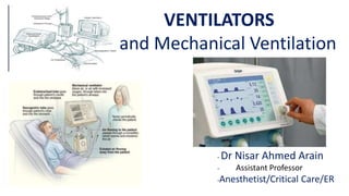
- 1. VENTILATORS and Mechanical Ventilation - Dr Nisar Ahmed Arain - Assistant Professor -Anesthetist/Critical Care/ER
- 3. -CONTENTS -1-Anatomy and Physiology of Respiratory system -2-Introduction about Mechanical Ventilation -3-History -4-Meaning of Mechanical Ventilation -5-Indications for Use -6-Types of Forms of Mechanical ventilation -7-Settings of Mechanical Ventilation -8-Modes of Mechanical Ventilation -9-Complications associated with Mechanical Ventilation -10-Nursing management of Ventilator Patients
- 4. -Anatomy and Physiology of Respiratory System
- 5. Introduction about Mechanical Ventilation --Mechanical Ventilation is typically used after an invasive intubation, a procedure wherein an endotracheal or tracheostomy tube is inserted into the airway. -- It is used in acute settings such as in the ICU for a short period of time during a serious illness. --It may be used at home or in a Nursing or re-habilitation institution, if patients have chronic illnesses that require long term Ventilation assistance.
- 6. -HISTORY --The Roman Physician Galen may have been the first to describe Mechanical Ventilation. --If you take a dead animal and blow air through its Larynx [Through a reed], you will fill its bronchi and watch its lungs attain the greatest distension. -- Vesalius too describes ventilation by inserting a reed or Cane into the trachea of the animals. --In 1908 George Poe demonstrated his Mechanical respirator by asphyxiating Dogs and seemingly bringing them back to life
- 7. -MEANING OF MECHANICAL VENTILATION -In Medicine, Mechanical Ventilation is a method to mechanically assist or replace of Spontaneous breathing
- 8. -INDICATIONS FOR USE OF VENTILATOR Common Medical indications for use include --Acute Lung injury (Including ARDS, Trauma) --Apnea with respiratory arrest, including cases from Intoxication --Chronic obstructive Pulmonary disease (COPD) --Acute Respiratory acidosis with partial pressure of carbon dioxide (pCO2) >50 mmHg and pH <7.25 which may be due to paralysis of the Diaphragm due to “Guillain Barre” syndrome, Myasthenia Gravis, Spinal Cord injury, or the effect of Anesthetic drugs and Muscle relaxant drugs --Increased work of breathing as evidenced by significant Tachypnea retractions, and other physical signs of Respiratory distress. --Hypoxemia with arterial partial pressure of Oxygen (PaO2) with supplemental fraction of inspired oxygen (FiO2) < 55 mmHg --Hypotension including Sepsis, shock, Congestive heart Failure --Neurological diseases such as Muscular Dystrophy Amyotrophic lateral sclerosis
- 9. -TYPES OR FORMS OF MECHENICAL VENTILATION --The two major types of Mechanical ventilation are Negative pressure and positive pressure ventilation --The main form of Mechanical ventilation, which works by increasing the pressure in the patient’s airway and thus forcing air into the lungs. Less common today are Negative pressure ventilators (For Example, the “Iron lung”) that create a negative pressure environment around the patient’s chest, thus sucking air into the lungs
- 10. --Types or Forms of Mechanical Ventilation -Negative pressure ventilation -Positive pressure ventilation
- 11. -Settings of Mechanical Ventilation --Mechanical Ventilator Settings Regulates the Rate, Depth and other characteristic's of ventilation Settings are based on the patient’s Status (ABG’s, Body weight, level of Consciousness and Muscle strength
- 12. -PARAMETERS OF MECHANICAL VENTILATION ARE FOLLOWING --RESPIRATORY RATE (f) :- Normally 10 to 20 b/m --TIDAL VOLUME (VT) :- 5 --- 15 ML / Kg --OXYGEN CONCENTRATION (FIO2) :-b/w 21--90% -- I : E Ratio :- 1: 2 --FLOW RATE :- 40– 100 L/ min --SENSITIVITY / TRIGGER :- 0.5 1.5 cm H2O --PRESSURE LIMIT :- 10---25 cm H2O --PEEP :- Usually, 5---10 cm H2O
- 13. -CONNECTION TO VENTILATORS --FACE MASK --AIR WAY --LARYNGEAL MASK --TRACHEAL INTUBATION --TRACHEOSTOMY
- 14. --MODES OF MECHANICAL VENTILATION --Controlled Mandatory Ventilation (CMV) --Assist—Control Mandatory Ventilation (ACV) --Synchronized Intermittent Mandatory Ventilation (SIMV) --Positive Expiratory End Pressure (PEEP) --Continuous Positive Airway Pressure (CPAP) --Pressure Support Ventilation (PSV)
- 16. --CONTROLLED MADATORY VENTILATION (CMV) -CONTROLLED MECHANICAL VENTILATION(CMV) --In this mode the ventilator provides a mechanical breath on a present timing Patients respiratory efforts are ignored. --This is generally uncomfortable for children and adults who are conscious and is usually only used in an unconscious patient. --It may also be used in infants who often quickly adapt their breathing pattern to the ventilator timing
- 17. -ASSIST – CONTROL MANDATORY VENTILATION (ACV) -Assist control (AC):-In this mode the Ventilator provides a mechanical breath with either a Pre-set tidal volume or Peak pressure, every time a patient initiates a breath. --Traditional control used only a Pre-set tidal volume. --When a Pre-set Peak pressure is used, this is also sometimes termed Intermittent Positive pressure Ventilation or IPPV. --However the initiation timing is the same-both provides a ventilator breath with patients every effort. --In most ventilators a back up minimum breath rate can be set in the event that the patient becomes apnoeic. --Although a maximum rate is not usually set, an alarm can be set if the ventilator cycles too frequently. --This can alert that the patient is Tachypneic or that the ventilator may be auto-cycling (A problem that results when the ventilator interprets fluctuations in the circuit due to the last breath termination as a new breath initiation attempt.
- 18. -SYNCHRONIZED INTERMITENT MANDATORY VENTILATION (SIMV) --In this mode the ventilator provides a pre-set Mechanical breath (Pressure or volume limited every specified number of seconds ( determined by dividing the respiratory rate into 60 – thus a respiratory rate of 12 results in a 5 second cycle time). -- Within that cycle time the ventilator waits for the patient to initiate a breath using either a pressure or flow sensor. -- When the ventilator senses the first patients breathing attempt within the cycle, it delivers the Pre-set ventilator breath. -- If the patient fails to initiate a breath , The ventilator delivers a Mechanical breath at the end of the breath cycle. --Additional spontaneous breaths after the first one within the breath cycle do not trigger another SIMV breath. However SIMV may be combined with pressure support. -- SIMV is frequently employed as a method of decreasing ventilatory support. (weaning) by turning down the rate, which requires the patient to take additional breaths beyond the SIMV triggered breath. --SYNCHRONIZED INTERMITENT MANDATORY VENTILATION (SIMV):-
- 19. -POSETIVE END EXPIRATORY PRESSURE --PEEP:- is functionally the same as CPAP, but refers to the use of an elevated pressure during the Expiratory Phase of the ventilatory cycle. After delivery of the set amount of breath by the ventilator, the patient then Exhales passively. -- The volume of gas remaining in the lung after a normal Expiration is termed “Functional Residual Capacity” (FRC). --The FRC is Primarily determined by the elastic qualities of the lung and chest wall. --In many lung diseases, leading to a decreased surface area for gas exchange and Intrapulmonary shunting with wasted oxygen inspired. -- Adding PEEP can reduce the work of breathing (at low levels) and help pressure FRC.
- 20. -CONTINUOUS POSETIVE AIRWAY PRESSURE (CPAP) --(CPAP)-=A continuous level of elevated pressure is provided through the patient circuit to maintain adequate oxygenation, decrease the work of breathing, and decrease the work of heart (such as in Left-sided heart failure --- CHF) --Note that no cycling of ventilator pressure occurs and the patient must initiate all breaths --In addition, no additional pressure above the CPAP pressure is provided during those breaths --CPAP may be used invasively through an endotracheal tube or tracheostomy or non-invasively with a face mask or nasal prongs
- 21. -PRESSURE SUPPORT VENTILATION -Pressure support ventilation (PSV). --When a patient attempts to breath spontaneously through an endotracheal tube, the narrowed diameter of the airway results in higher resistance to airflow, and thus a higher work of breathing. --PSV was developed as a method to decrease the work of breathing in-between ventilator mandated Breaths by providing an elevated pressure triggered by spontaneous breathing that “supports” ventilation during Inspiration
- 22. --COMPLICATIONS --HYPOTENSION --PNEUMOTHORAX --DECREASED CARDIAC OUTPUT --NOSOCOMIAL PNEUMONIA --POSETIVE WATER BALANCE --INCREASED INTRACRANIAL PRESSURE (ICP) --ALARMS TERNED OFF OR NON-FUNCTIONAL --SINUSITIS AND NASAL INJURY --MUCOSAL LESIONS --ASPIRATION ASPIRATION -G.I Bleeding -In appropriate Ventilation -Respiratory Acidosis OR Alkalosis -Thick Secretions -Patients discomfort due to Pulling OR Jarring of ETT or tracheostomy -High PaO2 -Low PaO2 -Anxiety and Fear -Dysrhythmia's or vagal reactions during or after Suctioning -Incorrect PEEP setting -Inability to tolerate Ventilator Mode
- 23. -TERMINOLOGIES USED IN MECHANICAL VENTILATION --APRV---airway-pressure-release-ventilation --ASB---assisted-spontaneous-breathing --ASV—assisted-spontaneous breathing --ASV---adaptive support ventilation ---a patented technology ---closed-loop-mechanical-respiration ---a further development of MMV ---can also stand for assisted spontaneous ventilation --ATC---automatic-tube-compensation --BIPAP---biphasic positive airway pressure --CMV---continuous mandatory ventilation --CPAP---continuous-positive-airway-pressure --CPPV---continuous-positive-pressure-ventilation --EPAP---expiratory-positive-airway-pressure
- 24. -TERMINOLOGIES USED IN MECHANICAL VENTILATION cont. --HVF---high-frequency-ventilation a-HFFI—high-frequency-flow-interruption b-HFJV---high frequency-jet-ventilation --HFOV---high-frequency-oscillatory-ventilation --HFPPV---high-frequency-positive-pressure-ventilation --ILV---independent-lung-ventilation also called separate sides positive pressure ventilation --IPAP---inspiratory positive airway pressure --IPPV---intermittent-positive-pressure-ventilation --IRV---inversed-ratio-ventilation (mechanical ventilation with switched respiration phases / time rate. --LEPPV—low-frequency-positive-pressure-ventilation —MMV---mandatory-minute-volume --NAVA---neutrally-adjusted-ventilatory-assistant
- 25. -NURSING MANAGEMENT --CHECK THE AIR AND OXYGEN CONNECTIONS --CONNECT THE VENTILATOR TUBES TO VENTILATOR -HOW TO KEEP THE VENTILATOR READY TO RECEIVE THE CASE
- 26. --CONNECT THE CHEST LUNG TO THE VENTILATOR TUBINGS --MAKE SURE THAT YOU CORRECTLY CONNECTED THE TUBING’S AND CHECK FOR ANY LOOSENESS -HOW TO KEEP VENTILATOR READY TO RECEIVE THE CASE
- 27. -HOW TO KEEP VENTILATOR READY TO RECEIVE THE CASE --CONNECT THE SERVO GUARD FROM THE PATIENT --CONNECT THE FILTER TO THE PATIENT
- 28. --HOW TO KEEP VENTILATOR READY TO RECEIVE THE CASE --CHECK THE TUBING’S FOR ANY LEAKAGE --CHANGE THE BACTERIA FILTER
- 29. -HOW TO KEEP VENTILATOR READY TO RECEIVE THE CASE --CHANGE THE BACTERIAL FILTER
- 30. -PLAN OF THE CARE FOR THE VENTILATED PATIENT -PATIENTS GOALS --Patients will have effective breathing pattern --Patients will have adequate gas exchange. --Patient’s Nutritional status will be maintained to meet body needs --Patient will not develop a pulmonary infection --patient will not develop problems related to immobility. --Patient and / or family will indicate understanding of the purpose for Mechanical ventilation
- 31. --NURSING INTERVENTIONS--1 --Observe changes in respiratory rate and depth Observe for the use of necessary muscles --Observe for tube misplacement – note and post cm. marking at lip / teeth after x ray confirmation --prevent accidental extubation by taping tube securely, by checking q.2h restarting / sedating as needed
- 32. -NURSING INTERVENTIONS--2 --Inspect thorax for symmetry of movement determines adequacy of breathing pattern; asymmetry may indicate “Hemothorax or Pneumothorax”. Measure Tidal volume and vital capacity --Asses for Pain --Monitor Chest X Rays --Maintain ventilator settings as ordered
- 33. -NURSING INTERVENTIONS--3 --Elevate head of bed from 60 to 90 degrees. This condition moves the abdominal contents away from the diaphragm, which facilitates its contraction --Monitor ABG’s. Determines acid base balance and need for oxygen. --Observe skin colour and capillary refill. Determine adequacy of blood flow needed to carry oxygen to the tissues
- 34. -NURSING INTERVENTIONS--4 --Observe for tube obstruction; suction; and ensure adequate humidification --Provide nutrition as ordered, e.g TPN lipids or parental feedings. --Use disposable saline irrigation units to rinse in-line suction; ensure ventilator tubing changed q. 7 days, in-line suction changed q. 24 h.; Ambu bags changes between patients and whenever become soiled
- 35. - NURSING INTERVENTIONS--5 --Asses for G.I problems. Preventative measures include relieving anxiety, antacids or H2 receptor antagonist therapy, adequate sleep cycles, adequate communication system. --Maintain muscle strength with active /active-assistive / passive ROM and prevent contractures with use of span-acid splints
- 36. -NURSING INTERVENTIONS--6 --Explain purpose / Mode / and all treatments. --Encourage patient to relax and breath with the ventilator. -- Explain Alarms; teach importance of deep breathing --Provide alternate method of communication --Keep call bell within reach of patient --Keep informed of results of studies / progress --Demonstrate confidence
- 37. --THINGS WHICH WE SHOULD NOT FORGET -SUCTIONING -MOUTH CARE -NEBULIZATION
- 39. -DEFINATION --A Medical ventilator is a machine designed to provide mechanical ventilation by moving breathable air into and out of the lungs, to deliver breaths to a patient who is physically unable to breath, or breathing in-sufficiently
- 40. -BAG IN A BOTTLE VENTILATORS --Most Anesthesia ventilators have a bellows in a box (Bag in a bottle, double circuit) design --The bellows is housed in a pressure chamber, and the inside of the bellows Is connected to the breathing system
- 41. -OXYLOG VENTILATORS WHAT IS HFOV --This is not to be confused with the high frequency jet ventilation (HFJV) which is rarely used --Its First established used is in neonatal ARDS --HFOV’s claim to fame is reduced of ventilator-induced Lung injury (VILI)
- 42. --DRAGER OXYLOG 3000 PLUS VENTILATOR
- 43. -DRAGER OXYLOG 3000 PLUS VENTILATOR IN USE
- 45. -BLEASE MEDICAL MANLEY MP3 VENTILATOR
- 46. -MANUAL JET VENTILATION FIXING
- 47. -FEATURES OF Manual Jet Ventilation --The manual jet ventilator consists of five permanently assembled parts 1-Hose to connect to high pressure (50 psi) oxygen supply with DISS connector (we keep an additional adapter for an Ohio-Quick-Connect outlet attached to the device 2-Flow Regulator 3-Valve with lever 4-Manometer 5-Hose to connect to patient side with Luer-Lock-Adapter
- 48. -HIGH FREQUENCY VENTILATION -High frequency ventilation is a type of Mechanical ventilation which utilizes a respiratory rate greater than four (4) times the normal value, and very small Tidal Volumes. -High frequency ventilation is thought to reduce ventilator-associated lung injury, especially in the context of ARDS and acute lung injury.
- 49. -HIGH FREQUENCY JET VENTILATOR
- 50. -THE BIRD VIP INFANT VENTILATOR
- 51. -TRANSPORT VENTILATORS:- -These are small and more rugged, and can be powered pneumatically or via AC or DC power sources
- 53. -INTENSIVE CARE VENTILATORS NEGATIVE PRESSURE VENTILATORS
- 54. -INTENSIVE CARE VENTILATORS NEGATIVE PRESSURE VENTILATORS --IRON LUNG VENTILATORS
- 55. -INTENSIVE CARE VENTILATORS NEGATIVE PRESSURE VENTILATORS
- 57. -THANK YOU -SAVE THE LIFE SAFELY