2. Singh K P. and Brill J A. 2014
2
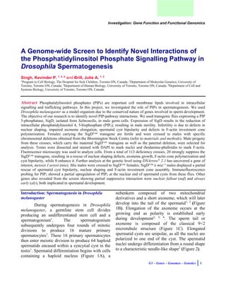
Figure 1. Drosophila spermatid development. A.
Early round spermatocytes contain a nebenkern
composed of two mitochondrial derivatives (mt), and
an elongating axoneme (ax). B. Elongation of the
axoneme and the mitochondrial derivative occur
simultaneously. C. Cross-section of the spermatid
axoneme, which is composed of a classical 9+2
microtubule. D. 64 spermatids get encased into a
syncytial cyst; polarity is established as all nuclei face
the one end of the cyst.
Figure 2. Changes in nuclear morphology of a
developing spermatid. Spermatid nuclei begin as
round and develop into a final characteristic needle-
like shape.
Additional research is required to understand the
mechanisms behind the drastic morphological
change of the spermatid nuclei. Once axonemal
elongation is complete, spermatids undergo
individualization, which involves the removal of
unnecessary organelles and cellular components
not required by mature sperm1, 6, 7
. During
individualization, investment cones containing
filamentous actin (F-actin) form around each
nucleus (Figure 3A) and migrate in synchrony
from the nuclear-end of the cyst towards the tail1
.
A cystic bulge or waste bag forms as the F-actin
cones migrate, which is later degraded1
(Figure
3B). Once individualization has occurred the
spermatids are able to enter the seminal vesicles
and are capable of fertilization1, 4
.
Studying spermatogenesis in Drosophila
provides us with a great model for understanding
microtubule assembly as the axoneme can reach
an approximate length of 2 mm, which is 40 times
longer than human sperm1, 3, 4
. In addition, we
also acquire a thorough understanding of F-actin
dynamics and changes in nuclear morphology1, 5
.
It has been previously described that
phosphatidylinositol phosphate signalling is
crucial for regulating the above-mentioned
processes in Drosophila spermatogenesis 3, 4
.
Phosphatidylinositol Phosphate Signalling in
Drosophila Spermatogenesis
Phosphatidylinositol phosphates, or PIPs
henceforth, are important cell membrane lipids
involved in intracellular signalling and trafficking
pathways8
. The goal of our research is to
investigate the role of PIPs in Drosphila
spermatogenesis. The transgenic flies used in our
experiments express varying levels of SigD a PIP
5-phosphatase, isolated from Salmonella3, 4, 14
.
SigD catalyzes the reverse reaction of PIP5-
kinase-1 reducing intracellular membrane bound
phosphatidylinositol 4, 5-bisphosphate or PIP2,
3. G3 – Genes – Genomes – Genetics 3
Figure 3. Filamentous actin (F-actin) investment cone
polymerization and migration. Once spermatids have
elongated, they undergo individualization. F-actin
investment cones migrate in synchrony from the
nuclear-end of the spermatid towards the tail,
removing unnecessary organelles and cellular
components not required by mature spermatids. A
cystic bulge or waste bag forms as the cones migrate,
which is degraded. A. DAPI and rhodamine-phalloidin
staining depicting F-actin investment cones associated
with nuclei. B. Migrating F-actin cones and formation
of the cystic bulge as indicated by arrows.
Figure 4. The phosphatidylinositol-phosphate signalling pathway. Phosphoinositol (PI) is phosphorylated by
PI4-kinase producing phosphatidylinositol 4-phosphate (PI4P), which is once again phosphorylated by PIP5-
kinase-1 producing phosphatidylinositol 4, 5-bisphosphate, or PIP2. SigD is a PIP5-phosphatase that catalyzes
the reverse reaction of PIP5-kinase-1, depleting membrane phosphatidylinositol 4, 5-bisphosphate or PIP2.
which is crucial during spermatid development 3, 4
(Figure 4). Phosphatidylinositol phosphate
signalling in Drosophila has been shown to be
involved in cellular growth and differentiation,
establishment of apical-basal polarity, actin
dynamics, as well as regulation of the cell cycle9,
10, 11, 12, 13
.
The Effects of SigD Transgenic Expression
during Drosophila Spermatogenesis
SigD Transgenic Flies have Reduced Levels of
PIP2
SigD, also known as SopB, is a protein
encoded by the Salmonella pathogenicity island 1,
which functions as a virulence factor for bacterial
survival14, 15
. SigD has the capacity to reduce
membrane PIP2, which is extensively associated
with the actin cytoskeleton; reduction causes
membrane ruffling16, 17
. This ruffling of the
membrane allows the pathogen to gain access into
epithelial cells, where it can replicate and escape
host immune responses14
. SigD acts as a
phosphoinositide phosphatase, specifically a PIP
5-phosphatase16
. To study the role of PIP2 in
Drosophila spermatogenesis, the SigD transgene
4. Singh K P. and Brill J A. 2014
4
was expressed under the regulation of the
spermatocyte specific β2-tubulin promoter18
. Two
different transgenic lines were used: one with a
high level of SigD expression in the testes
(SigDHigh
) and the second with a lower level of
expression (SigDLow
) 4
. SigDLow
flies carry the
transgene on the X chromosome resulting in
lower expression of the transgene, whereas
SigDHigh
flies carry SigD on the right arm of the
third chromosome, resulting in a higher
expression3, 4
. When the SigD transgene is
introduced into the Drosophila genome, distinct
phenotypes have been observed during
spermatogenesis3, 4
. Males carrying the SigD
transgene show sterility3, 4
.
Partial Reduction in PIP2 Levels During
Spermatogenesis Causes Male Infertility
Previous experiments using
immunofluorescence showed that PIP2 normally
associated with the plasma membrane of
spermatocytes is depleted in SigDLow
expressing
transgenic flies 4
. Spermatids of SigDLow
flies fail
to undergo nuclear elongation; they do not form
F-actin investment cones and consequently do not
undergo individualization3, 4
. Depletion of PIP2 in
SigDHigh
lines causes defects in meiotic
cytokinesis and a complete failure in spermatid
tail elongation3
, a more sever phenotype when
compared to SigDLow
. When an enzymatically
dead, or phosphatase-dead, variant of the SigD
(SigDdead
) was transgenically expressed, no
phenotype was observed confirming catalytic
activity of the functional SigD transgene3
. This
result confirmed that SigD is responsible for the
observed phenotypes as it depletes membrane
PIP2, and ultimately causes male sterility3, 4
.
Reduction in Membrane PIP2 Results in
Spermatid Cyst Bipolarity
Wild type elongating spermatid cysts are
unipolar, as all nuclei localize to one end of the
cyst1, 2, 3, 4
(Figure 5A). It has been previously
reported that a reduction in PIP2 levels results in
spermatid cyst bipolarity4
. Scattered nuclei are
also observed in the SigDLow
and SigDHigh
transgenic flies as a result of PIP2 depletion4
.
Bipolarity of the cyst occurs when nuclei appear
distributed evenly to each end of the cyst and
spermatid tails develop towards the center4
(Figure 5B). Experiments overexpressing sktl
(PIP5-kinase-1) in the SigDLow
transgenic
background resulted in a partial rescue of the
above-described phenotypes4
. Sktl is a PIP5-
kinase, which replenishes membrane PIP2
4
. This
further supports the importance of the PIP2 at the
membrane during spermatid development.
Reduction in Membrane PIP2 Leads to Defective
F-actin Assembly
PIP2 regulates actin dynamics in
association with downstream Rho family
GTPases, considered to be the master regulators
of the cytoskeleton4
. Membrane bound PIP2 is
shown to be involved in the regulation of
downstream effector proteins, such as the N-
WASP and ARP complexes that modulate the
actin cytoskeleton and its interactions7
. It has also
been reported that PIP2 links actin to the plasma
membrane through membrane linker proteins like
spectrin19, 20
. F-actin cone polymerization and
migration are essential for spermatids to undergo
individualization and become mature sperm4
(Figure 5A’). Expression of SigDLow
and SigDHigh
in Drosophila melanogaster testes reduces
membrane PIP2 leading to defective F-actin cone
polymerization and migration4
(Figure 5B’ and
C’). Since F-actin investment cones are required
for spermatid individualization, absence or
reduction of F-actin leads to spermatids incapable
of this process, rendering them non-functional4
.
The objective of our research is to perform
a genetic screen, testing deficiencies on the right
and left arms of the second and third chromosome
of the Drosophila melanogaster genome. We will
cross fertile-virgin females carrying the SigDLow
5. G3 – Genes – Genomes – Genetics 5
Figure 5. Effects of SigDLow
and SigDHigh
transgenic expression on spermatid development. A. Wild type
nuclei stained with DAPI, 64 nuclei are polarized to one end of the cyst. A’ Micrograph showing 64 F-actin
investment cones associated with nuclei. A’’ Merged micrograph showing long thin unipolar cysts. B.
Expression of SigDLow
causes defects in nuclear morphology, as well as bipolarity within the cysts, as
indicated by yellow arrows. B’ Lack of F-actin investment cones when SigDLow
is expressed. B’’ Merged
micrograph depicting truncated bipolar spermatid cysts. C. Expression of SigDHigh
leads to complete arrest
in axoneme elongation and nuclear morphology. C’ Complete lack of F-actin investment cones. F-actin is
detected in ring canals formed during incomplete cellular division. C’’ Merged image depicting a loss of
polarity within spermatid cysts.
transgene to Bloomington deficiency males and
select progeny carrying the maternal SigDLow
transgene and the paternal deficiency. We are
looking for dominant suppressers or enhancers of
nuclear shaping, spermatid cyst polarity, F-actin
cone polymerization and axoneme elongation. We
are interested in finding potential genes that
interact with the phosphatidylinositol phosphate
pathway. Performing a genetic screen has proven
in the past to be a reliable methodology in
understanding signal transduction pathways and
their interactions such as the sevenless and ras
signalling pathways, both involved in vital
intracellular signalling transduction processes24,
8. G3 – Genes – Genomes – Genetics 8
Figure 7. Example of a strong suppressor of F-actin cone polymerization, spermatid cyst bipolarity and axoneme
length. A. SigDLow
expressing virgin females were crossed to Df(3R)24971 males, progeny carrying the maternal
transgene and the paternal deficiency were selected for analysis. B. Phase micrograph showing length of
spermatid cysts, notice the length and size resemble wild type. C. DAPI fluorescence staining showing partial
rescue of bipolarity, nuclei are polarized to the leading edge of the cyst. D. Rhodamine-phalloidin fluorescence
staining showing completely formed F-actin investment cones. E. Merged micrograph. F. Flybase image
depicting the deleted region that the deficiency spans, the blue, red and white arrows represent genes,
highlighted in yellow is the deficiency tested.
An example of a strong suppressor is
Df(3R)24971, which consists of a deleted segment
from the right arm of the third chromosome
(deleted breakpoints, 3R:[83F1—84B2]). SigDLow
virgin females were crossed to Df(3R)24971
males, F1 progeny carrying the maternal SigDLow
transgene and paternal Df(3R)24971 were
selected for analysis (Figure 7A). DAPI
fluorescence staining marking nuclei showed a
partial rescue of bipolarity and nuclear shaping
defects (Figure 7C). A complete rescue of F-actin
cone polymerization was also observed (Figure
7D). Furthermore, the spermatid cysts displayed a
wild type characteristic length (Figure 7B/E).
Df(3R)24971 contained many deleted genes, in
order to precisely predict which gene or group of
genes is responsible for the suppression event we
have to narrow down this large interval by testing
smaller deficiencies that span the larger deletion
(Figure 7F).
Df(2R)24931 strongly enhances SigD associated
phenotypes
An example of an enhancer of nuclear
shaping and spreading was Df(2R)24931, a
deletion on the right arm of the second
chromosome (Deleted breakpoints, 2R: [52A10—
52D2]). Again, we crossed SigDLow
virgin
females
13. G3 – Genes – Genomes – Genetics 13
cytoskeleton
par-1 is
involved in
cellular
component
organization
and biogenesis
18401650.. 18480501 78 851 2R SUP px
CG6018
CG11362
Dnr1
5601375.. 5684102 82 727 3L SUP Eaf6
Blimp-1
Lin-28
Sse
Sif
Sif is predicted
to have
RacGEF
activity and
also contains a
PH domain,
where PIPs
and Rac/Rho
family
GTPases
interact
271425.. 327733 56 308 3L SUP Miple2
CG32845
Ttm2
RhoGEF3
Fwd
CG32344
Atac3
RhoGEF3 and
Fwd involved
in the PIP
pathway
11997642.. 12074922 77 280 3L SUP CG6828
Rols
CR43994
CG43993
CG6793
CG6793
CG43638
10743982.. 10920164 176 182 3R SUP Sdr
CR45598
CR45599
CG14861
RpL10Aa
14. Singh K P. and Brill J A. 2014
14
CR44944
Dpr9
CG6974
CG6966
Table 2. Results from heterozygous knockouts mutants tested with SigDLow
Meiosis I arrest, or mia, was observed as one of
two genes deleted in a deficiency (Df(3R)LT) that
was crossed with SigDLow
(Figure 10A/B). F1
testes once dissected and stained showed a strong
suppression of SigDLow
phenotypes (Figure 10C-
E). There was a partial rescue of bipolarity
(Figure 10D), nuclear shaping and F-actin cone
polymerization (Figure 10E). Mia encodes for a
transcription factor, which is involved in the
progression of the male cell cycle during
spermatogenesis21, 22
; it is also implicated in
cellular differentiation after meiosis23
. We go on
to further investigate the observed suppression of
SigDLow
, and to classify the mechanism of
interaction.
SigDLow
x mia-/+
flies show a partial up-regulation
of PIP2 at the membrane
Allele (-/+)
x SigDLow
Fluorescence Microscopic Analysis Results
nuf
TM3
(nuclear fallout)
-Partial rescue of bipolarity
-Partial rescue of F-actin investment
cones
-Cysts length resembles SigDLow
Weak
Suppressor
mia1 st e
TM6b
(meiosis I arrest)
-Rescue of bipolarity
-Rescue of nuclear morphology
-Rescue of F-actin investment cones
-Axoneme elongation resembles w1118
(wild type)
-Testis seem larger then wild type flies
Strong
Suppressor
(Results Figures
10/11)
aly5 red
TM6c
(always early)
-Some F-actin cones are seen through
the cysts
-Partial rescue of nuclear elongation
V. Weak
Suppressor
comr, cnbw
CyO
(cookie monster)
No Interaction No Interaction
sa’red
TM3
(spermatocyte arrest)
No Interaction No Interaction
w; nht cnbw
SM6a
(no hitter)
No Interaction No Interaction
16. Singh K P. and Brill J A. 2014
16
deplete membrane PIP2. By crossing flies, which
express SigDLow
with deficiency flies, it was
possible to detect which genes are vital in the
regulation of PIP2 at the membrane.
Further exploring Df(3R)24971 which
showed a strong suppression of SigD, we want to
know which gene or genes is or are resulting in
the suppression event. Most noticeable of the
suppression was the rescue of F-actin investment
cones, which were well shaped, as well as the
length of the cysts, which indicates complete
elongation of the axoneme. This perhaps indicates
that when these genes are present, they result in
down-regulation of PIP2 at the membrane, or an
alternate hypothesis is that they are altering the
function of our transgene.
When we analyze the results from
Df(2R)24931 which showed an enhancement of
the nuclear shaping defects, as the nuclei arrest in
a round shape as well as being scattered
throughout the cysts; and showed non-classical
cyst formation. This indicates to us that there is a
gene or group of genes that, under wild type
conditions are regulating PIP2 at the membrane.
Deletion of these genes is resulting in a worse
phenotype, which perhaps indicates that these
genes normally function to regulate PIP2 at the
membrane.
We observed a strong suppression of
SigDLow
when we crossed SigDLow
carrying virgin
female flies to heterozygous mutant knockouts of
mia. Results from SigDLow
x mia-/+
flies can act as
a control for the basis of the transgenic expression
of SigD. Both SigD and mia (and upstream
regulators boule and twine) are under the
regulation of the spermatocyte specific β2-tubulin
promoter29
, when the mia-/+
is knocked-out we
observed a strong suppression of SigDLow
phenotypes. This evidence confirms the
effectiveness of the SigD transgene in producing
the phenotypes previously described, as well as
the sensitivity of the screen, as a promoter control
was detected. As described in the results, there are
many other genes of great interest, which will be
tested in the future. We hope to find regulators of
the PIP pathway such as interactions that can
replenish membrane PIP2.
The results from the screen give us new
insight in genes involved in sperm development
in Drosophila. Due to the shared homology of
genes between Drosophila and humans, much of
the genetic and cellular process can be applied to
both models. The results from our screen present
new regulators, which affect the PIP pathway. It
is important to note that not only is the PIP
pathway involved in spermatogenesis in
Drosophila, rather the molecular properties of
cells, apical-basal polarity, actin dynamics and
changes in nuclear morphology are involved in
many cell based pathologies such as cancer. Many
variants of epithelial cancers are due to the loss of
apical-basal polarity, which is influenced by the
distribution of membrane bound
phosphatidylinositol 4, 5-bisphosphate (PIP2) and
phosphatidylinositol 3, 4, 5-trisphosphate (PIP3)
within the cell. Down-regulation of these vital
lipoproteins results in loss of cell-cell/cell-basal
lamina adhesion and activation of a
hypersensitive migratory behavior. Implications
from this screen may reveal novel players in
regulating cellular metastasis and migration of
cells.
Conclusion
Studying the phosphatidylinositol
phosphate signalling pathway in Drosophila
provides us with an excellent model of
understanding microtubule dynamics, F-actin
polymerization, cellular polarity and nuclear
morphology. Our results show the effectiveness
of performing a screen to detect novel interaction
of the phosphatidylinositol phosphate pathway.
The implications from this research will help us to
understand the vast network of intracellular
signalling transduction pathways that regulate
cellular homeostasis. We are better able to
understand the role of phosphatidylinositol
phosphates during spermatid development and
gain further knowledge of its interactions.
18. Singh K P. and Brill J A. 2014
18
14. Hapfelmeier S, Ehrbar K, Stecher B,
Barthel M, Kremer M, and Hardt WD.
(2004). Role of the Salmonella
Pathogenicity Island 1 Effector Proteins
SipA, SopB, SopE, and SopE2 in
Salmonella enterica Subspecies 1 Serovar
Typhimurium Colitis in Streptomycin-
Pretreated Mice. Infection and Immunity,
72:2 p. 795–809.
15. Norris FA, Wilson MP, Wallis TS, Galyov
EE, and Majerus PW. (1998). SopB, a
protein required for virulence of
Salmonella dublin, is an inositol
phosphate phosphatase. Proc. Natl. Acad.
Sci. USA 95, 14057-14059.
16. Marcus SL, Wenk MR, Steele-Mortimer
O. and Finlay BB. (2001). A synaptojanin-
homologous region of Salmonella
typhimurium SigD is essential for
inositolphosphatase activity and Akt
activation. FEBS Lett. 494, 201-207.
17. Terebiznik MR, Vieira OV, Marcus SL,
Slade A, Yip CM, Trimble WS, Meyer T,
Finlay BB and Grinstein S. (2002).
Elimination of host cell PtdIns (4,5) P2 by
bacterial SigD promotes membrane fission
during invasion by Salmonella. Nat. Cell
Biol. 4, 766-773.
18. Wong R, Hadjiyanni I, Wei HC, Polevoy
G, McBride R, Sem KP and Brill JA.
(2005). PIP2 hydrolysis and calcium
release are required for cytokinesis in
Drosophila spermatocytes. Curr. Biol. 15,
1401-1406.
19. Niggli V, Andreoli C, Roy C, and
Mangeat P. (1995). Identification of a
phosphatidylinositol-4, 5-bisphosphate-
binding domain in the N-terminal region
of ezrin. FEBS Lett. 376, 172–176.
20. Hirao M, Sato N, Kondo T, Yonemura S,
Monden M, Sasaki T, Takai Y and Tsukita
S. (1996). Regulation mechanism of ERM
(ezrin/radixin/ moesin) protein/plasma
membrane association: possible
involvement of phosphatidylinositol
turnover and Rho-dependent signaling
pathway. J. Cell Biol. 135, 37–51.
21. Fuller MT. (1998). Genetic control of cell
proliferation and differentiation in
Drosophila spermatogenesis. Semin. Cell
Dev. Biol. 9(4): 433--444.
22. Lin TY, Viswanathan S, Wood C, Wilson
PG, Wolf N, Fuller MT. (1996).
Coordinate developmental control of the
meiotic cell cycle and spermatid
differentiation in Drosophila
males. Development 122(4): 1331--1341.
23. White-Cooper H, Schafer MA, Alphey
LS, Fuller MT. (1998). Transcriptional
and post-transcriptional control
mechanisms coordinate the onset of
spermatid differentiation with meiosis I in
Drosophila. Development 125(1): 125—
134.
24. Simon MA, Bowtell DD, Dodson GS,
Laverty TR, Rubin GM. (1991). Ras1 and
a putative guanine nucleotide exchange
factor perform crucial steps in signaling
by the sevenless protein tyrosine kinase.
Cell 67: 701–716.
25. Therrien M, Morrison DK, Wong AM,
Rubin GM. (2000). A genetic screen for
modifiers of a kinase suppressor of Ras-
dependent rough eye phenotype in
Drosophila. Genetics 156: 1231–1242.
26. Georgiou M, Baum B. (2010). Polarity
proteins and Rho GTPases cooperate to
spatially organize epithelial actin-based
protrusions. J. Cell Sci. 123(7): 1089--
1098.
19. G3 – Genes – Genomes – Genetics 19
27. Jiang J, White-Cooper H. (2003).
Transcriptional activation in Drosophila
spermatogenesis involves the mutually
dependent function of aly and a novel
meiotic arrest gene cookie monster.
Development 130(3): 563--573.
28. Riggs B, Fasulo B, Royou A, Mische S,
Cao J, Hays TS, Sullivan W. (2007). The
concentration of Nuf, a Rab11 effector, at
the microtubule-organizing center is cell
cycle regulated, dynein-dependent, and
coincides with furrow formation. Mol Biol
Cell 18, 3313–3322.
29. Lin TY, Viswanathan S, Wood C, Wilson
PG, Wolf N and Fuller MT. (1996).
Coordinate developmental control of the
meiotic cell cycle and spermatid
differentiation in Drosophila males.
Development 122.
21. G3 – Genes – Genomes – Genetics 21
2. Add credentials: Username: dnarrowx Password: fwdgenebrill
3. Wait to be redirected to the homepage.
22. Singh K P. and Brill J A. 2014
22
4. Click: Databases.
5. Click: PHPMyAdmin.
23. G3 – Genes – Genomes – Genetics 23
6. Click: _dmelres in the left side panel.
7. Click: dmel_data.
24. Singh K P. and Brill J A. 2014
24
8. Click: Insert.
25. G3 – Genes – Genomes – Genetics 25
9. Data entry. In stk, add the Bloomington Stock Number of the deficiency. In sym, add the symbol of
the deficiency, arm for chromosome arm (ex. 3-Left), lft/rgt for the left and right breakpoints of the
deficiency, available through Flybase. Res_type for results: SUP for suppressor and ENH for enhancer,
true implies a strong interaction (strong suppressor or strong enhancer), false means no interaction or
weak interactions (Remember to focus on the strong interactions, but don’t dismiss weak interactions,
in the comments sections mention your observations). Add your initials in Ins and comments.
27. G3 – Genes – Genomes – Genetics 27
3. Click the plus (+) symbol beside the E and S to load data, select current arm for data, which is
specific for a single chromosome arm.
4. Data load, if suppressor for 2L is selected data in the suppressors column will load (if enhancer
is selected, column will now load enhancers under same column, column title will then change
to enhancers). Non-interacting regions of the chromosome arm will also load under the inactive
column.
28. Singh K P. and Brill J A. 2014
28
5. Click the play arrow at the bottom of the collapse column, this will output intervals where
inactive and active regions have combine, and in cancelled out areas where there is no
interaction and outputs intervals where a potentially interacting gene is located.
29. G3 – Genes – Genomes – Genetics 29
6. By clicking the play arrow under the remaining column, the outputs are intervals of the current
chromosome arm that have not been tested by any deficiency. These data indicate either no
deficiency available in the area, or areas that where there was no deficiency available.
7. Take the intervals generated by the collapse column and input them into the GBrowse section
of Flybase. This will allow you to see what genes and or deficiencies span this area.
30. Singh K P. and Brill J A. 2014
30
8. Smaller interval. Intervals where there are less then 8-10 genes are good indicators for further
analysis, larger intervals with many genes need to be narrowed down further, all deficiencies
that span this area should be looked into.
9. To see what deficiencies cover this region, you can view the spanning aberrations by clicking
the spanning aberrations line.
10. Load all intervals into an excel file and list genes/deficiencies available, as well as comments
regarding genes (alleles available, type of allele – RNAi or transposable elements),
molecular/biological function, expression patters (testis specific expression patterns).
11. To order Bloomington Deficiency Flies please contact Dr. Lacramioara Fabian
(lala.fabian@utoronto.ca) or Gordon Polevoy (gordon.polevoy@sickkids.ca).