1. Annexin II Is a Major Component of Fusogenic Endosomal Vesicles
Neff Emans,* Jean-Pierre Gorvel,* Carmen Walter,* Volker Gerke,r Roland KeUner,* Gareth Griffiths,*
and Jean Gruenberg*
*European Molecular Biology Laboratory, D-6900 Heidelberg, Germany; and ~Max-Planck Institute for BiophysicalChemistry,
Am Fassberg, D-3400 Goettingen, Germany
Abstract. We have used an in vitro assay to follow
the proteins transferred from a donor to an acceptor
upon fusion of early endosomes. The acceptor was a
purified early endosomal fraction immunoisolated on
beads and the donor was a metabolically-labeled early
endosomal fraction in suspension. In the assay, both
fractions were mixed in the presence of unlabeled
cytosol, and then the beads were retrieved and
washed. The donor proteins transferred to the acceptor
were identified by two-dimensional gel electrophoresis
and autoradiography. Approximately 50 major proteins
were transferred and this transfer fulfilled all criteria
established for endosome fusion in vitro. However,
only a small subset of proteins was efficiently trans-
ferred, if donor endosomes were briefly sonicated
to generate small (0.1 #m diam) vesicles before the as-
say. These include two acidic membrane proteins, and
three alkaline peripheral proteins exposed on the cyto-
plasmic face of the membrane. Partial sequencing and
Western blotting indicated that one of the latter com-
ponents is annexin II, a protein known to mediate
membrane-membrane interactions. Immunogold label-
ing of cryosections confirmed that annexin n is pres-
ent on early endosomes in vivo. These data demon-
strate that annexin II, together with the other four
proteins we have identified, is a major component of
fusogenic endosomal vesicles, suggesting that these
proteins are involved in the binding and/or fusion
process.
T
HE pathways of membrane traffic are now well estab-
lished, as are some of the mechanisms involved in
sorting and transport of proteins between different
compartments. Recent advances have come predominantly
from genetic analysis in yeast (Schekman, 1992) and from
the in vitro reconstitution of individual steps of the biosyn-
thetic or the endocytic pathway (Balch, 1989; Goda and
Pfeffer, 1989; Gruenberg and Howell, 1989; Rothman and
Orci, 1992). In mammalian cells, several proteins required
for membrane transport have been identified and character-
ized (Gruenberg and Clague, 1992), including NSF and as-
sociated proteins (Rothman and Orci, 1992), coat proteins
(Pearse and Robinson, 1990; Serafini et al., 1991; Duden et
al., 1991)and regulatory components belonging to the GTP-
binding protein super-family (Bourne et al., 1990; Balch,
1992).
The mechanisms which mediate first the formation of
close contacts between membranes and then fusion itself are
still unclear. Studies on fusion of secretory granules or en-
veloped viruses have both led to the view that a pore or a
collar may be formed at the site of contact via the oligomer-
J.-E Gorvel's present address is Centre d'Immunologie de Marseille-
Luminy, Case 906, 13288 Marseille Cedex 9, France.
Addresscorrespondenceto J. Gruenberg at European Molecular Biology
Laboratory, Postfach 10.2209, D-6900 Heidelberg, Germany.
ization of specific proteins (Almers, 1990; Bentz et al.,
1990). Lipid flow would then be initiated along the extended
hydrophobic surfaces of these proteins. The hemagglutinin
of influenza virus envelope contains an amphipathic a-helix,
which becomes exposed upon fusion (Wiley and Skehel,
1987); a similar motif has also been found in other viral fu-
sion proteins and in a protein implicated in sperm-egg fusion
(Blobel et al., 1992). Until now, this domain has not been
found in any of the proteins that have been implicated in in-
tracellular membrane transport. However, the protein com-
plex consisting ofNSF and associatedproteins, which are re-
quired for intra-Golgi transport, is proposed to regulate
intracellular fusions, although its precise role is not known
(Rothman and Orci, 1992).
In this paper, we modified our cell-free fusion assay
(Gruenberg and Howell, 1986, 1987; Gruenberg et al.,
1989) to identify proteins that may mediate binding/fusion
of early endosomal membranes. This analysis was possible
because early endosomes display a striking tendency to un-
dergo homotypic fusion with each other in vitro (Davey et
al., 1985; Gruenberg and Howell, 1986, 1987;Braell, 1987;
Diaz et al., 1988; Woodman and Warren, 1988; Gruenberg
et al., 1989). This process is highly specific (Gruenberg et
al., 1989; Bomsel et al., 1990) and microtubule-indepen-
dent (Bomseletal., 1990). It is regulated byphosphorylations
(Tuomikoski et al., 1989; Thomas et al., 1992; Woodman
9 The Rockefeller University Press, 0021-9525193/03/1357/13 $2.00
The Journal ofCell Biology, Volume 120, Number6, March 1993 1357-1369 1357
onFebruary22,2011jcb.rupress.orgDownloadedfrom
Published March 15, 1993
2. et al., 1992), N-ethyl-maleimide-sensitive factor (NSF)~
(Diaz et al., 1989) and GTP-binding proteins (Gorvel et al.,
1991; Colombo et al., 1992; Lenhard et al., 1992). This high
and specific fusion activity in vitro may indicate that, in vivo,
early endosomes, likeother cellular organdies, are organized
in a dynamic network connected by fusion/fission events
(Gruenberg and Howell, 1989). Recent in vivo observations,
in fact, showthat endosomes can be organized in an extensive
and dynamic tubular network (Hopkins et al., 1990), which
may be part of the early endosome (Tooze and Hollinshead,
1991).
Our approach was to study the transfer of early endosomal
proteins from a donor to an acceptor membrane upon fusion.
As acceptor, we used an early endosomal fraction immobi-
lized on a solid support by immunoisolation (Gruenberg and
Howell, 1986, 1987;Gruenberg et al., 1989). The donor was
either a metabolically-labeled, early endosomal fraction in
suspension, or the same fraction which had been briefly
sonicated to generate small vesicles. The proteins trans-
ferred in the fusion assay were then identifiedby high resolu-
tion two-dimensionalgel electrophoresis (Bravo, 1984; Celis
et al., 1990) and autoradiography. Essentially all early en-
dosomal proteins were transferred from an intact donor (=50
major species), as expected for a homotypic fusion. The
small GTP-binding protein rab5, which localizes to early en-
dosome (Chavrier et al., 1990; Chavrier et al., 1991) and
regulates early endosome fusion in vitro (Gorvel et al., 1991)
and endocytosis in vivo (Bucci et al., 1992), was identified
amongst the transferred proteins.
The number of transferred proteins could be significantly
reduced when the donor was briefly sonicated before the as-
say. More importantly, only five proteins were then trans-
ferred with high efficiency(two membrane proteins and three
peripheral proteins). One of the latter components was found
to be annexin II, which we could detect on the surface of
early endosomes in vivo. Proteins of the annexin family, in
particular annexin II (see, Burgoyne, 1988; Gerke, 1989a),
have been previously implicated in membrane-membrane
interactions, although their physiological functions are still
debated (see, Siidhof et al., 1982; Crompton et al., 1988;
Burgoyne and Geisow, 1989; Zaks and Creutz, 1990a; Pol-
lard et al., 1990a; Lin et al., 1992). Our data suggest that
annexin II, together with the other four proteins we have
identified, mediates membrane-membrane interactions be-
tween early endosomes in the assay.
Materials and Methods
Cells and Viruses
Monolayers of BHK cells were grown and maintained as described (Gruen-
berg et al., 1989). For each experiment, 6 • 10-cm Petri dishes were plated
16 h before use. Cells were metabolically labeled with 0.2 mCi/dish
[35S]Met for 16 h in medium containing low Met (1.5 mg/liter). To
depolymerize microtubules, cells were preincubated with 10/~M nocoda-
zole for 1 h at 37~ and nocodazole remained present in all incubations
(Gruenberg et al., 1989). HRP was internalized by fluid phase endocytosis
as described in Gruenberg et al. (1989) and Gorvel ct al. (1991). Vesicular
stomatitis virus (VSV) was produced as previously described (Gruenberg
and Howell, 1985). All manipulations of the cells were at 4~ except when
indicated.
1. Abbreviations used in thispaper: bHRP, biotinylatedHRP; NEM, N-ethyl-
maleimide; NSF, NEM-sensitive factor; PNS, post-nuclear supernatant;
VSV, vesicular stomatitis virus; VSV-G, glycoprotein G of vesicular stoma-
titis virus envelope.
G-Protein Implantation and Internalization into
Early Endosomes
We have previously described our protocol to introduce the transmembrane
spike glycoprotein G of vesicular stomatitis virus (VSV-G) into the plasma
membrane by low pH-mediated fusion of the viral envelopewith the plasma
membrane (Gruenberg and Howell, 1985, 1986, 1987; Gruenberg et al.,
1989). Briefly, we added 50 #g VSV to 1.3 x 10⁷ cells (one 10-cm dish);
8 gg total VSV were fused with the plasma membrane, corresponding to
a density of =70 VSV-G molecules/gin2 membrane surface area. Upon
warming the cells up to 37~ for 5 rain, the VSV-G molecules were rapidly
internalized into early endosomes, essentially as a synchronous wave
(Gruenberg and Howell, 1986, 1987; Gruenherg et al., 1989). We used six
dishes in a typical experiment.
Fractionation ofEndosomes on a Flotation Gradient
After homogenizationofthe cells and preparation of a post-nuclear superna-
tant (PNS) (Gruenberg et al., 1989; Gorvel et al., 1991), the first fraction-
ation step was the same flotation gradient we used previously (Gorvel et al.,
1991). Briefly, the PNS was brought to 40.6% sucrose and 0.5 mM EDTA,
loaded at the bottom of an SW 60 tube, sequentially overlaid with 1.5 ml
16% sucrose in D20, 3 mM imidazole pH 7.4 containing 0.5 mM EDTA,
then 1.0 ml 10% sucrose in D20, 3 mM imidazole pH 7.4 containing 0.5
mM EDTA and finally 0.5 ml homogenization buffer (250 mM sucrose, 3
mM imidazole pH 7.4). The gradient was run for 60 rain at 35,000 rpm
using a SW 60 rotor. Early endosomeswere collectedat the 16-10% sucrose
interface. The fraction was then split into four equal aliquots and immedi-
ately processed for immunoisolation. Protein determination in the fractions
was as described by Bradford (1976).
Immunoisolation
We have previously described our protocol to immunoisolate endosomal
fractions, using the cytoplasmic domain of VSV-G as antigen (Gruenberg
and Howell, 1985, 1986, 1987; Gruenberg et al., 1989). To analyze the pro-
tein composition of the acceptor, the fractions .were prepared from
metabolically-labeled cells. The endosomal fractions recovered from the
flotation gradient were used as input for subsequent immunoisolation. The
immuno-adsorbent was prepared by binding =5/~g of a monoclonal anti-
body against a cytoplasmic epitope of VSV-G (P5D4; Kreis, 1986) to 1 mg
anti-mouse polyacrylamide heads, as described (reviewed in Howell et al.,
1989). Approximately 20/zg protein of the fraction recovered from the gra-
dient were mixed with 1 nag immuno-adsorbent in 1 ml PBS-contalning
5 mg/ml BSA (PBS-BSA), and rotated end-over-end for 2 h at 4~ The
beads with bound cellular materials were then recovered by centrifugation
at 2500 g for 5 rain, and washed once with PBS-BSA and once with PBS.
The beads were processed for two-dimensional gel electrophoresis and the
gels analyzed by autoradiography.
In the cell-free assay, the acceptor fraction was prepared with the same
protocol, but the cells were not metabolically labeled. The immunoisolated
fraction was then immediately added to the assay, as described below.
Quantification ofEndocytic VesicleFusion In Vitro
We have quantified the occurrence of early endosome fusion in vitro with
the cell-freeassay we have established (Gruenberg et al., 1989; Tuomikoski
et al., 1989; Bomsel et al., 1990; Gorvel et al., 1991). The assay uses two
early endosomal fractions prepared after separate internalization of avidin
and biotinylated HRP (bHRP) into two cell populations. (a) bHRP present
in the fluid phase was cointernalized with the implanted VSV-G protein for
5 rain at 37~ The cells were homogenizedand fractionated using the gra-
dient (see above). Early endosomes were collected from the 16-10% inter-
face and immunoisolated as above. After washing once with PBS-BSA, 1
mg beads with bound bHRP-labeled fraction was resuspended in 50/tl
homogenization buffer and directly used in the cell-free assay. (b) Avidin
was prebound at 4~ to cell surface biotinylated proteins under conditions
where it retained free biotin binding sites, providing a membrane-attached
marker in the fusion assay (as described in Gorvel et al., 1991). The com-
plex avidin-hiotinylatedprotein was then internalized into early endosomes
for 5 rain at 37~ and the avidin remaining at the cell surface was quenched
with excess biotinylated insulin. Avidin-labeledearly endosomes were then
fractionated using the flotation gradient, and one 50-pl aliquot (15 ttg pro-
tein) was directly used in the fusion assay. In some experiments, avidin-
labeled early endosomal elements were fragmented by sonication for 1 •
7 s on ice with a probe-type sonicator. In the fusion assay, the two fractions
were combined in the presence of 50/~1cytosol (5 mg/rrd protein), adjusted
The Journal of Cell Biology, Volume 120, 1993 1358
onFebruary22,2011jcb.rupress.orgDownloadedfrom
Published March 15, 1993
3. to 12.5 mM Hepes (pH 7.4), 1.5 mM MgOAc, 3 mM imidazole (pH 7.4),
1 mM DTT, 50 mM KOAc, and complemented with 8 /~1 of an ATP-
regenerating system (1:1:1 mixture of 100 mM ATP brought to pH 7.0 with
KOH, 800 mM creatine phosphate, and 4 mg/ml creatine phosphokinase)
and 8/~1 of I mg/ml biotin-insulin stock solution as a quenching agent. To
deplete ATE 1.6/~1of apyrase (1,200 U/ml) replaced the ATP-regenerating
system. The assay as well as the quantification of the avidin-bHRP complex
formed during fusion were carried out as described (Gruenberg et al., 1989;
Bomsel et al., 1990; Gorvel et al., 1991).
TransferofProteinsduringFusion ofEndocytic Vesicle
In Vitro
To identify the proteins transferred between early endosomal elements upon
fusion in vitro, essentially the same assay was used. One early endosomal
fraction was freshly prepared from unlabeled cells after internalization of
VSV-G for 5 min at 37"C and fractionation using the flotation gradient. The
early endosomes were then immunoisolated from the 16-10% interface and
resuspended in 50 ~tlhomogenizationbuffer. In the transfer assay, this frac-
tion is referred to as acceptor fraction. The second fraction, referred to as
donor, was prepared from cells metabolically labeled with [35S]Met and
was directly collected from the 16-10% interface ofthe gradient. The fusion
assay was carded out as above. To identify the proteins transferred during
fusion, the immuno-adsorbent plus bound material was washed and then
processed for two-dimensional gel electrophoresis. The gels were analyzed
by autoradiography. In some experiments, the donor was fragmented by a
brief sonieation step (1 x 7 s) before the transfer assay. To identify the
membrane-associatedproteins of the sonieated donor, the fraction was then
centrifuged at 150,000 g for 45 min and then pellets and supernatants were
processed for two-dimensionalgel analysis. Extraction in Triton X-114 was
as described by Bordier (1981).
Two-dimensionalGelElectrophoresis
A combination of IEF and SDS-PAGE was used to resolve proteins in two
dimensions as described by Bravo (1984). The samples were solubilized in
9.8 M urea, 4% w/v NP40, 2% v/v ampholines pH 7-9 and 100 mM DTT.
The tube gels used for the IEF gels were 25 cm long and 2.5 mm in internal
diameter. IEF gels were run at 1,200 V for 17 h. The pH gradient was linear
between pH = 4.5 and 7.4. The second dimension resolving gels were 15%
(wt/vol) acrylamide, 0.075 % (wt/vol) N-N' methylene-bis-acrylamide and
the stacking gels were 5% (wt/vol) acrylamide, 0.25% (wt/vol) N-N'
methylene-bis-acrylamide. After electrophoresis the gels were fixed and
prepared for autoradiography using Entensify (Dupont, New England Nu-
clear Research Products, Boston, MA).
Sequencing
The spot corresponding to protein b was excised from six two-dimensional
gels, minced, electroeluted in one dimensionand analyzed as previously de-
scribed (Kurzchalia et al., 1992). Briefly, the protein was digested with
trypsin and the resultingpeptides extracted from the gel slice were separated
by reverse phase HPLC. As a control, we used spots cut out from nonrele-
vant regions of the same two-dimensional gels. The elution profiles of the
specific and control digests were compared to select specific peptides,
which were analyzed by Edman degradation using a sequencer (model
477A, Applied Biosystems, Inc., Foster City, CA).
Electron Microscopy
To provide an electron-dense marker of early endosomes, the cells were in-
cubated for 5 min at 37"C in the presence of 5 nm BSA-gold (Ludwig et
al., 1991). The cells were then washed in ice-cold PBS, and processed for
cryosections and immunogold labeling as described by Grifliths et al.
(1984).
Results
Our initial goal was to reconstitute in vitro the transfer of
proteins that occurs upon fusion of a donor with an acceptor
endosome, and then to use this assay to identifyproteins that
may be involved in the process. We used early endosomes
immunoisolated on a solid support as an acceptor fraction
and metabolically-labeled early endosomes in suspension as
a donor fraction. In the assay, fusion would result in the
transfer of metabolically-labeled proteins from the donor to
the unlabeled acceptor. The acceptor can then be retrieved,
washed and analyzedby two-dimensional gel electrophoresis
and autoradiography, to identify donor proteins that have
been transferred.
Preparationof theAcceptor Fraction
As antigen for immunoisolation of the acceptor, we used the
cytoplasmic domain of the spike VSV-G. The G-protein was
first implanted into the plasma membrane by low pH-
mediated fusion of the viral envelope with the plasma mem-
brane (White et al., 1980; Gruenberg and Howell, 1985) and
then internalized into early endosomes by incubation for 5
rain at 37~ (Gruenberg and Howell, 1986, 1987;Gruenberg
et al., 1989). The cells were homogenized and a PNS was
prepared. The PNS was then fractionated using a step flota-
tion gradient, which separates early endosomes containing
the small GTP-binding protein rab5, from the plasma mem-
brane and from late endosomes containing rab7 and the
cation-independent mannose-6-phosphate receptor (Gorvel
et al., 1991). The early endosomal fraction recovered from
the gradient was enriched =13-fold. As a second step, early
endosomes containing the VSV-G protein were immunoiso-
lated using beads with a bound antibody recognizing the
VSV-G cytoplasmic domain (Gruenberg and Howell, 1985,
1986, 1987; Gruenberg et al., 1989; Howell et al., 1989;
Thomas et al., 1992). Early endosomes could be prepared
with a yield of 12% and an enrichment of 73x over the ho-
mogenate, using HRP cointernalized with the G-protein as
a marker of the early endosome content.
PurifiedAcceptor FractionFuseswithDonor
MembranesIn Vitro
Using our cell-free assay (Gorvel et al., 1991), we then in-
vestigated whether the purified early endosomal acceptor re-
tained its fusion activity in vitro. In these experiments, the
lumenal content of acceptor early endosomes was labeled
with bHRP instead of HRE The donor fraction was an early
endosomal fraction recovered from the flotation gradient
(enrichment =13 x, see above) and contained endosomes la-
beled with tetravalent avidin attached to the lumenal side of
the endosomal membrane via proteins originally biotinylated
at the cell surface (Gorvel et al., 1991). If fusion occurred
upon incubation of acceptor and donor membranes in the as-
say, a complex was formed between avidin and bHRP. The
immunoisolated fraction was then retrieved by centrifuga-
tion and, after washing, the avidin-bHRP complex was im-
munoprecipitated with anti-avidin antibody and the enzy-
matic activity of bHRP was quantified (see Gorvel et al.,
1991 and references therein). Table I shows that fusion oc-
curred with a high efficiency(=60%). In agreement with our
previous studies, fusion was inhibited by low concentrations
of GTP'yS (Table I), low temperatures, 1 mM N-ethyl-
maleimide (NEM) or in the absence of ATP or cytosol (not
shown).
Protein TransferduringEarlyEndosomeFusion
In Vitro
Since fusion activity was high after purification, the acceptor
and donor fractions were then used to study the transfer of
proteins during fusion. Both fractions were prepared as
above, except that avidin and bHRP were omitted and that
Emans et al. Annexin H in Endosome Fusion 1359
onFebruary22,2011jcb.rupress.orgDownloadedfrom
Published March 15, 1993
4. Table I. Fusion E~ciency of Early Endosomal Fractions
before and after Fragmentation
OD bHRP Efficiency
%
(A) Total amount 0.206 + 0.004 100
(B) Fusion with intact EE 0.126 + 0.005 61 + 3
+GTP',r 0.002 4- 0.003 1 + 2
(C) Fusion with fragmented EE 0.042 4- 0.006 20 4- 3
+GTPyS 0.001 4- 0.002 1 4- 1
Fusion was measuredby the formationof a complexbetween avidinand
bHRP,whichhad beenseparatelyinternalizedintotheearlyendosomes(EE)
oftwocellpopulations.Afterinternalization,avidinwasboundtothelumenal
sideoftheendosomalmembranevia biotin,whereasbHRPdistributedwithin
the endosomalcontent.ThebHRP-labeledfractionwaspreparedusinga gra-
dient followedby immunoisolation(acceptor)and the avidin-labeledfraction
was recoveredfromthe gradient(donor).(.4)The eti~ciencyof avidin-bHRP
complex formationduring fusionis indicatedas a percentageof the total
amount of complexformedin the presenceof detergent(Grnenberget al.,
1989). The complexwasthenimmunoprecipitatedwithanti-avidinantibodies
and the activityofhHRPquantified(ODbHRP).(B) Fusionwithintactearly
endosomes. Subcellularfractionsprepared fromavidin- and bHRP-labeled
ceilswerecombinedinthefusionassayinthepresenceof5 mg/mlcytosoland
ATPand incubatedfor 45 minat 37~ Whenindicated10gM GTP'rSwas
present. (C) Fragmentedearlyendosomes.As in B, butafterbriefsonication
of the avidin-labeledfraction.
the donor was prepared from cells which had been labeled
with p~S]Met for 16 h. In the cell-free assay, acceptor and
donor were mixed in the presence of unlabeled cytosol, the
donor membranes being thus, the only 35S-labeled compo-
nent in the reaction. At the end of the experiment, the beads
were retrieved and washed, and the bound cellular materials
were analyzed by high resolution two-dimensional gel elec-
trophoresis (Bravo, 1984; Celis et al., 1990) and autoradiog-
raphy.
Fig. 1 A shows the pattern of 35S-labeled donor proteins
transferred to the unlabeled acceptor in the assay. This trans-
fer fulfilled the different criteria established for the fusion of
early endosomes in vitro. Transfer was abolished at 4~ in
the absence of ATP or cytosol and in the presence of 1 mM
NEM (not shown) or 10 ~tM GTP3,S (Fig. 1 B) (see Gruen-
berg and Howell, 1989; Mayorga et al., 1989). Transfer was
also abolished with antibodies against the small GTP binding
protein rab5 (not shown), as expected for early endosome fu-
sion (Gorvel et al., 1991). As an additional control, we used
a metabolically-labeled late endosome fraction recovered
from our gradient (Gorvel et al., 1991) as donor. No transfer
was then detected (not shown), in agreement with our previ-
ous findings that early and late endosomes do not directly
fuse with each other (Gruenberg et al., 1989; Bomsel et al.,
t990; Gorvel et al., 1991).
As shown in Fig. 1A, '~,50 major proteins were transferred
in the assay, which distributed over the entire pH range dur-
ing isoelectric focusing in the first dimension and over appar-
ent mobilities from 14 to 200 kD after electrophoresis in the
second dimension. In this and in the following experiments,
several proteins that were detected in the controls (Figs. 1
B, 2 B, and 4 B) appeared somewhat enriched in the specific
fractions (Figs. 1 A, 2 A, and 4 A), including actin (small
star) and a few heat-shock proteins, identified by their typi-
cal mobilities (Celis et al., 1991). The significance of this ob-
servation is not clear. However, these proteins, like all pro-
teins detected in the control experiments, were disregarded
in our analysis. Long thin arrows and letters in Fig. 1 A indi-
Figure 1. Identification of early endosomal proteins transferred
from donor to acceptor after fusion. (A) The acceptor early en-
dosomal fraction was prepared by immunoisolation from unlabeled
cells, and the donor early endosomal fraction was prepared from
cells metabolically labeled with [3sS]Met using a flotation gra-
dient. In the assay, both fractions were mixed in the presence of un-
labeled cytosol and ATE and then incubated for 45 rain at 37~
At the end of the assay, the fraction bound to the solid support was
retrieved, washed and analyzed by isoelectric focusing in the first
dimension (direction of electrophoresis from left to right) followed
by SDS-PAGE in the second dimension (direction of electrophore-
sis from top to bottom). The gels were then analyzed by autoradi-
ography. The arrows point at typical examples of transferred pro-
teins. The position of rab5 is indicated by a large star next to the
corresponding arrow. The small star shows the position of actin,
identified by its mobility on two-dimensional gels. The molecular
weight markers are indicated by solid dots (14.3, 30, 46, 69, 97, and
200 kD). (B) Same as A, but in the presence of 10/~M GTP'yS.
The Journalof Cell Biology,Volume120, 1993 1360
onFebruary22,2011jcb.rupress.orgDownloadedfrom
Published March 15, 1993
5. A
w
Isonicatlon
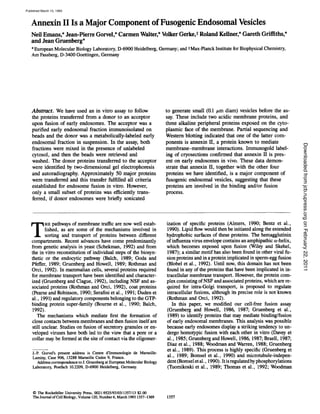
OOO000Figure 3. Outline of donor sonication. The sonication of a donor
early endosomal element is outlined. Some early endosomal com-
ponents (solid boxes) may not be very abundant or present in
specialized regions of the membrane, in contrast to others (solid
circles). When small vesicles are generated by sonication, the
former components may be present in a subpopulation of vesicles,
but not the latter ones.
Figure2. Protein composition ofthe earlyendosome acceptor frac-
tion. (A) The acceptor early endosomal fraction was prepared by
immunoisolation as in Fig. 1 (except that the cells had been
metabolically-labeled with [35S]Met)and then analyzed in two-
dimensional gels followedby autoradiography. Directions of elec-
trophoresis and labeling of typical proteins are as in Fig. 1. (B)
Same as A, but the specific antibody was omitted during im-
munoisolation.
cate typical examples of transferred proteins, which will be
discussed below. Because a large collectionof donor proteins
were transferred in the assay, we compared them with the
protein pattern of the acceptor fraction itself.
Comparison with the Protein Composition
of the Acceptor
The acceptor fraction was prepared as above from cells
which had been labeled with [35S]Met for 16 h, and then
analyzed in two-dimensional gels (Fig. 2 A). Approximately
50 major proteins could be identified which were absent
from the control, and their pattern closely resembled that ob-
tained after transfer (compare Fig. 1 A with Fig. 2 A). Some
differences could be observed when comparing autoradio-
grams from several experiments. In the experiment shown in
Figs. 1A and 2 A, a group of acidic proteins (40-50 kD) were
more abundant after transfer (Fig. 1 A) than after direct im-
munoisolation (Fig. 2 A), whereas a few high molecular
weight proteins exhibited the opposite distribution. The do-
nor proteins transferred in vitro (Fig. 1A) were, however, es-
sentially identical to those of the acceptor (Fig. 2 A).
It is difficult to compare the patterns we obtained with the
two-dimensional endosomal patterns obtained by other
groups (Baenziger and Fiete, 1986; Schmid et al., 1988;
Beaumelle et al., 1990). The conditions used to prepare the
fractions, to label the cellular materials, and to run the iso-
electric focusing gels were different. However, proteins with
similar mobilities can be identified, particularly in the low
molecular weight region (see Beaumelle et al., 1990).
We could identify the small GTP-binding protein rab5
among both the acceptor proteins (Fig. 2 A, large star) and
the donor proteins that are transferred (Fig. 1A, large star),
in good agreement with the fact that rab5 localizes to early
endosomes (Chavrier et al., 1990, 1991) and regulates early
endosome fusion in vitro (Gorvel et al., 1991) and endocyto-
sis in vivo (Bucci et al., 1992). To localize rab5 in the gels,
[35S]Met-labeled acceptor membranes were prepared from
cells overexpressing rab5. The gels were then blotted onto
nitrocellulose and the overexpressed rab5 was identified af-
ter p2P]GTP overlay (Gorvel, J.-P., M. J. Clague, L.
Huber, P. Chavrier, O. Steele-Mortimer, and J. Gruenberg,
submitted for publication). The rab5 protein migrated to an
alkaline position (pI = 9.0) relative to other rab proteins (L.
Huber and K. Simons, EMBL, unpublished observation).
These experiments indicate that acceptor and donor mem-
branes have essentially the same protein composition and
that both contain rab5.
Protein Transfer From Sonicated Donor Vesicles
We reasoned that molecules mediating endosome-endosome
interactions may be heterogeneously distributed in the plane
of the membrane (see outline Fig. 3). If donor endosomes
were fragmented into small vesicles, these proteins would be
present in a sub-population of vesicles, which would remain
fusion competent. In our assay, we would then expect these
proteins to be more efficiently transferred (if they are re-
Emanset al. AnnexinHinEndosomeFusion 1361
onFebruary22,2011jcb.rupress.orgDownloadedfrom
Published March 15, 1993
6. Figure4. Identification of proteins transferred after fragmentation
of the early endosomal donor fraction. (,4) The transfer assay was
carried out in parallel from the same early endosomal fractions as
in Fig. 1, except that in this experiment the metabolically-labeled
donor fraction was fragmented by sonication for 7 s before the
transfer assay. The sample was then analyzed as in Fig. 1 and the
autoradiogram was exposed for the same length oftime. Only a sub-
set of early endosomal proteins was transferred after fragmenta-
tion. Letters indicate early endosomal proteins transferred with the
same efficiency from sonicated or intact donors (compare with Fig.
1). Rab5 is indicated by a star. Arrowheads point at proteins trans-
ferred with a reduced efficiency. Both arrowheads and arrows point
at the proteins indicated by long arrows in Figs. 1and 2. Star (actin)
is as in Figs. 1 and 2. (B) Same as A, but in the presence of 10 #M
GTP'yS.
quired for the process), than other endosom_al proteins. To
fragment the donor, we used a single 7-s sonication pulse.
An analysis by electron microscopy and flow cytometry
showed that a homogeneous population of small vesicles of
=100 nm diana had been generated (not shown). Fusion ac-
tivity measured as above with membrane-bound markers
was retained after sonication, albeit with a reduced efficiency
(Table I), possibly indicating that only a subpopulation of
avidin-labeled vesicles remained fusogenic (see below).
Figure5. Soluble and membrane-associated proteins of the soni-
cared donor fraction. The donor fraction was prepared from
metabolically-labeled cells (as in Fig. 1), briefly sonicated (as in
Fig. 4), centrifuged at 150,000 g for 45 rain. Pellets and superna-
tants were then analyzed in two-dimensional gels. Letters indicate
the same proteins as in Figs. 1, 2, and 4. Whereas proteins c, d,
and e were only membrane-associated, a fraction of proteins a and
b were released after sonication. Proteins not transferred after soni-
cation are indicated by small double arrows. Metabolically-labeled
tab5 could only be identified in immunoisolated fractions or after
transfer (largestar in Figs. 1, 2, and 4), but not in the complete
donor fraction (Figs. 5 and 6). (,4) Supernatant (soluble proteins).
(B) Pellet (membrane-associated proteins).
As shown in Fig. 4 A, protein transfer occurred after soni-
cation, arid retained all characteristics of endosome fusion,
including GTPTS-sensitivity (Fig. 4 B). The number of
transferred proteins was significantly reduced, as predicted
(compare with Fig. 1 A), and each one of them, including
rab5, was identified in the acceptor (Fig. 2 A) and in the
fused donor (Fig. 1 A). These data indicate that fusogenic
donor vesicles generated by sonication contain a defined sub-
set of proteins.
Protein Transfer In Vitro Is Selective
We first investigated whether the number of transferred pro-
The 3oumalof Cell Biology,Volume120, 1993 1362
onFebruary22,2011jcb.rupress.orgDownloadedfrom
Published March 15, 1993
7. teins was reduced after sonication, because lumenal proteins
were lost. In the absence of sonication, both internalized
HRP, a fluid phase marker, and 35S-labeled proteins were
only associated with the pellet after high speed centrifuga-
tion of endosomal fractions recovered from the gradient.
When the same experiment was repeated after sonication,
>95 % of internalized HRP was found in the supernatant, but
only a relatively small fraction (30%) of the 35S-labeled
proteins (data not shown).
We then analyzed in two-dimensional gels the high speed
pellets and supernatants obtained after centrifugation of the
sonicated 35S-labeled donor. As shown in Fig. 5, the major-
ity of the donor proteins which remained associated with
vesicles after sonication were clearly not transferred to the
acceptor membranes in the assay (examples are indicated by
small double arrows in Fig. 5 B). (The fraction containing
the donor exhibits a protein pattern distinct from that of the
immunoisolated acceptor, because the former is enriched
15x in early endosomes and the latter 73 x.) To extend these
observations, intact 35S-labeled donors were extracted in
Triton X-114 (Bordier, 1981). Detergent and aqueous phases
were then analyzed in two-dimensional gels. Many putative
transmembrane proteins present in the detergent phase were
not detected amongst the proteins transferred after sonica-
tion (smalldoublearrowsin Fig. 6). As expected, high speed
pellets obtained after sonication (Fig. 5 B) and Triton X-114
extracts (Fig. 6 B) did not exhibit identical protein composi-
tions (see below and Table II), although overall patterns were
comparable (particularly for proteins with a molecular
weight ~<30 kD).
These experiments demonstrate that many membrane-
associated proteins of the sonicated donor were not trans-
ferred in the assay. Thus, protein transfer after sonication
was not caused by random fusion of donor membranes but
occurred in a selective manner. The proteins which are then
transferred include both membrane and peripheral proteins,
and are presumably present within a subpopulation of donor
vesicles which remain fusogenic in the assay.
A Subset of Proteins Is Transferred with High
EJ~ciency after Sonication
Our prediction was that proteins involved in membrane-
membrane interactions should be transferred with high
efficiency from a sonicated donor. In fact, a small group of
five proteins was not only efficientlytransferred, when com-
pared to other proteins, but this efficiency was essentially
identical to that observed if the donor was intact (compare
Fig. 4 A with Fig. 1 A; all proteins that could be detected
in any of the controls were disregarded). These five proteins
are indicated by letters (a to e) in Figs. 1, 2, 4, and 6, whereas
examples of proteins transferred with lower efficiencyare in-
dicated by arrowheads in Fig. 4 A. Proteins a, b and c mi-
grate as a series of spots, possibly reflecting post-transla-
tional modifications. Rab5 itselfmay not belong to this small
group, although it is transferred with a relatively high
efficiency.
The state of membrane-association of these five proteins
is summarized in Table II. They include two acidic putative
membrane proteins of =40 kD (d) and =200 kD (e), and
three groups of alkaline peripheral proteins of =48 (a), 38
(b), and 14 kD (c). The latter proteins (a-c) are likely to be
exposed on the cytoplasmic face of endosomes, because they
Figure6. Triton X-114extraction of the sonicated donor fraction.
The donor fraction was prepared from metabolically-labeled ceils
as described in the legendof Fig. 1, extracted in Triton X-114(Bor-
dier, 1981), and analyzed in two-dimensional gels. Letters are as
in Fig. 4. Protein b and d partitioned exclusivelyinto the water and
the detergent phase, respectively. In contrast, proteins a, c, and e
were found in both phases. Proteins not transferred aftersonication
are indicated by small double arrows. As in Fig. 5, metabolically-
labeled rab5 could notbe identifiedin the complete donor fraction.
However,rab5 is associated to the cytoplasmic face of endosomes
via addition of an aliphatic chain and partitions into the detergent
phase ofTriton X-114(Gorvel et al., 1991).(A)Aqueousphase. (B)
Detergent phase.
could be released by salt treatment (not shown). Because
these proteins are efficientlytransferred from a sonicated do-
nor (transfer efficiency is identical both with and without
sonication) in contrast to most other donor proteins, we be-
lieve that they are strong candidates for being components
that mediate the interactions between endosomal membranes
in the assay.
Annexin H Belongs to the Subset of Proteins
Transferred with High EJ~iency In Vitro
The spot corresponding to protein b in two-dimensional gels
of donor membranes was excised from the gel and digested
with protease, and the resulting peptides were separated by
Emans et al. Annexin 11 in Endosome Fusion 1363
onFebruary22,2011jcb.rupress.orgDownloadedfrom
Published March 15, 1993
8. TableII. Proteins Transferred with the Same Efficiency with
or without Sonication of the Donor
Protein MW IP Sonication Triton X-114 pH 11
a 48 alk m/s m/s s
b 38 alk m/s s s
c 14 alk m m/s s
d 40 ac m m m
e 200 ac m m/s m
The table summarizes the properties of the five proteins transferred with the
same efficiency with or without sonication. They are indicated by the same let-
ters as in the figures. Fig. 5 shows the high speed pellet (B) and supernatant
(A) after sonication of the donor; Fig. 6 shows the detergent (B) and water
phase (A) after Triton X-114 extraction of the donor (pH 11 treatment of the
donor was carried out in 0.1 N NaOH for 30 min on ice, not shown). Proteins
a to c migrated as a few spots with the same apparent molecular weight, possi-
bly reflecting post-translational modifications. MW, approximate molecular
weight; IP, isoelectricpoint; alk, alkaline; ac, acidic; m, membrane-associated
(or detergent-associated for Triton X-114); s, soluble; m/s, both membrane-
associated and soluble.
HPLC and sequenced. The entire process was repeated, to
limit the possibility of an error, and the characteristic pep-
tides are indicated in Fig. 7. Both rounds resulted in the
identification of a single protein, annexin II, when searching
the SWlSSPROT database with the FASTA (GCG package)
and SCRUTINEER (Sibbald and Argos, 1990) programs.
Protein b and annexin II also share the same apparent molec-
ular weight. Moreover, the predicted isoelectric point of an-
nexin II, obtained using the PEPTIDESORT program (GCG
package), is in good agreement with that of protein b.
We could unambiguously establish that protein b is an-
nexin II, and not another member of the annexin family, be-
cause both peptides indicated in Fig. 7 correspond to mo-
tives completely conserved in chick, mouse, bovine, and
human annexin II proteins (amino acids 233-240 and
251-260; see Gerke et al., 1991, for annexin II sequence
comparison), but not in other annexins. Finally, to demon-
strate that protein b is an annexin, we used an antibody
against a conserved annexin motif (cp2; Gerke, 1989b) to
blot our two-dimensional gels. As shown in Fig. 8, the anti-
body only recognized protein b. We, therefore, conclude that
protein b is annexin II, or a very closely related protein, be-
cause both share common sequences, as well as physical and
immunological characteristics.
Annexin H Localizes to Early Endosomal Membranes
In Vivo
Having established that protein b was annexin II, we then
studied its subcellular distribution in vivo after immunogold-
labeling on cryosections. We used a rabbit polyclonal anti-
body raised against purified annexin II, which only recog-
nized annexin II on Western blots (not shown). As previously
reported (see Gerke, 1989a), annexin II was present on the
plasma membrane (Figs. 9 and 10). The gold particles were
often distributed unevenly: while some areas were heavily
labeled, others were essentially devoid of label (compare the
two adjacent areas indicated by large arrows in Fig. 9 B).
Small but significant amounts of gold were associated with
clathrin-coated pits and vesicles (Fig. 9, C-E). In contrast,
caveolae-like structures were usually unlabeled (Figs. 9, A-B
and 10 A).
We then investigated whether annexin II was also present
in early endosomes. These were unambiguously identified in
233 240 Figure7. Sequencesofprotein
... S Y S P Y D M L... b peptides. The amino acid
sequences oftwo peptides ob-
251 260 tainedatiertwoseparaterounds
...L E N A F L N L V Q... ofproteolyticdigestionofpro-
tein b are indicated. Eachpep-
tide corresponds to a stretch of amino acids, which is completely
conservedin the sequencesofbovine,human, mouse, and chick an-
nexin II, and absent from the sequenceof other members ofthe an-
nexin family in the samespecies. The first and last amino acids of
each peptide is numbered after the sequence in these species.
cryosections after the internalization of 5 nm BSA-gold from
the medium for 5 min at 37~ as previously described (Lud-
wig et al., 1991). Annexin II was found to be abundant on
the surface of early endosomes containing 5 nm BSA gold
(Figs. 9 B, and 10, A-C), although labeling of the plasma
membrane was even higher (see Fig. 9 B and 10, A and B).
In early endosomes, tubular or cisternal regions of the mem-
brane appeared to be preferentially labeled, whereas little,
if any, labeling was associated with the vesicular regions (see
Fig. 10, A and B). These observations demonstrate that the
protein we have identified with our in vitro assay as an early
endosomal component presumably involved in membrane-
membrane interactions, indeed localizes to early endosomes
in vivo.
Discussion
Recent studies on the mechanisms of membrane transport
have revealed the existence of complex regulatory mecha-
nisms, including GTP-dependent switches mediated by
members of the GTP-binding protein super-family as well as
post-translational modifications (Gruenberg and Clague,
1992; Schekman, 1992). However, relatively little is known
about proteins that may facilitate membrane-membrane in-
teractions and/or catalyze the fusion step itself. In yeast, the
small GTP-binding proteins Sec4p (Goud et al., 1988) and
Yptlp (Oka et al., 1991; Rexach and Schekman, 1991; Segev,
1991) may be required for vesicle targeting at two different
steps of secretory membrane transport. During intra-Golgi
transport in mammalian cells, coat proteins are presumably
involved in the docking of Golgi vesicles (see Orci et al.,
1989) and a complex formed by NSF and its associated pro-
teins is necessary for fusion (Rothman and Orci, 1992).
NSF, and its homologue Secl8p in yeast, is also required in
other steps of membrane transport. Finally, proteins which
may contribute to the docking/fusion of synaptic vesicles
with the plasma membrane have been identified (Thomas
and Betz, 1990; Petrenko et al., 1991; Bennett et al., 1992).
In the present study, our goal was to identify proteins that
may mediate interactions occurring between early endoso-
mal membranes. Our approach was to use an in vitro assay
that measures the transfer of proteins from a metabolically-
labeled donor to an unlabeled acceptor immunoisolated on
beads. In the assay, both fractions are mixed in the presence
of unlabeled cytosol, the donor fraction thus being the only
labeled component. If fusion occurs, metabolically-labeled
proteins, originally present in donor endosomes, become
components of acceptor endosomes. The beads can be re-
trieved and washed, and the transferred proteins analyzed by
high resolution two-dimensional gels and autoradiography.
The Journal of Cell Biology, Volume 120, 1993 1364
onFebruary22,2011jcb.rupress.orgDownloadedfrom
Published March 15, 1993
9. Figure 8. Western blotting of
protein b with anti-annexin
antibodies. PNSs prepared
from unlabeled cells were
mixed with donor fractions
prepared from metabolically-
labeled cells (seeFig. 1). The
mixture was analyzed in two-
dimensional gels and trans-
ferred to nitrocellulose. The
part ofthe gel containingpro-
tein b was blotted with anti-
anncxinantibodies(cp2,Gerke,
1989b). The nitrocellulose
sheet was then exposed onto
x-ray film to reveal the posi-
tion of protein b (arrow). A
showsthe blot and Bthe auto-
radiogram.
This transfer assay fulfillsthe differentcriteria that have been
previously established for the fusion of early endosomes. It
is inhibitedby antibodies against the small GTP-binding pro-
tein rab5 (Gorvel et al., 1991). In addition, no transfer is de-
tected between early and late endosomes, in agreement with
the fact that these two compartments do not directly fuse
with each other in vitro (Gruenberg et al., 1989; Bomsel et
al., 1990; Gorvel et al., 1991).
Early Endosomal Proteins are Transferred upon Fusion
We observed that the donor proteins which are transferred
upon fusion and the proteins of the immunopurifiedacceptor
exhibit essentially the same, relatively complex composition
(=50 major species). Both contain the small GTP-binding
protein tab5, consistent with its localization to early endo-
somes (Chavrier et al., 1990, 1991; Gorvel et al., 1991). Be-
cause the same proteins are detected by either method (in-
direct donor-to-acceptor transfer or direct analysis of the
acceptor), we believe that the significance of both observa-
tions is considerably strengthened and that these proteins are
likely to be bona fide constituents of early endosomes.
Moreover, this protein pattern is significantlydifferent from
that of purified late endosomes, which are enriched in the
small GTP-binding protein rab7, the mannose-6-phosphate
receptor and lysosomal membrane glycoproteins (Gorvel et
al., 1991; F. Aniento, N. Emans, and J. Gruenberg, manu-
script in preparation). These observations strongly suggest
that acceptor and donor compartments in the assay share the
same protein composition (see Gruenberg et al., 1989).
A Subset ofl~ve Early Endosomal Proteins Are
Transferred with High Efficiency
It is highly unlikely that the majority of the early endosomal
proteins detected in the autoradiograms are involved in
membrane-membrane interactions. When sonicatingthe do-
nor into small (0.1 #m diam) vesicles before the assay, the
number of transferred proteins is significantly decreased.
These proteins can allbe identifiedamongst early endosomal
proteins, and their transfer retains all criteria established for
early endosome fusion in vitro. Moreover, these proteins
only represent a small fraction of the total donor membrane-
associated proteins, ruling out the possibilitythat random fu-
sion events occurred after sonication.
Most strikingly, only a small set of five proteins is then
transferred with an efficiencyidentical to that observed with
an intact donor (Table II). These proteins include two puta-
tive trans-membrane proteins and three groups of peripheral
membrane proteins. Salt extractions suggest that the latter
proteins are exposed on the cytoplasmic face of early endo-
somes. Rab5 itself may not belong to this subset, although
it is transferred with a relatively high efficiencyafter sonica-
tion. Further studies are clearly required to elucidatethe role
of rab5 in early endosome recognition/fusion in vitro (Gor-
vel et al., 1991) and to determine whether rab5 interacts,
perhaps transiently, with one of the five proteins we have
identified. Because these five proteins are selectively trans-
ferred with high efficiencyfrom sonicated donor vesicles, we
believe that they are strong candidates for being components
of the mechanism controlling membrane-membrane inter-
actions.
Annexin H May Mediate Endosome
Membrane Interactions
Partial sequencing of protein b (one of the five proteins we
have identified; Figs. 1, 2, and 4) reveals that it is annexin
II, or a very closely related protein. In addition, both pro-
teins share physical and immunological characteristics, as
well as biochemical properties. Protein b, which behaves as
a peripheral membrane protein, remains membrane as-
sociated during both fractionation and in vitro fusion, and
annexin II is known to be very tightly associated to mem-
branes at physiological concentrations of Ca++(Powell and
Glenney, 1987; Drust and Creutz, 1988; Blackwood and
Ernst, 1990). Annexin II also forms a heterotetramer (cal-
pactin I) consisting of two annexin II molecules and two light
chains of 11 kD (Gerke and Weber, 1985). In our assay, we
Emansr al. AnnexinHinEndosomeFusion 1365
onFebruary22,2011jcb.rupress.orgDownloadedfrom
Published March 15, 1993
10. Figure 9. Localizationof annexin II to the plasma membrane. Annexin II (small arrowheads) was localized to the plasma membrane after
immunogold labeling of cryosections with a polyclonalantibody raised against purified annexin II followedby 9 nm gold coupled to protein
A. Clathrin-coated pits (C-E, small arrows) were labeled with anti-annexin II antibodies, in contrast to caveolae-like structures (A and
B, large arrowheads). Plasma membrane labeling was often unevenly distributed (compare areas indicated by large arrows in B). In B,
early endosomes (E) were identified after incubation of the cells for 5 min at 37~ in the presence of 5 run BSA gold (small arrows).
Bar, 0.1 #m.
The Journalof Cell Biology,Volume120, 1993 1366
onFebruary22,2011jcb.rupress.orgDownloadedfrom
Published March 15, 1993
11. Figure 10. Localization of annexin II to early endosomes. Labeling of annexin H (small arrov:heads) on cryosections was as in Fig. 9.
To identify early endosomes, the cens were incubated for 5 rain at 37~ in the presence of 5 nm BSA gold (small arrows), as in Fig.
9 B. Annexin II was present on cistemal and tubular membranes of early endosomes (E), whereas little, if any, labeling was detected
on vesicular regions (for example, V in A). In B, a clathrin-coated region of the early endosome is indicated by a large arrowhead. (P)
Plasma membrane. (Large arrowhead in A) Unlabeled caveolae-like structure. Bar, 0.1 ~m.
onFebruary22,2011jcb.rupress.orgDownloadedfrom
Published March 15, 1993
12. do not know whether monomeric or heterotetrameric an-
nexin II is involved. However, protein c, which is transferred
with high efficiency, exhibits a molecular weight in the range
expected for the light chain, and, as the light chain, behaves
as a peripheral protein.
The role of Ca++ itself in the endosome recognition/fu-
sion process is far from clear. Ca++chelators do not affect
endosome fusion (Diaz et al., 1988; Wessling-Resnick and
Braell, 1990). Under these conditions, however, annexin II
remains associated to endosomal membranes (unpublished
observation). In fact, extremely low free Ca++ concentra-
tions (<10-8 M) are needed for the association of the an-
nexin II-lightchain complex to phospholipid vesicles (Powell
and Glenney, 1987). Chelators may, therefore, not suffice to
cause the release of the membrane-associated annexin mole-
cules. Annexin II may also remain membrane associated via
interactions with other components of the endosomal mem-
branes, possibly including one of the proteins we have
identified, or via post-translational modifications. It is well
established that annexin II can be phosphorylated, in partic-
ular by the p60s~ kinase (Glenney, 1987; Gerke, 1989a),
and recently p60~-s~has been localized to endosomal mem-
branes (Kaplan et al., 1992).
Annexin II has been previously implicated in membrane-
membrane interactions. The protein causes phospholipid
vesicles and chromaffin granules to aggregate and to fuse
(Drust and Creutz, 1988; Blackwood and Ernst, 1990). In
addition, annexin II seems to be involved in the interactions
of chromaffin granules with the plasma membrane (Ali et
al., 1989), a process dependent on phosphorylation via pro-
tein kinase C (Sarafian et al., 1991). The precise function of
annexin II is, however, not clear. From these data and from
our observations, it is tempting to speculate that annexin II
may be required for the formation of tight interactions be-
tween endosomal membranes, a step preceding the occur-
rence of fusion. Annexin II may also be directly involved in
the fusion process itself, possibly in conjunction with other
factors including NSF (Diaz et al., 1989; Rothman and Orci,
1992).
The studies mentioned above suggest that annexin II is in-
volved in the fusion of secretory vesicles with the plasma
membrane, whereas our work suggests that the same mole-
cule, or a closely related species, is involved in early endo-
some-early endosome interactions. The subcellular distri-
bution of annexin II which we have observed (plasma
membrane and endosomes), is consistent with the participa-
tion of the protein in both pathways. However, we also ob-
served some immunogold labeling of late endosomes and
tubular membranes in the vicinityof the Golgi complex, pre-
sumably the TGN (not shown). Because our antibody only
recognized annexin II on Western blots, these observations
may suggest that annexin II is also involved in other path-
ways. Alternatively, our polyclonal antibody may recognize,
albeit with a reduced efficiency, some epitopes common to
other members of the annexin family. Several lines of evi-
dence, in fact, suggest that other annexins are also involved
in membrane traffic. Annexin VII (synexin) has been impli-
cated in membrane interactions (Creutz et al., 1978)and can
promote liposome fusion (Pollard et al., 1990a and b) as can
annexin I (Blackwood and Ernst, 1990; Oshry et al., 1991).
In addition, annexin VII (synexin), annexin IV and annexin
VI (p67) can all cause chromaffin granule aggregation in
vitro (Zaks and Creutz, 1990b). Finally, the latter protein
has also been recently implicated in the formation of coated
vesicles at the plasma membrane (Linet al., 1992). It will
clearly be important to establish to what extent annexin pro-
teins may have specific functions in different steps of mem-
brane transport. Our future studies will attempt to elucidate
the role of annexin II, and the other four proteins we have
identified, in the process of early endosome-early endosome
interactions.
We wish to thank P. Blundell, European Molecular Biology Laboratory
(EMBL) and R. Bravo (Princeton University, Princeton, NJ) for their help
in setting up two-dimensionalgels, A. Summerfield (EMBL) for the photo-
graphic work, T. Houthaeve (EMBL) for technical assistance with protein
sequencing, and G. Smith (EMBL) for his help with flow cytometry. We
are also grateful to Ariel Blocker and Robert Parton for critically reading
the manuscript.
J.-P, Gorvel was supported by a fellowship from the Alexander-von-
Humbolt Stiftung (Federal Republic of Germany).
Received for publication 22 October 1992 and in revised form 26 Novem-
ber 1992.
References
All, S. M., L J. Geisow, and R. D. Burgoyne. 1989. A role for calpactin in
calcium-dependent execytosis in adrenal chromaffin cells. Nature (Lond.).
340:313-315.
Almers, W. 1990. Exocytosis. Annu. Rev. PhysioL 52:607-624.
Balch, W. E. 1989. Biochemistry of interorganelle transport. s Biol. Chem.
264:16965-16968.
Balch, W. E. 1992. From G minor to G major. Curr. Biol. 2:157-160.
Baenziger, J. U., and D. Fiete. 1986. Separation of two populations of endo-
cytic vesicles involved in receptor-ligand sorting in rat hepatocytes. J. BioL
Chem. 261:7445-7454.
Beanmelle, B. D., A. Gibson, and C. R. Hopkins. 1990. Isolation and prelimi-
nary characterization of the major membrane boundaries of the endocytic
pathway in lymphecytes. J. Cell Biol. 111:1811-1823.
Bennett, M. K., N. Calakos, and R. H. Scheller. 1992. Syntaxin: a synaptic
vesicle protein implicated in docking of synaptie vesicles at presynaptic ac-
tive zones. Science (Wash. DC). 257:255-259.
Bentz, J., H. Ellens, and D. Alford. 1990. An architecture for the fusion site
of influenza bemagglutinin. FEBS (Fed. Fur. Biochem. Soc.) Left. 276:1-5.
Blackwood, R. A., and J. D. Ernst. 1990. Characterization of Ca2(+~-
dependentphospholipid binding, vesicle aggregation and membrane fusion
by annexins. Biochem. J. 266:195-200.
Blobel, C. P., T. G. Wolfsberg, C. W. Turck, D. G. Mytes, P. Pfimakoff, and
L White. 1992. A potential fusion peptide and an integrin ligand domain in
a protein active in sperm-egg fusion. Nature (Loud.). 356:248-252.
Bomsel, M,, R. G. Parton, S. A. Kuznetsov, T. A. Schroer, andJ. Gruenberg.
1990. Microtubule- and motor-dependentfusion in vitro between apical and
basolateral endocytic vesicles from MDCK cells. Cell. 62:719-731.
Bordier, C. 1981. Phase separation of integral membrane proteins in Triton
X-114 solution. J. Biol. Chem. 256:1604-1607.
Bourne, H. R,, D. A. Sanders, and F. McCormick. 1990. The GTPase super-
family: a conserved switch for diverse cell functions. Nature (Lond.).
348:125-132.
Bradford, M. M. 1976. A rapid and sensitive method for the quantitation of
microgram quantities of protein utilizing the principle of protein-dye bind-
ing. Anal. Biochem. 72:248-254.
Braell, W. A. 1987. Fusion between endocytic vesicles in a cell-free system.
Proc. Natl. Acad. Sci. USA. 84:1137-1141.
Bravo, R. 1984. Two-dimensional gelelectrophoresis:a guide for thebeginner.
In Two-dimensional Gel Electrophoresis of Proteins, L E. Celis and R.
Bravo, editors. Academic Press, Orlando, Florida. 3-36.
Bucci, C., R. G. Parton, I. H. Mather, H. Stunnenberg, K. Simons,B. Hoflack,
and M. Zerial. 1992. The small GTP-ase rub5 functions as a regulatory fac-
tor in the early endecytic pathway. Cell. 70:715-728.
Burgoyne, R. D. 1988. Calpactin in exocytosis? Nature (Loud.). 331:20.
Burgoyne, R. D., and M. J. Geisow. 1989. The annexin family of calcium-
binding proteins. Cell Calcium. 10:1-10.
Cells, J. E., B. Gesser, H. H. Rasmussen, P. Madsen, H. Leffers, K. Dejgaard,
B. Honore, E. Olsen, G. Ratz, J. B. Lanridsen, B. Basse, S. Mouritzen, M.
Hellerup, A. Andersen, E. Walbum, A. Cells, G. Bauw, M. Puype, J. van
Damme, and J. Vandekercldaove. 1990. Comprehensive two-dimensional
gel protein databases offera global approach to the analysis of human ceils:
The transformedamnioncells(AMA) masterdatabase and its link to genome
DNA sequence data. Electrophoresis. 11:989-1071.
The Journal of Cell Biology, Volume 120, 1993 1368
onFebruary22,2011jcb.rupress.orgDownloadedfrom
Published March 15, 1993
13. Chavrier, P., R. G. Patton, H. P. Hauri, K, Simons, and M. Zerial. 1990.
Localisation of low molecular weight GTP binding proteins to exocytic and
endocytic compartments. Cell. 62:317-329.
Chavrier, P., J. P. Gorvel, E. Stelzer, K. Simons,J. Gruenberg, and M. Zerial.
1991. Hypervariable C-terminal domain of rab proteins acts as a targetting
signal. Nature (Lond.). 353:769-772.
Colombo, M. L, L. S. Mayorga, P. J. Casey, and P. D. Stahl. 1992. Evidence
of a role for heterotrimeric GTP-binding proteins in endosome fusion.
Science (Wash. DC). 255:1695-1697.
Creutz, C. E., C. L Pazoles, and H. B. Pollard. 1978. J. Biol. Chem.
253:2858-2866.
Crompton, M. R, S. E. Moss, and M. J. Crumpton. 1988. Diversity in the
lipocortin/calpactin family. Cell. 55:1-3.
Davey, J., S. M. Hurttey, and G. Warren. 1985~Reconstitutionof an endocytic
fusion event in a cell-free system. Cell. 43:643-652.
Diaz, R., L. Mayorga, and P. D. Stahl. 1988. In vitro fusionof endosomesfol-
lowing receptor-mediated endocytosis. J. Biol. Chem. 263:6093-6100.
Diaz, R., L. S. Mayorga, P. J. Weidman, J. E. Rothman, and P. Stahl. 1989.
Vesicle fusion following receptor-mediated endocytosis requires a protein
active in Golgi transport. Nature (Lond.). 339:398--400.
Drust, D. S., and C. E. Creutz. 1988. Aggregation of chromaflln granules by
calpactin at micromolar levels of calcium, Nature (Lond.). 331:88-91.
Duden, R., G. Griffiths, R. Frank, P. Argos, and T. E. Kreis. 1991. B-COP,
a protein associatedwith non-clathrincoated vesiclesand the Golgi complex,
shows homology to/3-adaptin. Cell. 64:649--665.
Gerke, V., and K. Weber. 1985. Calcium-dependentconformational changes
in the 36-kDa target of Rous sarcoma vires tyrosine kinase. J. Biol. Chem.
260:1688-1695.
Gerke, V. 1989a. Tyrosine protein kinase substrate p36: a member of the an-
nexin family of Ca2+/phospholipid-binding proteins. Cell. Motil. Cytoskele-
ton. 14:449-454.
Gerke, V. 1989b, Consensuspeptide amibodiesreveal a widespread occurrence
of Ca~+/lipid-binding proteins of the annexin family. FEBS (Fed. Fur. Bio-
chem. Soc.) Left. 258:259-262.
Gerke, V., W. Koch, and C. Thiel. 1991. Primary structure and expression of
the Xenopus laevis gene encoding annexin II. Gene. 104:259-264.
Glenney, I. R. 1987. Calpactins: calcium-regulated membrane skeletal pro-
teins. Bioessays. 7:173-175.
Goda, Y., and S. R. Pfeffer. 1989. Cell-free systemsto studyvesicular transport
along the secretory and endocytic pathways. FASEB (Fed. Am. Soc. Exp.
Biol.) 3:2488-2494.
Gorvel, J.-P., P. Chavrier, M. Zerial, and L Gmenberg. 1991. rab5 controls
early endosome fusion in vitro. Cell. 64:915-925.
Goud, B., A, Salminen, N. C. Walworth, and P. J. Novick. 1988. A GTP-
binding protein required for secretion rapidly associateswith secretory vesi-
cles and the plasma membrane in yeast. Cell. 53:753-768.
Griffiths, G., A. MeDowell, R. Back, and J. Dubochet. 1984. On the prepara-
tion of eryosections for immunocytochemistry. J. Ultrastruct. Res. 89:
65-78.
Gmenberg, L, and K. E. Howell. 1985. Immunoisolationof vesictesusinganti-
genie sites either located on the cytoplasmic or the exoplasmic domain of
an implanted viral protein. A quantitative analysis. Eur. J. Cell Biol.
38:312-321.
Gruenberg, J., and K. E. Howell. 1986. Reconstitutionof vesiclefusionsoccur-
ring in endocytosiswith a cell-free system. EMBO (Eur. Mol. Biol. Organ.)
J. 5:3091-3101.
Gruenberg, J., and K. E. Howell. 1987. An internalized transmembraneprotein
resides in a fusion-competent endosome for less than 5 min. Proc. Natl.
Acad. Sci. USA. 84:5758-5762.
Gruenberg, J., and K. E. Howell. 1989. Membrane traffic in endocytosis: In-
sights from cell-free assays. Annu. Rev. Cell Biol. 5:453--481.
Gmenberg, J., G. Griffiths, and K. E. Howell. 1989. Characterization of the
early endosome and putative endocytic carrier vesicles in vivo and with an
assay of vesicle fusion in vitro. J. Cell Biol. 108:1301-1316.
Gmenberg, J., and M. J. Clague. 1992. Regulation of intracellular membrane
transport. Curt. Opin. Cell Biol. 4:593-599.
Hopkins, C. R., A. Gibson, M. Shipman. and K. Miller. 1990. Movement of
internalized ligand-receptorcomplexesalong a continuousendosomal reticu-
lum. Nature (Lond.). 346:335-339.
Howell, K. E., R. Schmid, J. Ugelstad, and L Gruenberg. 1989. Immunoisola-
tion using magnetic solid supports: subcellular fractionation for cell-free
functional studies. Methods Cell Biol. 31A:266-292.
Kaplan, K. B., J. R. Swedlow, H. E. Varmus, and D. O. Morgan. 1992. Local-
ization of pp60c-src with endosomal membranes in mammalian fibroblasts.
J. Cell Biol. 118:321-334.
Kreis, T. 1986. Microinjected antibodies against the cytoplasmic domain of
vesicular stomatitis vires glycoprotein block its transport to the cell surface,
EMBO (Fur. Mol. Biol. Organ.) J. 5:931-941.
Kurzchalia, T. V., J.-P. Gorvel, P. Dupree, R. Parton, R. Ketlner, T.
Houthaeve, L Gmenberg, and K. Simons. 1992. Interactions of tab5 with
cytosolic proteins. J. Biol. Chem. In press.
Lenhard, J. M., R. A. Kahn, and P. D. Stahl. 1992. Evidence for ADP-
ribosylation factor (ARF) as a regulator of in vitro endosome-endosomefu-
sion, J. Biol. Chem. 267:13047-13052.
Lin, H. C., T. C, Stidhof, and R. G. W. Anderson. 1992. Annexin VI is re-
quired for budding of clathrin-coated pits. Cell. 70:283-291.
Ludwig, T., G. Griffiths, and B. Hoflack. t991. Distribution of newly synthe-
sizedlysosomalenzymesin the endocytiepathwayof normal rat kidneycells.
J. CellBiol. 115:1561-1572.
Mayorga, L. S., R. Diaz, and P. D. Stahl. 1989. Regulatory role for GTP-
binding proteins in endocytosis. Science OVash. DC). 244:1475-1477.
Oka, T., S.-I. Nishikawa, and A. Nakano. 1991. Reconstitution of GTP-
binding Sarl protein function in ER to Golgi transport. J. Cell Biol.
114:671-679.
Orci, L., V. Malhotra, M. Amherdt, T. Semfini, and J. E. Rothman. 1989. Dis-
section of a single round of vesicular transport: sequential intermediates for
intercisternal movement in the Golgi stack. Cell. 56:357-368.
Oshry, L., P. Meers, T. Mealy, and A. I. Tauber. 1991. Annexin-mediated
membrane fusion of human neutrophil plasma membranesand phospholipid
vesicles. Biochim. Biophys. Acta. 1066:239-244.
Pearse, B., and M. S. Robinson. 1990. Clathrin, adaptors and sorting. Annu.
Rev. Cell Biol. 6:151-172.
Petrenko, A. G., M. S. Perin, B. A. Davietov, Y. A. Ushkaryov, M. Geppert,
and T. Siidhof. 1991. Binding of synaptotagminto the c~-latrotoxin receptor
implicates both in synaptic vesicle exocytosis. Nature (Lond.). 353:65-68.
Pollard, H. B., A. L. Bums, and E. Rojas. 1990a. Synexin (annexin VII): a
cytosolic calcium-binding protein which promotes membrane fusion and
forms calcium channels in artificial bilayer and natural membranes. J.
Membr. Biol. 117:101-112.
Pollard, H, B., A. L. Bums, and E. Rojas. 1990b. Synexin, a new member of
the annexingene family, is a calcium channel and membrane fusionprotein.
Prog. Ctin. Biol. Res. 349:159-172.
PoweU, M. A., and L R. Glenney. 1987. Regulationof calpactin I phospholipid
binding by calpactin I light chain binding and phospborylation by p60v~.
Biochem. J. 247:321-328.
Rexach, M. F., and R. W. Schekman. 1991. Distinctbiochemical requirements
for the budding, targeting and fusion of ER-derived transport vesicles. Z
Celt Biol. 114:219-229.
Rothrnan, J. E., and L. Orci. t992. Molecular dissection of the secretory path-
way. Nature (Lond.). 355:409-415.
Sarafian, T., L. A. Pradel, I. P. Henry, D. Aunis, and M. F. Bader. 1991. The
participation of annexin II (calpactin I) in calcium-evoked exocytosis re-
quires protein kinase C. J. Cell Biol. 114:1135-1147.
Schekman, R. 1992. Genetic and biochemical analysis of vesicular traffic in
yeast. Curt. Opin. Cell Biol, 4:587-592.
Segev, N. 1991. Mediation of the attachment or fusion step in vesicular trans-
port by the GTP-binding Yptl protein. Science (Wash. DC). 252:1553-
1556.
Serafini, T., G, Stenbeck, A. Brecht, F. Lottspeich, L. Orci, L E. Rothman,
and F, Wieland. 1991. A coat subunit of Golgi-derived non-clathrin-coated
vesicleswith homology to the clathrin-coatedvesicle coat protein B-adaptin.
Nature (Lond.). 349:215-220.
Schmid, S. L., R. Fuchs, P. Male, and I. Mellman. 1988. Two distinct sub-
populations of endosomes involved in membrane recycling and transport to
lysosomes. Cell. 52:73-83.
Sibbald, P. R., and P. Argos. 1990. Scrutineer: a computer program that flexi-
bly seeks and describes motifs and profiles in protein sequence databases,
Comput. Appl. Biosci. 6:279-288.
S•dhof, T. C., J. H, Walker, and J. Obrocki. 1982. Calelectrin self-aggregates
and promotes membrane aggregation in the presence of calcium. EMBO
(Eur. Mol. Biol. Orgar,) J. 1:1167-1170.
Thomas, L., and H, Betz. 1990. Synaptophysinbinds to physophilin, a putative
synaptic vesicle plasma membrane protein. Z Cell Biol. 111:2041-2052.
Thomas, L., P. Clarke, M. Pagano, and J. Gmenberg. 1992. Inhibition of
membrane fusion in vitro via cyclin B but not cyclin A. J. Biol. Chem.
267:6183-6187.
Tooze, J., and M, Hollinshead. 1991. Tubular early endosomal networks in
AtT20 and other cells. J. Cell Biol. In press.
Tuomikoski, T., M. Felix, M. Doree, and J. Gruenberg. 1989. Inhibition of
endocytic vesicle fusionin vitro by the cell-cycle control protein kinasecdc2.
Nature (Lond.). 342:942-945.
Wessling-Resnick,M., and W. A. Braetl. 1990, Characterization of the mecha-
nism of endocytic vesicle fusion in vitro. J. Biol. Chem. 265:16751-16759.
Wiley, D. C., andJ. J. Skehel. 1987. The structure and function of the hemag-
glutinin membrane glycoprotein of influenza virus. Annu. Rev. Biochem.
56:365-394.
White, J., J. Kartenbeck, and A. Helanins. 1980. Fusionof Semliki Forest virus
with the plasma membrane can be induced by low pH. J. Cell Biol. 87:
264-272.
Woodman, P. G., and G. Warren. 1988. Fusionbetween vesicles from the path-
way of receptor-mediated endocytosisin a cell-free system. Eur. J. Biochem.
173:101-108.
Woodman, P., D. I. Mundy, P. Cohen, and G. Warren. 1992. Cell-free fusion
of endocytic vesicles is regulated by phosphorylation. J. Cell BioL t 16:
331-338.
Zaks, W. L, and C. E, Creutz. 1990a. Evaluation of the annexins as potential
mediators of membrane fusion in exocytosis. J. Bioenerg. Biomembr. 22:
97-120.
Zaks, W. J., and C. E. Creutz. 1990b. Annexin-chromaffingranule membrane
interactions: a comparative study of synexin, p32 and p67. Biochim. Bio-
phys. Acta. 1029:149-160.
Emans et al. Annexin lI in Endosome Fusion 1369
onFebruary22,2011jcb.rupress.orgDownloadedfrom
Published March 15, 1993

![et al., 1992), N-ethyl-maleimide-sensitive factor (NSF)~
(Diaz et al., 1989) and GTP-binding proteins (Gorvel et al.,
1991; Colombo et al., 1992; Lenhard et al., 1992). This high
and specific fusion activity in vitro may indicate that, in vivo,
early endosomes, likeother cellular organdies, are organized
in a dynamic network connected by fusion/fission events
(Gruenberg and Howell, 1989). Recent in vivo observations,
in fact, showthat endosomes can be organized in an extensive
and dynamic tubular network (Hopkins et al., 1990), which
may be part of the early endosome (Tooze and Hollinshead,
1991).
Our approach was to study the transfer of early endosomal
proteins from a donor to an acceptor membrane upon fusion.
As acceptor, we used an early endosomal fraction immobi-
lized on a solid support by immunoisolation (Gruenberg and
Howell, 1986, 1987;Gruenberg et al., 1989). The donor was
either a metabolically-labeled, early endosomal fraction in
suspension, or the same fraction which had been briefly
sonicated to generate small vesicles. The proteins trans-
ferred in the fusion assay were then identifiedby high resolu-
tion two-dimensionalgel electrophoresis (Bravo, 1984; Celis
et al., 1990) and autoradiography. Essentially all early en-
dosomal proteins were transferred from an intact donor (=50
major species), as expected for a homotypic fusion. The
small GTP-binding protein rab5, which localizes to early en-
dosome (Chavrier et al., 1990; Chavrier et al., 1991) and
regulates early endosome fusion in vitro (Gorvel et al., 1991)
and endocytosis in vivo (Bucci et al., 1992), was identified
amongst the transferred proteins.
The number of transferred proteins could be significantly
reduced when the donor was briefly sonicated before the as-
say. More importantly, only five proteins were then trans-
ferred with high efficiency(two membrane proteins and three
peripheral proteins). One of the latter components was found
to be annexin II, which we could detect on the surface of
early endosomes in vivo. Proteins of the annexin family, in
particular annexin II (see, Burgoyne, 1988; Gerke, 1989a),
have been previously implicated in membrane-membrane
interactions, although their physiological functions are still
debated (see, Siidhof et al., 1982; Crompton et al., 1988;
Burgoyne and Geisow, 1989; Zaks and Creutz, 1990a; Pol-
lard et al., 1990a; Lin et al., 1992). Our data suggest that
annexin II, together with the other four proteins we have
identified, mediates membrane-membrane interactions be-
tween early endosomes in the assay.
Materials and Methods
Cells and Viruses
Monolayers of BHK cells were grown and maintained as described (Gruen-
berg et al., 1989). For each experiment, 6 • 10-cm Petri dishes were plated
16 h before use. Cells were metabolically labeled with 0.2 mCi/dish
[35S]Met for 16 h in medium containing low Met (1.5 mg/liter). To
depolymerize microtubules, cells were preincubated with 10/~M nocoda-
zole for 1 h at 37~ and nocodazole remained present in all incubations
(Gruenberg et al., 1989). HRP was internalized by fluid phase endocytosis
as described in Gruenberg et al. (1989) and Gorvel ct al. (1991). Vesicular
stomatitis virus (VSV) was produced as previously described (Gruenberg
and Howell, 1985). All manipulations of the cells were at 4~ except when
indicated.
1. Abbreviations used in thispaper: bHRP, biotinylatedHRP; NEM, N-ethyl-
maleimide; NSF, NEM-sensitive factor; PNS, post-nuclear supernatant;
VSV, vesicular stomatitis virus; VSV-G, glycoprotein G of vesicular stoma-
titis virus envelope.
G-Protein Implantation and Internalization into
Early Endosomes
We have previously described our protocol to introduce the transmembrane
spike glycoprotein G of vesicular stomatitis virus (VSV-G) into the plasma
membrane by low pH-mediated fusion of the viral envelopewith the plasma
membrane (Gruenberg and Howell, 1985, 1986, 1987; Gruenberg et al.,
1989). Briefly, we added 50 #g VSV to 1.3 x 10⁷ cells (one 10-cm dish);
8 gg total VSV were fused with the plasma membrane, corresponding to
a density of =70 VSV-G molecules/gin2 membrane surface area. Upon
warming the cells up to 37~ for 5 rain, the VSV-G molecules were rapidly
internalized into early endosomes, essentially as a synchronous wave
(Gruenberg and Howell, 1986, 1987; Gruenherg et al., 1989). We used six
dishes in a typical experiment.
Fractionation ofEndosomes on a Flotation Gradient
After homogenizationofthe cells and preparation of a post-nuclear superna-
tant (PNS) (Gruenberg et al., 1989; Gorvel et al., 1991), the first fraction-
ation step was the same flotation gradient we used previously (Gorvel et al.,
1991). Briefly, the PNS was brought to 40.6% sucrose and 0.5 mM EDTA,
loaded at the bottom of an SW 60 tube, sequentially overlaid with 1.5 ml
16% sucrose in D20, 3 mM imidazole pH 7.4 containing 0.5 mM EDTA,
then 1.0 ml 10% sucrose in D20, 3 mM imidazole pH 7.4 containing 0.5
mM EDTA and finally 0.5 ml homogenization buffer (250 mM sucrose, 3
mM imidazole pH 7.4). The gradient was run for 60 rain at 35,000 rpm
using a SW 60 rotor. Early endosomeswere collectedat the 16-10% sucrose
interface. The fraction was then split into four equal aliquots and immedi-
ately processed for immunoisolation. Protein determination in the fractions
was as described by Bradford (1976).
Immunoisolation
We have previously described our protocol to immunoisolate endosomal
fractions, using the cytoplasmic domain of VSV-G as antigen (Gruenberg
and Howell, 1985, 1986, 1987; Gruenberg et al., 1989). To analyze the pro-
tein composition of the acceptor, the fractions .were prepared from
metabolically-labeled cells. The endosomal fractions recovered from the
flotation gradient were used as input for subsequent immunoisolation. The
immuno-adsorbent was prepared by binding =5/~g of a monoclonal anti-
body against a cytoplasmic epitope of VSV-G (P5D4; Kreis, 1986) to 1 mg
anti-mouse polyacrylamide heads, as described (reviewed in Howell et al.,
1989). Approximately 20/zg protein of the fraction recovered from the gra-
dient were mixed with 1 nag immuno-adsorbent in 1 ml PBS-contalning
5 mg/ml BSA (PBS-BSA), and rotated end-over-end for 2 h at 4~ The
beads with bound cellular materials were then recovered by centrifugation
at 2500 g for 5 rain, and washed once with PBS-BSA and once with PBS.
The beads were processed for two-dimensional gel electrophoresis and the
gels analyzed by autoradiography.
In the cell-free assay, the acceptor fraction was prepared with the same
protocol, but the cells were not metabolically labeled. The immunoisolated
fraction was then immediately added to the assay, as described below.
Quantification ofEndocytic VesicleFusion In Vitro
We have quantified the occurrence of early endosome fusion in vitro with
the cell-freeassay we have established (Gruenberg et al., 1989; Tuomikoski
et al., 1989; Bomsel et al., 1990; Gorvel et al., 1991). The assay uses two
early endosomal fractions prepared after separate internalization of avidin
and biotinylated HRP (bHRP) into two cell populations. (a) bHRP present
in the fluid phase was cointernalized with the implanted VSV-G protein for
5 rain at 37~ The cells were homogenizedand fractionated using the gra-
dient (see above). Early endosomes were collected from the 16-10% inter-
face and immunoisolated as above. After washing once with PBS-BSA, 1
mg beads with bound bHRP-labeled fraction was resuspended in 50/tl
homogenization buffer and directly used in the cell-free assay. (b) Avidin
was prebound at 4~ to cell surface biotinylated proteins under conditions
where it retained free biotin binding sites, providing a membrane-attached
marker in the fusion assay (as described in Gorvel et al., 1991). The com-
plex avidin-hiotinylatedprotein was then internalized into early endosomes
for 5 rain at 37~ and the avidin remaining at the cell surface was quenched
with excess biotinylated insulin. Avidin-labeledearly endosomes were then
fractionated using the flotation gradient, and one 50-pl aliquot (15 ttg pro-
tein) was directly used in the fusion assay. In some experiments, avidin-
labeled early endosomal elements were fragmented by sonication for 1 •
7 s on ice with a probe-type sonicator. In the fusion assay, the two fractions
were combined in the presence of 50/~1cytosol (5 mg/rrd protein), adjusted
The Journal of Cell Biology, Volume 120, 1993 1358
onFebruary22,2011jcb.rupress.orgDownloadedfrom
Published March 15, 1993](data:image/gif;base64,R0lGODlhAQABAIAAAAAAAP///yH5BAEAAAAALAAAAAABAAEAAAIBRAA7)