HMCS Vancouver Pre-Deployment Brief - May 2024 (Web Version).pptx
2 GEB302_DMMK_Clone-PCR.pdf
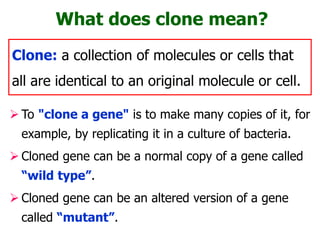
1. What does clone mean?
To "clone a gene" is to make many copies of it, for
example, by replicating it in a culture of bacteria.
Cloned gene can be a normal copy of a gene called
“wild type”.
Cloned gene can be an altered version of a gene
called “mutant”.
Clone: a collection of molecules or cells that
all are identical to an original molecule or cell.
2. In 1983, Polymerase Chain Reaction (PCR) was first
developed by Kary Mullis (USA). He was awarded the
Nobel prize in chemistry along with Michael Smith for
his work on PCR.
Since its introduction, the PCR has revolutionized the
method of DNA analysis in both research and clinical
laboratories.
Since PCR is a repetitive DNA synthesis reaction, it can
amplify DNA from as little material as a single cell, and
from very old tissue isolated from Egyptian mummies, a
frozen mammoth and insects trapped in ancient amber.
Tools of genetic engineering: PCR
3. Components of PCR reaction
Template DNA.
Primers.
Thermostable DNA polymerase.
- Taq DNA polymerase
dNTPs.
- dATP, dTTP, dCTP, dGTP
PCR Buffer (Mg++).
Thermo-cycler.
Thermus aquaticus is the source
of Taq DNA polymerase.
4. 2. Annealing: The reaction mixture is
cooled down. Primers anneal to the
complementary regions in the DNA
template strands, and double strands
are formed again between primers and
complementary sequences.
3. Extension: The DNA polymerase
synthesizes a complementary strand.
The enzyme reads the opposing strand
sequence and extends the primers by
adding nucleotides in the order in
which they can pair. The whole process
is repeated over and over.
1. Denaturation: DNA fragments are heated at high tempera-
tures, which reduce the DNA double helix to single strands.
These strands become accessible to primers.
PCR procedures: steps
6. PCR procedures: conditions
Complete denaturation of the DNA template.
Optimal annealing temperature. The annealing step
is critical for high PCR specificity.
Optimal extension temperature.
Number of PCR cycles.
Final extension step.
Contamination of the DNA
must be prevented by
separating the areas for
DNA extraction and PCR.
7. PCR primer design
PCR amplification is performed routinely and thousands of
PCR protocols have been developed.
Since PCR is both a thermodynamic and an enzymatic
process, factors such as primer design and the reaction
chemistry used are very critical for high specificity in PCR.
Guidelines for the design and use of primers
Length 18 - 30 nt
GC content 40 - 60%
Tm information Similar Tm for all primer pairs
Estimating optimal
annealing temperature
Usually 5°C below the
calculated Tm
8. The Tm is defined as the temperature in degree Celsius, at
which 50% of all molecules of a given DNA sequence are
hybridized into a double strand, and 50% are present as
single strands.
What is Tm?
The Tm is affected by a
number of factors:
• Concentration of DNA.
• Concentration of ions
in the solution, most
notably Mg+ and K+.
• Length of DNA and
type of sequence.
9. How to predict Tm?
There are several methods to calculate a theoretical Tm,
based on different physical models of what is happening in
the hybridization or melting process.
2+4 rule of thumb method:
This very simple method assigns 2°C to each A-T pair and
4°C to each G-C pair. The Tm then is the sum of these
values for all individual pairs in a DNA double strand. This
takes into account that the G-C bond is stronger than the
A-T bond.
Tm= (wA+xT)*2 + (yG+zC)*4
where w, x, y, z are the number of the bases A,
T, G, C in the sequence, respectively.
Note that the 2+4 rule is valid for a small length range
only, about 20-40 nt. It is very easy to compute, but is of
course very inaccurate.
Where possible, this method should be avoided.
10. Predicting Tm: Linear regression method
A more sophisticated method is the linear regression based on
the length of the DNA molecule and the GC ratio. Based on
empirical data, a number of linear regression terms for the Tm
have been proposed.
One term, from Bolton and McCarthy, PNAS 84:1390 (1962), as
presented in Sambrook, Fritsch and Maniatis, Molecular
Cloning, (1989, CSHL Press), is:
Tm = 81.5 + 16.6(log10[Na+]) + 0.41*(%GC) – 600/length
where [Na+] is the molar sodium concentration, (%GC) is the
GC ratio, and length is the length of the sequence.
Note that these formulae are just approximations, as they do
not take into account stacking effects and consider nucleotide
properties only in the form of an averaged GC ratio.
11. Methylation-specific PCR (MSP)
MSP enables the methylation status of target DNA to be
determined after sodium bisulfite treatment.
The method requires two sets of primers: one set that
anneals to unchanged cytosines (i.e., methylated
cytosines in the genomic DNA) and another set that
anneals to uracil resulting from bisulfite treatment of
cytosines (not methlyated in the genomic DNA).
Amplification products derived
from the primer set for unchanged
sequences indicates the cytosines
were methylated and were thus
protected from alteration.
12. Nested PCR
Two sets of primers are used in
two successive reactions.
In the first PCR, one pair of
primers is used to generate DNA
products, which will be the target
for the second reaction.
In second PCR, another pair of
primers whose binding sites are
located (nested) within the first set
is used, thus increasing specificity.
Nested PCR is often more
successful in specifically amplifying
long DNA products and used to
detect pathogens.
13. Assembly PCR
Also known as Polymerase
Cycling Assembly (PCA).
It involves an initial PCR
with primers that have an
overlap and a second PCR
that uses 1st PCR products
as template to generate the
final full-length product.
This technique is useful to
crate mutant libraries using
degenerate primers.
14. Differential display PCR
Differential display PCR is based on reverse transcription PCR (RT-
PCR), and is used to compare and identify differences in mRNA (and
therefore gene) expression patterns between two cell lines or
populations.
In this technique, first-strand cDNA synthesis is primed with a primer
complementary to ~13 nucleotides of the poly(A) tail of mRNA and
the adjacent 2 nucleotides of the transcribed sequence.
After reverse transcription and PCR amplification, amplified
products are visualized using gel electrophoresis. The banding
patterns observed can be compared to identify differentially
expressed cDNAs in the 2 populations.
Invented in the 1990s, the technique fast became a key tool in gene
expression analysis. However, it has been more recently superseded
by microarrays and qRT-PCR.
15. Quantitative Real-Time PCR (qRT-PCR)
Real-time progress of DNA amplification by measuring the
release of fluorescent "flashes" during amplification. A
computer measures the rate of "flashing" in 96 simultaneous
experimental PCR reactions relative to a control reaction.
Fluorescent dyes, such as
SYBR Green, or
fluorescence-containing
DNA probes, such as
FRET probes, are used
to measure the amount of
amplified product as the
amplification progresses.
16. The first application of PCR was for genetic testing,
where a sample of DNA was analyzed for the presence
of genetic disease mutations.
PCR analysis is also essential to pre-implantation
genetic diagnosis, where individual cells of a developing
embryo are tested for mutations.
PCR can also be used as part of a sensitive test for
tissue typing, vital to organ transplantation.
Many forms of cancer involve alterations to oncogenes.
By using PCR-based tests to study these mutations,
therapy regimens can sometimes be individually
customized to a patient.
Application of PCR: Medical applications
17. Diagnosis of the middle ear infection known as otitis media.
PCR technique has been employed to detect bacterial DNA in
children's middle ear fluid, signaling an active infection even
when culture methods failed to detect it.
Lyme disease, the painful joint inflammation caused by
bacteria transmitted by tick bites, can be diagnosed by
detecting the disease organism's DNA contained in joint fluid.
PCR is the most sensitive and specific test for Helicobacter
pylori, the disease organism now known to cause almost all
stomach ulcers.
PCR techniques have also been employed to detect three
different sexually transmitted disease organisms such as
herpes virus and papilloma viruses as well as chlamydia from
a single swab sample.
Diagnostic applications
18. The development of PCR-based genetic fingerprinting
protocols has seen widespread application in forensics.
The genetic fingerprinting can uniquely discriminate any
person from the entire population of the world.
Minute samples of DNA from single dried blood spot, saliva
on cigarette butt, semen, etc. can be isolated from a crime
scene, and compared to that from suspects, or from a DNA
database of earlier evidence or convicts.
Less discriminating forms of DNA fingerprinting are used in
parental testing, where an individual is matched with their
close relatives.
- DNA from unidentified human remains can be tested,
and compared with that from possible parents.
- Similar testing can be used to confirm the biological
parents of an adopted (or kidnapped) child.
- The actual biological father of a newborn can also be
confirmed (or ruled out).
Forensic applications
19. PCR has been applied to many areas of research in
molecular genetics, DNA sequencing and genetic
mapping.
PCR allows rapid production of short pieces of DNA,
even when nothing more than the sequence of the
two primers is known.
A common application of PCR is the study of
patterns of gene expression. Tissues (or even
individual cells) can be analyzed at different stages
to see which genes have become active, or which
have been switched off.
PCR can also be used in phylogenetic analysis.
Research applications
20. PCR can exclude suspects but
cannot prove guilt
Even when evidence such as semen and blood stains
are years old, PCR can make unlimited copies of the
tiny DNA amounts remained in stains for investigation.
DNA profiling is only one of many pieces of evidence
that can lead to a criminal conviction, but it has proved
invaluable in demonstrating innocence.
Sometimes, seemingly strong DNA evidence does not
lead to a conviction.
Dozens of cases have involved people who have spent
years in jail for crimes they did not commit until PCR
exonerated them.
21. Vector is a DNA molecule into which exogenous
DNA is integrated for cloning and that has the ability
to replicate in a suitable host cell.
Vectors are used to assist in the transfer, replication
and sometimes expression of a specific DNA
sequences in a target cell.
Vectors may be plasmids, a bacteriophage, cosmids,
bacterial artificial chromosomes and yeast artificial
chromosomes.
Tools of Genetic Engineering:
Cloning Vectors
22. Properties of vector
A vectors must possess the following properties:
1. Vectors must have origin of replication to replicate
autonomously in the cell population as the host organism
grows and divides.
2. Vectors must have unique sites for many restriction
enzymes called multi-cloning site (MCS) into which DNA
insert can be cloned without disrupting essential function.
3. Vectors must be fairly small, low molecular weight DNA
molecules to facilitate their isolation and handling.
4. Vectors must have some selectable marker that will enable
the recombinant vector to be selected from large population
of cells that have not taken up foreign DNA.
23. Vector types
Plasmids - are found naturally in bacteria and replicate inside
the bacterial cell.
Bacteriophage - replicate in E. Coli in the lytic or lysogenic
mode.
Cosmids - They are hybrid vectors of phage and plasmids.
Bacterial artificial chromosomes (BACs) - are based on the F
factor of E. coli that confers the ability to conjugate.
Yeast artificial chromosomes (YACs) - were primarily used in
genome sequencing projects.
Vector Insert size (kb)
Plasmid <10 kb
Bacteriophage 10-20 kb
Cosmids 33-50 kb
BACs 75-300 kb
YACs 100-1000 kb
24. Vectors based on plasmids
Plasmids are circular, dsDNA molecules that replicate indep-
endently and are separated from a cell’s chromosomal DNA.
The independent replication of plasmids is due to the pres-
ence of certain sequences acting as the origin of replication.
The size of the plasmids varies from less than 1.0kb to more
than 200kb.
Most of the plasmids are not required for the survival of in
which they reside.
In many cases, however, they are essential under certain
environment, such as in the presence of antibiotics.
Smaller plasmids are much desirable for
gene cloning experiments.
Examples of plasmid vectors are pBR322
(4.4kb), pBR345 (0.7kb), pMB9 (5.8kb), etc.
25. Plasmid vectors (contd.)
The smaller plasmids use the DNA replicative enzymes of
the host cells and larger plasmids carry genes that code
for special enzymes necessary for their replication.
Under certain conditions, some plasmids may integrate into
bacterial chromosome where they replicate along with the
bacterial chromosome. These types of plasmids are called
episomes or integrative plasmids.
pBR322 was one of the first versatile plasmid vectors deve-
loped; it is the ancestor of many of the common plasmid
vectors used in research laboratories.
pBR322 contains an origin of replication (ori) and a gene
called rop that helps to regulate the number of copies of
plasmid DNA in the cell.
26. (4.4kb)
Origin of replication (ori).
Selectable marker (ampR & tetR).
MCS (ClaI & HindIII).
Fairly small in size (~4.4kb).
Nomenclature of pBR322
p- denotes plasmid.
BR- indicates the
laboratory of Bolivar
and Rodriguez.
322- indicates a
distinguished number.
pBR322 is an artificial plasmid, which was derived from
three different but naturally occurring plasmids.
27. pBR322 was constructed by using three different naturally
occurring plasmids. The ampicillin resistance gene was
derived from RSF2124 and tetracycline resistance gene was
taken from pSC101. The origin of replication was obtained
from pMB1.
Another 3 RE sites fall within the origin region and therefore
can not be used for cloning purposes.
Several pBR derivative vectors with multiple cloning sites
have been constructed to enhance cloning versatility.
pBR322 has 21 unique
restriction (RE) sites.
But only 11 RE sites can
be used to insertionally
inactivation of the
antibiotic resistance gene.
28. The number of molecules of a plasmid in a single bacterial
cell is termed as copy number. It ranges from 1 to more
than 50 per cell.
Plasmid can be categorized on the basis of number of copies
per cell as,
1. Relaxed plasmids, which are normally maintained at
multiple copies per cell and
2. Stringent plasmids, which have a limited number of
copies per cell.
Plasmid can also be classified as conjugative plasmids
and non-conjugative plasmids, depending on whether or
not they carry a set of transfer gene called the tra genes.
These tra genes promote bacterial conjugation.
Generally conjugative plasmids are of high molecular weight
and are present as 1-3 copies per cell, whereas nonconjuga-
tive plasmids have low molecular weight and are present in
multiple copies, i.e., 20-25 copies per cell.
Copy number of plasmids
30. Vectors based on bacteriophage
The cloning of single genes is usually best carried out using
plasmids, since the insert will rarely be larger than about 2kb.
But, for cloning of larger pieces of DNA (e.g. during gene
library construction), these plasmids are not suitable as larger
inserts increase plasmid size, making the transformation
inefficient.
Larger molecules can be injected in host bacterial cell by viral
particles (bacteriophages). Commonly used bacteriophages are
M13, f1, fd and Lambda () phage.
phage genome is 49kb in length, and the central 20 kb is
only used for lysogeny; it can be replaced by foreign DNA
through ligation of arms with insert using DNA ligase followed
by in vitro packaging into phage particles using cell extracts
that contain pieces of phage heads and tails. The final
preparation is used to infect the new E. coli cells.
31. Bacteriophage based vectors
Phage lambda can do two
different things when it enters
the cell:
– lytic cycle: it can start
reproducing itself
immediately, which
produces about 200 new
phages in 15 minutes and
kills the cells.
– lysogenic cycle: the
lambda DNA can integrate
into the host chromosome
and remain dormant for
many generations. When
given the proper signal, the
integrated DNA (prophage)
leaves the chromosome
and enters the lytic cycle.
34. Cosmid cloning vector
Cosmids are hybrid DNA molecules and can live in a dual
living system. They combine essential elements of a plasmid
and lambda phages.
Their plasmid part enables them to replicate as it has origin
of replication and also helps in selection due to the presence
of marker genes.
Their lambda part (cos sequences) allows them to be
packaged in a phage coat and to be transduced to a recipient
by the lambda infection machinery. Since it has no genes for
viral proteins, viral particles are not formed in the host.
Recombinant cosmid is injected into the bacterial cells where
they arrange into a circle and replicates as a plasmid without
host cell lysis.
Foreign DNA fragments up to 50kb can be cloned using a
cosmid vector that can be maintained and recovered just as
plasmids.
37. Bacterial Artificial Chromosome (BAC)
The F factor of E.coli is capable of handling large segments
of DNA.
Recombinant BACs are introduced into E. coli by electropo-
ration (a brief high-voltage current).
Once rBACs are in the cell, they replicate like an F factor.
Has a set of regulatory genes, OriS, and repE which control
F-factor replication, and parA and parB which limit the
number of copies to one or two.
A chloramphenicol resistance gene, and a cloning segment.
BACs can hold up to 300 kbs (e.g. pBAC108L, pBeloBac11).
39. parA and parB (maintain
single copy number).
Cm (Chloramphenicol)
marker.
OriS, and repE
(control F-factor
replication).
Genetic map of pBeloBAC11
40. YACs can hold up to 1000 kbs (1 Mb) DNA segments. They are
also called mini-chromosome.
YACs are linear DNA segments that contain all the molecular
components required for replication in yeast.
YACs have been designed to replicate as plasmids in bacteria
when no foreign DNA is present. Once a fragment is inserted,
YACs are transferred to the yeast cells where they replicate as
eukaryotic chromosomes.
Initially, YAC was used for investigation of the maintenance of
chromosomes in the cell. Later on, it was used as vectors for
carrying very large cloned fragments of DNA.
YACs have also been used for physical mapping of human
chromosome in “Human Genome Project”.
Yeast Artificial Chromosome (YAC)
41. A typical YAC consists of centro-
mere element (CEN) for chro-
mosomal segregation during cell
division, telomere and origin of
replication (ori) that were
isolated and joined to E. coli
plasmids (e.g. pYAC3).
The pYAC3 vector contains E.
coli origin of replication (oriE)
and a bacterial selectable
marker (ampR) together with
yeast selectable markers (TRP1,
SUP4 and URA3) and autono-
mously replication sequence
(ARS) which acts as yeast ori.
YAC (contd.)
43. A shuttle vector is a vector constructed so that it can
propagate in two different host species. Therefore, DNA
inserted into a shuttle vector can be tested or manipulated
in two different cell types. It has two origins of replication, each
of which is specific to a host.
Since shuttle vectors replicate in two different hosts, they are
often known as bifunctional vectors.
One of the most common types of shuttle vectors is the yeast
shuttle vector, which have components that allow for replication
and selection in both E. coli and yeast cells.
Almost all commonly used Saccharomyces cerevisiae vectors
are shuttle vectors.
There are also adenovirus shuttle vectors, which can propagate
in E. coli and mammals.
Shuttle Vector
44. The E.coli component of a yeast shuttle vector includes an
origin of replication and a selectable marker such as
antibiotic resistance and beta-lactamase.
The yeast component of a yeast shuttle vector includes
autonomously replicating sequence (ARS), a yeast
centromere (CEN) and a yeast selectable marker such as
URA3 (a gene that encodes an enzyme for uracil synthesis).
Example of Shuttle Vector: pHV14,
pEB10, pHP3, etc. replicate both in
Bacillus subtilis and E. coli.
pJDB219 is another shuttle vector
that can replicate in E.coli and
Yeast (Saccharomyces cerevisiae ).
Shuttle vector (contd.)
45. Expression Vectors are the vectors that contain suitable
expression signals to have maximum gene expression.
Expression vector are designed for the expression of protein
product coded by that inserted gene.
Expression vectors could be either prokaryotic or eukaryotic.
The following expression signals are introduced into expression
vectors to get maximum protein expression:
Insertion of a strong promoter.
Insertion of a strong termination codon.
Adjustment of distance between promoter and cloned
gene.
Insertion of transcription termination sequence.
Insertion of a strong translation initiation sequence.
Expression Vectors
46. Prokaryotic Expression Vectors
Expression vectors for bacterial hosts are generally plasmids
that have been engineered to contain appropriate regulatory
sequences for transcription and translation such as strong
promoters, ribosome-binding sites, and transcription
terminators.
Eukaryotic proteins can be made in bacteria by inserting a
cDNA fragment into an expression vector. Large amounts of
a desired protein can be purified from the transformed cells.
In some cases, these proteins can be used to treat patients
with genetic disorders.
For example, human growth hormone, insulin and several
blood coagulation factors have been produced using rDNA
technology and prokaryotic expression vectors.
48. Prokaryotic cells may be unable to produce functional
proteins from eukaryotic genes even when all the signals
necessary for gene expression are present because many
eukaryotic proteins must go through post-translational
modified.
Several expression vectors that function in eukaryotes have
been developed (e.g. pcDNA4/HisMax, pSV240).
These vectors contain eukaryotic origins of replication,
marker genes for selection in eukaryotes, transcription and
translation control regions, and additional features required
for efficient translation of eukaryotic mRNA, such as
polyadenylation signals and capping sites.
Eukaryotic Expression Vectors
50. 1. Transformation is a process by which exogenous genetic materials
are introduced into bacterial cells.
For transformation to happen, bacteria must be in a state of
competence. Competent bacterial cells are now commercially
available or they can be prepared in the laboratory.
Introduction of foreign DNA into eukaryotic cells is often
called transfection.
2. Conjugation method refers to the transfer of genetic material between
two bacterial cells via direct contact.
3. Transduction is the injection of foreign DNA into the host bacterium
by a bacteriophage virus.
4. Electroporation is another method where the cells are briefly shocked
with an electric field that creates holes in the cell membrane through
which the plasmids are entered. After the electric shock, the holes are
rapidly closed by the cell's membrane-repair mechanisms.
Processes of rDNA transfer into bacteria
51. After the introduction of rDNA into suitable host cells, it is
essential to identify those cells which have received rDNA
molecules. This process is called selection or screening.
The methods used for screening of recombinants in E. coli
are
direct selection,
insertional inactivation,
blue-white selection
PCR amplification,
gel electrophoresis,
DNA sequencing, and
colony hybridization (nucleic acid hybridisation).
Identification of host cells containing rDNA
52. In direct selection, cells are grown under conditions in which
only transformed cells can survive; all the other cells die.
If the plasmid containing rDNA has selectable marker
(ampR), the recombinants will only grow and form colonies
on medium containing ampicillin.
This procedure can not confirm whether the recombinants
growing on such medium contain religated plasmid vector or
contain recombinant plasmid with foreign DNA molecules
because ampR gene is present in both cell types.
In this case, the transformed cells have to be individually
tested for the presence of the desired recombinant DNA,
which can be accomplished by colony screening technique
using PCR.
Direct selection
53. Insertional inactivation
A piece of DNA is inserted into the unique PstI site in pBR322.
This insertion disrupts the gene coding for a protein that
provides ampicillin resistance to the host bacterium.
Hence, the chimeric plasmid will no longer survive when plated
on a medium that contains ampicillin.
56. Colonies are overlaid with a DNA-binding membrane
such as nylon.
Colonies are transferred to membrane, then lysed, and
DNA is denatured.
Membrane is placed in a heat-sealed bag with a solution
containing the labeled radioactive probe; the probe
hybridizes with denatured DNA from colonies.
Membrane is rinsed to remove excess probe, then dried;
X-ray film is placed over the filter for autoradiography.
Using the original plate, cells are picked from the colony
that hybridized to the probe.
Cells are transferred to a medium for growth and further
analysis.
Colony hybridization (contd.)
57. An ampicillin & tetracycline resistant plasmid, pBR322, is
cleaved with PstI, which cleaves within the ampicillin resistance
gene. The cut plasmid is ligated with PstI digested Drosophila
DNA to prepare a genomic library, and the mixture is used to
transform E. coli K12.
(a) Which antibiotic should be added to the medium to select cells
that have incorporated a plasmid?
Answer to the following questions
(b) If recombinant cells were plated on
medium containing ampicillin or
tetracycline and medium with both
antibiotics, on which plates would
you expect to see growth of bacteria
containing plasmids with Drosophila
DNA inserts?
(c) How can you explain the presence
of colonies that are resistant to
either ampicillin or tetracycline?
58. Common host and model organisms used in molecular biotechnology
59. Common host and model organisms used in molecular biotechnology