Acute pancreatitis (AP) can range from mild to severe. Mild AP is treated conservatively with nothing by mouth, hydration, pain control, and blocking pancreatic secretion. Severe AP requires intensive care for volume resuscitation, inhibiting pancreatic secretion, antiprotease therapy, pain management, and detoxification. Diagnosis involves enzyme levels, imaging, and severity scales. Differential diagnosis excludes other abdominal issues. Prognosis depends on severity and multi-organ failure. Treatment aims to stop the inflammatory process and prevent infection.
2. Terminology
• Acute pancreatitis — acute aseptic inflammation of the
pancreas with demarcation, with the pancreocytes’ necrosis
and ferment autoagression with gland’s necrosis and
secondary infection addition.
• Pancreonecrosis = destrucrive pancreatitis = necrotic
pancreatitis
• Abbreviations:
AP – acute pancreatitis
PG – pancreatic gland
3. Some statistics
• From 3 to 6% of urgent abdominal pain cases.
• 3rd place after acute appendicitis and acute
cholecystitis.
• Lethality from 3 to 9 %, within destructive forms
from 40 to 70 %.
• the most difficult problem of the today’s
abdominal surgery situations (lack of some
pathogenesis’ issues, no strict consensus on
treatment in every case).
13. Endocrine secretion
α cells
glycogen
blood sugar’s
elevation
glucagon
β cells
glucose
blood sugar’s
decrease
insulin
D cells
inhibits insulin
secretion
control of the blood
sugar level
somatostatin
14. Etiology: duct system depressurization
1. Gallstones disease (30-40%), including terminal region of the bile duct
pathology (choledocholythiasis, odditis, cholangitis, big duodenal
papilla’s stricture)
2. Alcoholism (30-75%). mechanism:
а. Pancreatic excretory function’s stimulation by alcohol.
б. Pancreatic secret’s evacuation alternation because of the Oddie’s
sphinctor’s spasm due to the duodenum’s irritation (morphine-like
action).
3. Trauma of the pancreas: penetrating wounds, closed wounds,
intraoperative wounds.
4. Gastrointestinal tract diseases (stomach and duodenum’s ulcers with
penetration, chronic duodenal passability alternation)
5. Blood flow alternation – chronic mesenteric ischemia (atherosclerosis
alternation of the pancreatic vessels), centralisation of the blood
flow
15. Etiology
6. Endocrine issues.
a. Hyperparathyroidism – hypercalcemia – pancreatic proteolitic
enzymes activation.
b. Hypothyroidism
c. Lipid metabolism alterantions – hyperlipidemia, including hereditary
(Friderichsen’s disease)
d. Protein metabolism alternation, insufficient protein input
7. Chronic infections (B and C hepatitis viruses, epidemic parotitis, CMV)
8. Pancreatotoxic medications (sulfanylamines, tetracyclins, non steroid
anti-inflammatory treatmets, furosemid, estrogens,
immunosupressants, steroids)
9. Rare anomalies (pancreatic development anomalies,
hyperaminoacidurie, allergic and auto allergic reactions).
19. Pathogenesis
Hypertension or trauma depressurization
of the duct system pancreatic enzymes outflow
in the interstitium
Lipolytic cascade (fat necrosis)
acidosis
Proteolytic enzyme’s activation
Proteolytic cascade (hemorragic necrosis)
systemic vascular alternations
20. Classification
I. Oedematous (intersitial) panreatitis.
II. Steril pancreonecrosis.
- based on the necrosis characteristics:
fatty, hemorragic, mixed;
- based on lesion abundance:
little sources, big sources, subtotal;
- based on localisation:
head, queue, generalized.
III. Infected pancreonecrosis.
21. • Complications:
I. Parapancreatic infiltrate.
II. Pancreatic abscess.
III. Peritonitis: fermentative (abacterial), bacterial.
IV. Septic flegmona of the peritoneal fat: parapancreatic,
paracolic, pelvic.
V. Arrosive hemorrage.
VI. Mechanical jaundice.
VII. Pseudocyst: steril, infected.
VIII. Inner and exterior digestive fistulas.
26. Periods of the Pancreatitis progression
1. Hemodynamic alterations period (pancreatogenic
shock), first 3 days
2. Period of multyorganic failure (shock organs
syndrome), 4-10 days
3. Period of sequestration and suppurative
complications, 10 and more days:
3.1. aseptic destructive complications
3.2. septic destructive complications (abdominal
pancreatogenic sepsis)
28. Clinic of the AP
Mondor’s triade
• Pain
• Vomiting
• Meteorism
29. Oedemous pancreatitis
Mild form of pancreatitis (interstitious oedema
of the pancreatic gland)
• 80 % of patients with AP
• Lethality less than 1 %
31. Clinic of the AP
• Sudden strong constant pain in the epigastric region with
left hypochondrium irradiation, left loin region (1-3 days)
• uncessant vomiting, without relief
• Pale skin, lips cyanosis
• Tachycardia within 5-6 hours
• Possible hyperthermia (SIRS)
• Leucocytosis if satisfactory hemodynamics
32. Clinic of the AP
• Kerte’s symptom (epigastric pain
and rigidness)
• Voskresensky symptom (aortic
pulsations lessen in the
epigastrium)
• Mayo-Robson symptom (strong
pain in the left vertebro-costal
angle)
• Peritoneal symptoms (peritoneal
irritation because of the
hemorragic peritoneal infusion)
33. Symptoms of severe acute pancreatitis
• Begins with an unstable hemodynamics, up to collapse
(arterial pressure<50 mm Hg)
• Strong pain symptoms. Pain localises in the epigastrium
with the irradiation to the left hypochondrium, under left
scapula
• Uncessant pain, without relief
• Signs of the dynamic intestinal blockage, intestinal paralysis,
abdominal distention
34. Symptoms of the severe AP
• Pain during superficial palpation of the abdomen, muscular
contraction of the anterior abdominal wall , especially in the
epigastric region
• Diminishing sound during percussion in the in the declivious
regions (effusion)
• Leucocytosis with PNN, inflammation markers
• Blood and urine high levels amylase, lipase, tripsine (during
first two days), high enzymes level in the peritoneal
exsudate
35. «Colour» symptoms
Skin pallor with cyanotic spots in severe cases:
• Mondor’s symptom (violet spots on the face and body)
• Grey-Turner symptom (cyanotix spots on the lateral walls of the
abdomen)
• Gruenwald’s symptom (periombilical cyanotic spots)
• Holsted’s symptom (cyanosis on the abdominal skin)
• Cullen’s symptom (icteric colouring of the periombilical region)
36. Mechanism of the process’ spreading
Exsudation type
1.Anterior (peritonitis);
2.Posterior
(retroperitoneonecrosis);
3.Mixed type.
40. Diagnosis of the AP
• 1. Clinical symptoms’ evaluation.
• 2. Determination of the activity of the enzymes in the blood: amylase
(lipase), in urine(amylase), peritoneal exsudate
• 3. Evaluation and control of the hemodynamic’s determinants -
arterial pressure, heart rate, shock index Альговера
• 4. Dynamics of the homeostatic determinants
• 5. Scales of severity (Ranson, Glasgow-Imrie)
• 6. Complexe instrumental diagnosis:
1) abdominal US
2) X-rays of the thorax and abdomen
3) CT
4) laparocentesis
5)laparoscopy
41. Ultra-sound diagnosis - method of choice
• Increased and fuzzy gland
• effusion in the , peritoneal space
45. CT severity scale (Balthazar)
• A) Normal pancreas (0 points);
• В) local or diffuse lesion of the pancreas with hypodense inclusions in
its tissue with fuzzy contours, pancreactic’s duct enlargement (1
point);
• С) Pancreatic tissues metamorphosis , analogic to the B-stage, with
the inflammatory metamorphosis in the peripancreatic cellulose (2
points);
• D) Metamorphoses + rare liquid formations out of the pancreatic
tissue (3 points);
• Е) D metamorphoses + two or more formations out of the pancreas or
abscess (4 points).
46. Laboratory diagnosis
Enzymes in the blood flow – Hyperfermentemia (first
2-3 days)
1.Blood amylase increases,diastase in the urine;
2.Blood lipase increases
3.Blood tripsine increases.
More specific and more expensive
in total necrosis there is no fermentemia
47. • Bilirubin, transaminases
- Hyperbilirubinemia if biliary pancreatitis (mechanical
jaundice);
- Parenchymatous hyperbilirubinemia (toxic liver
lesion); cytolysis
- Increased transaminases (ASAT, ALAT)
Very important detox role of the liver
Calcium level determination (falls) – prognostic
marker, necrosis marker.
Protein, urea, creatinine, etc. – evaluation of organ’s
failure
Laboratory diagnosis
48. Evaluation of severity of AP
Very important diagnostic and prognostic stage
Prognosis
Choice of the treatment strategy
Specific stratification scales: Ranson, Glasgow-Imrie,
Apache II.
Scales of the multi organ’s failure: SAPS, SAPS II,
SOFA, MODS и тд.
To quantify the severity of the AP (in points) using
clinical and lab analysis
53. If AP diagnosis suspected -
hospitalisation in the surgery
department
If biliary AP – urgent endoscopic
papillotomia
54. • Two main goals of the treatment
1. To stop the process:
1.1 Recuperation of the duct system function;
1.2 Blockage of the pancreatic secretion.
2. To prefer the aseptic process.
56. Mild pancreatitis
Basic conservative treatment
In the surgery department
1. No alimentation (table 0)
2. Local hypothermia (ice on the abdomen)
3. Nasogastric sond (aspiration)
4. Infusion therapy and microcirculation lesions corrections in
the pancreas:
- infusion therapy 30 ml for 1 kg of boy wight + forced diuresis
during 24-48 hours
- rheological treatment (реополиглюкин, трентал)
5. Pancreatic secretion blockage: М-холинолитики,
блокаторы Н2 рецепторов, ИПП, 5-фторурацил
6. Spasmolytics
57. Mild pancreatitis
Basic conservative treatment
In the surgery department
1. 7. Desensibilising treatment (супрастин, димедрол,
пипольфен)
2. 8. Bile excretion control.
3. (if choledocholythiasis, stricture) –
4. ESPT,
5. lithoextraction
7. 9. If no effect during 6 hours = diagnosis of the severe AP
=> hospitalisation in the reanimation department
58. Severe pancreatitis
Intensive conservative treatment
In the reanimation department
1. Basic treatment
2. Treatment inhibiting the pancreatic secretion
- sandostatin
during 3-5 days
3. Antifermentive therapy
- aprotinin
During 3 days
59. Severe pancreatitis
Intensive conservative treatment
In the rea
4. 4. Painkillers and antiparalysis therapy
- epidural anaesthesia
- General blood volume normalisation with normalisation
of the central veinous pressure and hematocritis
60. In reanimation
5. 5. Transfusion:
- Hemodilution (40-60 ml/kg a day) for hypovolemia
extinction;
- crystalloid solutions (0,9 % NaCl, glucose + insuline,
лактасол, salt solutions (45-80 ml/kg), Ringer’s solution;
- colloid plasma remplacement (полиглюкин,
реополиглюкин)
- correction КОС (5% р-р гидрокарбоната натрия из расчёта
2,5-3 мг/кг)
61. 7. 6. Detox therapy:
- - Forced diuresis (фуросемид, маннитол, сорбитол);
under diuresis control (не менее 4-5 мл/кг в час);
contraindications: acute renal failure, chronic renal
failure, acute cardiovascular failure, acute myocardial
infarction.
- - Extracorporal detox methods (hemosorbtion,
hemofiltration, plasmapheresis, plasmosorbtion, thoracic
canal’s dreinage).
- - Intestinal lavage through nasointestinal sond with salt
electrolyte solution
- - Peritoneal dyalysis during laparoscopy
62. - punction aspiration of the omental liquid with US control
8. 7. Nutritive support (if no intestinal paralysis)
- glucose-salt solutions
- nutritive solutions (берламин, нутризон,
нутрилан)
9. 8. Prophylactic antibiotherapy
- меропенем, имипенем
- цефалоспорины III-IV поколений
- фторхинолоны II-III поколений + метронидазол
63. I. AP during first 24 hours. Strategy
• 1. Effective blockage of the pancreatic secretion (table
0, nasogastric sond, secretoblocators);
• 2. Massive infusion-detox therapy (не <50 мл/кг) –
recuperation МЦР;
• 3. Antioxydant and antihypoxy therapy;
• 4. If biliary AP – occlusion’s treatment (ЭПСТ,
lithoextraction).
• Goal: to achieve the failure phenomenon
65. II. Sterile necrosis. Surgical strategy minimally-
invasive surgery
• Pancreatogenic peritonitis – laparoscopy.
• Parapancreatic liquid content – punction under US
control.
• Earl open operation –100% infection chance,
lethality - 50%.
66. Open operation indications
• 1. Absence of the minimally invasive surgery
possibility
• 2. Absence of the therapy’s effectiveness and
progressive patient’s condition deterioration (total
necrosis or not adequate therapy) – desperation
surgery
67. III. Infected necrosis = abdominal sepsis
• +
• Active surgical strategy:
• 1. minimally invasive methods (punction
drainage under US control);
• 2. open surgery
• GOAL: Ubi pus ibi incisio
69. The goal of the surgical treatment – surgical
detoxication
1. Hemorragic exsudate’s removal from the abdominal cavity
in order to prevent the enzymatic toxicity, abdominal
cavity’s drainage.
2. Omentum’s drainage with pancreas’ decompression by
parietal peritoneum’s section around the gland.
3. Rational drainage of the retroperitoneal space for the
purulent source’s sanation from toxic products of the
inflammation and necrolysis
77. 7 — angle section
(Ryo-Branco)
8 — left
transrectal section
9 — lumbar section fot the
body’s and queue’s access
10 — lumbar section for the
gland’s head surgery
Хирургические доступы к поджелудочной железе
86. Classification
Complications of the AP:
I. Parapancreatic infiltrat.
II. Pancreatic abscess.
III. Peritonitis: fermentative (abacterial), bacterial.
IV. Septic flegmona of the retroperitoneal cellulose:
parapancreatic, paracolic, pelvic.
V. Erosive hemorrage.
VI. Mechanical jaundice.
VII. Psudocyst: sterile, infected.
VIII. Internal and external digestive fistulas.
87. Complications of the AP
Complications of the pancreatogenic toxemia
I. Pancreatogenic shock
II. Shock organ syndrome:
1. Encephalopathy
2. Acute cardiovascular failure
3. Acute respiratory failure
4. Acute liver failure
5. Acute renal failure
6. Enteral failure
91. Infection of the pancreonecrosis
1. Endogene way:
• duodenopancreatic reflux (unlikely);
• Bacterial translocation of the intestins (intestins –
sepsis promotor). Earlier the gastro-intestinal
tract begins to be functionnal, less the risk of the
infection: intestinal resistance to the colonisation,
abusive intestinal colonisation syndrome
2. Exogene way:
Surgical intervention (any). Risk increases with the
operation’s size.
92. Pancreatogenic effusion in the peritoneal cavity (ferment
ascitis-peritonitis)
• Peritoneal symptoms with growing symptoms of the
peritoneal irritation.
• Liquid in the peritoneal cavity during US.
• Strategy – laparoscopic drainage of the abdominal
cavity
93. Limited effusion
1. Acute parapancreatic effusion (before 2 months, no
capsule);
2. Pseudocyst (more than 2 months, capsule
formation).
• US diagnosis
• Firstly, conservative strategy (complexe AP treatment).
• Disappears spontaneously (30-50%).
• If no resorption, growth, infection signs – intervention
needed (US-controlled punction laparotomy).
94. Parapancreatic infiltrat
• Inflammatory tumor, conglomerate of losely fixed to one
another tissues around the pancreas
• with participation of parietal peritoneum, big omentum,
stomach, transverse colon, lig. gastrocolicum, retroperitoneal
cellulose.
• Infiltrat – limitation, protection reaction
1. Formation (loose) of infiltrat – 3-4 days;
2. Formed (dense) infiltrat – after 5 days.
95. Parapancreatic infiltrate
Symptoms:
• Less abdominal pain;
• Better general patient’s condition;
• Dense, not painful, fixed formation in the epigastrium,
• Infiltrate’s size can vary a lot, can occupy all the upper
abdominal region;
• There can be positive local symptoms and peritoneal
symptoms - negative;
• Constant SIRS symptoms with the decreasing tendency
96. Parapancreatic infiltrate
• Diagnosis: US, CT.
• Treatment – AP treatment.
• Results:
1. Resolution: pain descreases, infiltrate dissapears,
normalisation of the body temperature –
Diminishing of the AP symptoms.
2. Infection operation (minimally invasive
open interventions)
97. Limited infected effusion
1. Pancreatic abscess.
2. Infected psudocyst.
3. Septic flegmona of the retroperitoneal cellulose:
parapancreatic, paracolic, pelvic.
symptoms, diagnosis, treatment (in the acute
appendicitis lesson).
Retroperitoneal flegmona - dissection and
drainage out of the peritoneum (lumbotomy).
98. Erosive hemorrage
Into the suppurative cavity
conditions:
• around the pancreas there is
• a lot of main
vessels;
• massive destructive
process;
• Surgical intervention earlier.
Symptoms: general (hypotony, tachycardia, etc) and local
(wound hemorrhage) blood loss symptoms.
Strategy: urgent intervention – tamponnade or closing,
vessel’s ligature.
99. Digestive fistulas
• Pancreaic fistulas;
• Intestinal fistulas (stomach,
duodenum, small intestin,
colon – colon transversum)
Teason – erosion of the empty organ’s wall during
suppurative necrotising process
Symptoms: presence of the empty organ’s content
in the wound . Degradation of the wound process
(from duodenum - active pancreatic ferments, colon –
agressive flora). Loss of the empty organ’s content –
progressive weight loss.
Editor's Notes
"Острый панкреатит - наиболее ужасное из всех острых заболеваний органов брюшной полости. Внезапность начала, беспрецедентное по тяжести страдание, вызванное этой болезнью, и летальность, ею обусловленная, позволяют назвать ее наиболее устрашающей из всех возможных катастроф"
B. Lord Moynihan (1925)
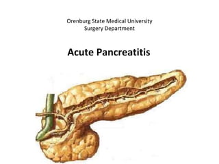
Поджелудочная железа, pancreas, располагается забрюшинно в верхнем отделе брюшной полости. Длина ее 14—18 см, ширина в области головки 5—8 см, в средней части — 3,5—5 см, толщина — 2—3 см. Железа подразделяется на головку, caput pancreatis, расширенную часть, лежащую справа от позвоночника, тело, corpus pancreatis, и хвост, cauda pancreatis, суживающийся в направлении селезенки. Головка железы уплощена; в ней различают переднюю и заднюю поверхности, facies anterior et posterior. У нижнего края головки располагается крючковидный отросток, processus uncinatus, длиной 2—5 см, шириной 3—4 см. Форма отростка непостоянна, чаще всего клиновидная или серповидная. На границе между головкой и телом имеется борозда, incisura pancreatis, в которой проходят верхние брыжеечные сосуды. Тело железы имеет призматическую форму, поэтому в нем различают три поверхности: переднюю, facies anterior, заднюю, facies posterior, и нижнюю, facies inferior. Поверхности отделены друг от друга верхним, передним и нижним краями, margo superior, anterior et inferior. Поджелудочная железа прилежит к позвоночному столбу и крупным сосудам забрюшинного пространства; тело ее несколько выступает в вентральном направлении, образуя сальниковый бугор.
Передняя поверхность железы покрыта брюшиной и соприкасается с задней стенкой желудка, от которого она отделена узкой щелью — полостью сальниковой сумки. Задняя поверхность прилежит к забрюшинной клетчатке, органам и крупным сосудистым стволам, расположенным в ней. Головка поджелудочной железы помещается в С-образном изгибе двенадцатиперстной кишки. Вверху она прилежит к нижней и задней поверхностям верхней части двенадцатиперстной кишки. В некоторых случаях железистая масса прикрывает также частично переднюю или заднюю поверхность нисходящей части двенадцатиперстной кишки.