SlideShare a Scribd company logo
1 of 87
Under the esteemed guidance of ;-
Prof. Dr. SUHAIL MAJID JAN (HOD)
Dr. ROOBAL BEHAL (Associate Professor)
By:-
MUMTAZ ALI PG (3nd Year)
‘’DEPARTMENTOF PERIODONTICS AND ORAL IMPLANTOLOGY’’
GOVNERNMENTDENATLCOLLEGE& HOSPITAL, SRINAGAR.
“SEMINAR’’
STANDARD IMPLANT
SURGICAL PROCEDURES
“No one should die
with their teeth
sitting in a glass of
water”
Per-Ingvar Branemark
May 3, 1929 – December 20,2014
INTRODUCTION
DEFINITION
HISTORICAL BACKGROUND
DENTAL IMPLANT COMPONENTS.
STANDARD IMPLANT SURGICAL PROCEDURE
General principles of implant surgery
• Patient preparation
• Implant site preparation
• One stage versus two stage implant surgeries
CONTENTS
 Two stage “submerged”
Implant placement
• Flap designs, incisions and reflection
• Implant site preparation
• Flap closure and suturing
• Post operative care
• Second stage exposure surgery
 One stage “non-submerged” implant
placement
• Flap designs, incisions and elevation
• Implant site preparation
• Flap closure and suturing
• Postoperative care
 Conclusion
INTRODUCTION
Dental implants ;- designed to
provide a foundation for
replacement of teeth that look,
feel, and function like natural
teeth.
Partial and removable
prosthesis may not bring
satisfactory results.
Goal of modern dentistry ;- restore
normal contour, function, comfort,
esthetics, speech and health of a
patient.
This leads to increased need
and use of implant and implant
supported prosthesis.
DEFINITION
Any object or material, such as an alloplastic substance
or other tissue, which partially or completely inserted or
grafted into body for therapeutic, diagnostic, prosthetic
or experimental purposes.
Can be defined as a substance that is placed into the jaw
to support a crown or fixed or removable denture.
- Charles M Weiss
A prosthetic device or alloplastic material implanted into
oral tissues beneath the mucosal or periosteal tissues
and/or within the bone to provide retention and support
for fixed or removal prosthesis.
- Edward J Fredrickson
IMPLANT
DENTAL
IMPLANT
DENTAL
IMPLANT
 Archeological findings showed that the ancient Egyptian and South American civilizations already
experimented with re-implanting lost teeth with hand-shaped ivory or wood substitutes.
 In the 18th century lost teeth were sometimes
replaced with extracted teeth of other human
donors.
 The implantation process was probably
somewhat crude and the success rates
extremely low due to the strong immune
reaction of the receiving individual.
HISTORY
2500 BC - Ancient Egyptians -
gold ligature.
500 BC - Etruscan population -
gold bands incorporating pontics.
500 BC - Phoenician population - gold
wire.
300 AD - Phoenician population - Carved
Ivory teeth.
600 AD - Mayan population -
implantation of pieces of shell.
1911 - Greenfield –iridoplatinum
basket soldered with 24 carat gold.
1943 –Dahl- subperiosteal type of
implant
Late 1970s and Early 1980s -
Tatum - custom
blade implants of Titanium alloy
Early 1980s - Tatum - Titanium
root form implant
Modern Historical Developments
The first Subperiosteal Implant was placed in 1948 by Gustav Dahl
The Endosteal Blade Implant, introduced independently in 1967 by
Leonard Linkow and Ralph and Harold Roberts
After 1980s –hollow basket Core vent
implant Niznick et al
-Screw vent implant
-Screw vent implant with
Hydroxyapatite coating
- Implant with titanium plasma
spray
 The quantum leap in Oral Implantology was
achieved in 1952 in Sweden by PER INGVAR
BRANEMARK (Father of Modern dental
implantology)
 He founded the phenomenon of Osseointegration.
 Dr. Branemark's research shifted more towards the use of titanium appliances
in human bone, including the use of titanium screws as bone anchors for lost
teeth.
 In 1982, the Toronto Conference on Osseointegration in Clinical Dentistry laid
down the first parameters on what is to be considered successful implant
treatment within the stringent confines of the scientific community.
Classification Of Implant by Charles. A. Babbush
There are five main types:
1.ENDOSTEAL IMPLANT
 An implant which is placed into the alveolar bone and/ or basal bone of
the mandible or maxilla
 Transects only one corticle plate
 Most commonly used
Blade implant
It consists of thin plates in
the form of blade
embedded into the bone
Root form
implant
Designed to mimic the
shape of the tooth For
directional load
distribution
Ramus frame
implant
Horse shoe shaped stainless
steel device Inserted from
one retromolar pad to other
2. SUBPERIOSTEAL IMPLANT
 Placed directly beneath the periosteum overlying the bony cortex
 Do not penetrate into the jawbone.
 Consists of non-Osseo integrated framework that rests on the
surface of the jaw or beneath the mucoperiosteum.
 Can be bilateral or unilateral
3. TRANSOSTEAL IMPLANT
 Other names- staple bone implant
Mandibular staple implant
Transmandibular implant
 Combines the subperiosteal and endosteal components
 Penetrates both cortical plates
 very similar to a nut and bolt arrangement
 Used in mandibles only
 penetrate the entire jaw to emerge opposite the entry site, usually at the bottom of the
chin.
4. INTRAMUCOSAL IMPLANTS
 Inserted into oral mucosa
 Mucosa is used as attachment site for metal inserts
•Described by Dr CHARLES WIESS
•Complete encapsulation of
implant with soft tissue
•Soft tissue interface could
resemble highly vascular
periodontal fibers of natural
dentition
•Described by
BRANEMARK
•Direct contact between bone &
surface of loaded implant
•Bio active materials that
stimulate formation of bone
are used
Cylindrical dental implants
• In the form of cylinder
• Depends on coating or surface conditioning to provide microscopic
retension & bonding to bone
• pushed or tapped into prepared bone site
• Straight, tapered or conical
Threaded dental implants
• The surface is threaded, to increase surface area of implant
• This results in distribution of forces over greater peri-implant bone
volume
Perforated dental implants
• are made of inert micro porous membrane material (mixture of cellulose
acetate) in intimate contact with & supported by layer of perforated
metallic sheet material (pure titanium)
Plateau dental implant
• Plateau shaped implant with sloping shoulder
Solid dental implant
• They are of circular cross section without vent or hollow in the body
Vented dental implant
• It is hydroxyapetite coated cylinder with patented vertical groove
connecting to apical vents designed to facilitate seating and allow bone in
growth to prevent rotation
Hollow dental implant
Hollow design in apical portion
Systematically arranged perforations along sides of implant
Increased anchoring surface
• Depending on the materials used:
Metallic implants
[titanium, titanium alloy, cobalt chromium molybdenum alloy]
Non- metallic implants [ceramics,
carbon]
• According to loading
• Immediate(<2weeks)
• Early(2weeks -2mts)
• Delayed (>3mts)
• According to method of placement
• Tapping system
• Threading system
 Based on the surface
Machined surface
Sand blasted
Acid etched
HA coating
Plasma spray
Bioactivesurface
Oxidizedsurface
Combination of one/more
PARTS OF DENTAL IMPLANT
Implant Body or
Fixture: the component
that is placed within the
bone during first stage of
surgery.
Abutment
Is the part of implant, which resembles
a prepared tooth, and is designed to be
screwed into the implant body via
Abutment screw
It is the primary component, which
provides retention to the prosthesis
Crown:
replicate the original teeth to
provide a biting surface and
aesthetic appearance:
Crown: Material Used:
Porcelains (metal supported or
metal free) or metal normally
gold)
Abutment: Materials Used:
Titanium.
Implant Body or Fixture:
Materials Used: Titanium &
titanium oxide
--Crestal
Module
--Body
--Apex
OTHER IMPLANT
COMPONENTS
Healing Screw
During the healing phase, this screw is normally placed in the superior surface of the body.
functions -Facilitates the suturing of soft tissue
over the edge of the implant.
Healing Caps
dome-shaped screws. Length ranges from 2-10mm. Project through
the soft tissue into the oral cavity Function –prevent overgrowth of
tissues around the implant during healing phase.
Impression posts/coping:
Is a small stem that facilitates the transfer of
the intraoral location (of the implant or the
abutment) to a similar position on the cast.
They are screwed into implant body during
impression making.
Analogue or Implant Replica
Analogues are used by laboratory technicians to replicate
implants and their position in a patient’s mouth.
The analogue,screwed onto the impression coping, is set
into the plaster model during casting
Patient preparation
Implant site
preparation
One stage Vs two stage implant
surgery
GENERAL PRINCIPLES OF IMPLANT SURGERY
1. Explanation of risks and benefits to the patient.
2. Written / Informed consent
3. Local or General Anesthesia depending on patient’s needs.
PATIENT
PREPARATION
1. Implants must be sterile and made of a biocompatible material (e.g., titanium).
2. Implant site preparation should be performed under sterile conditions.
3. Implant site preparation should be completed with an atraumatic surgical technique that
avoids overheating of the bone during preparation of the recipient site.
4. Implants should be placed with good initial stability.
5. Implants should be allowed to heal without loading or micro- movement (i.e., undisturbed
healing period to allow for osseointegration) for 2 to 4 or 4 to 6 months, depending on the
bone density, bone maturation, and implant stability.
BASIC PRINCIPLES OF IMPLANT THERAPY TO
ACHIEVE OSSEOINTEGRATION
1. Patient drape
2. Rinsing or swabbing the mouth with chlorhexidine gluconate for 1 to 2 minutes
immediately before the procedure.
3. Atraumatic implant site preparation.
4. Avoid damage to bone or vital structures
5. Copious irrigation to avoid heating and debris removal.
6. The implant must be placed in healthy bone.
7. The surgical site should be kept aseptic.
SURGICAL SITE PREPARATION
 Good operating light
 Good high volume suction
 A dental chair which can be adjusted by foot controls
 A surgical drilling unit which can deliver relatively high speeds (up to 3000 rpm) and
low drilling speeds (down to about 10 rpm) with good control of torque
 An irrigation system for keeping bone cool during the drilling process
 The appropriate surgical instrumentation for the implant system being used.
 Sterile drapes, gowns, gloves, suction tubing etc.
 The appropriate number and design of implants planned plus an adequate stock to
meet unexpected eventualities during surgery
OPERATIVE REQUIRMENTS
 The surgical stent
 The complete radiographs including tomographs
 A trained assistant
 A third person to act as a get things in between to and from the sterile and
non-sterile environment.
 Light handles should be autoclaved or covered with sterile aluminum foil.
 The instrument tray and any other surfaces which are to be used are
covered in sterile drapes.
ONE STAGE SURGICAL
TECHNIQUE
VS
TWO STAGE SURGICAL
TECHNIQUE
In the one-stage approach, the implant or the
abutment emerges through the mucoperiosteum/
gingival tissue at the time of implant placement.
ONE STAGE TECHNIQUE
ONE STAGE SURGICAL TECHNIQUE
ADVANTAGES ONE STAGE TECHNIQUE
 Easier Mucogingival management around the
implant.
 Patient management is simplified because a
second stage exposure surgery is not necessary.
 In the two-stage approach, the top of the implant and
cover screw are completely covered with the flap
closure.
 Implants are allowed to heal, without loading or micro
movement, for a period of time to allow for osseointegration.
 The implant must be surgically exposed following an
undisturbed healing period.
TWO STAGE SURGICAL TECHNIQUE
 In the second-stage (exposure) surgery, the implant
is uncovered and a healing abutment is connected
to allow emergence of the implant/abutment
through the soft tissues, thus facilitating access to
the implant from the oral cavity.
 The restorative dentist then proceeds with the
prosthodontic aspects of the implant therapy
(impressions and fabrication of prosthesis) after
soft tissue healing.
 2-4 months – Dense cortical bone, good initial stability.
 4-6 monts – Loose trabecular bone,Grafted sites, lesser implant stability.
 Situations that require simultaneous bone augmentation procedures at the
time of implant placement because membranes can be covered by primary
flap closure, which will minimize postoperative exposure.
 Prevents movement of the implant by the patient, who may inadvertently bite on
the healing abutment during the healing period (one-stage protocol).
 Mucogingival tissues can be augmented if desired at the second-stage surgery
in a two-stage protocol.
ADVANTAGES OF TWO STAGE TECHNIQUE
The first stage ends by;-
 Suturing
 So the implant remains submerged and isolated from the oral cavity.
 Mandible implants – 2 to 4 months
 Maxillary implants – 4 to 6 months
 Longer periods –
 less dense bone
 Less initial implant stability
 Shorter periods –
 More dense bone
 Altered surface microtopography
TWO STAGE “SUBMERGED” IMPLANT PLACEMENT
In second stage
The implant is uncovered and a healing abutment is connected to
allow emergence of the implant through the soft tissue, thus
facilitating access to the implant from the oral cavity.
1. Flap Design, Incisions, And Elevation
 Vary slightly depending on the location and
objective of the planned surgery.
 Crestal
 The incision is made from along the crest of the
ridge, bisecting the existing zone of keratinized
mucosa
 Adv. Easy to manage, results in less bleeding, less
edema, faster healing.
 Suturing placed generally do not interfere with the
healing.
 Remote
 The incision is made some distance from the planned
osteotomy site.
 Layer suturing is indicated to minimize the
bone graft exposure.
TWO STAGE “SUBMERGED” IMPLANT PLACEMENT.
 A mucoperiosteal (full-thickness) flap is reflected
up to or slightly beyond the level of the
mucogingival junction, exposing the alveolar
ridge of the implant surgical sites.
 Elevated flaps may be sutured to the buccal
mucosa or the opposing teeth to keep the
surgical site open during the surgery.
 The bone at the implant site(s) must be
thoroughly debrided of all granulation tissue.
2. IMPLANT SITE PREPARATION
 Once the flaps are reflected and the bone is prepared (i.e.,
all granulation tissue removed and knife-edge ridges
flattened), the implant osteotomy site can be prepared.
 A series of drills are used to prepare the osteotomy site
precisely and incrementally for an implant.
 A surgical guide or stent is inserted, checked for proper
positioning, and used throughout the procedure to
direct the proper implant placement.
Sequence of drills used for standard-
diameter (4.0-mm) implant site
osteotomy preparation:
Round,2mm twist, pilot,3mm twist,
and countersink.
Bone tap (not shown here) is an
optional drill that is sometimes used
in dense bone before implant
placement.
COUNTERSINK
ROUND 2mm TWISTED PILOT 3mm TWISTED
ROUND BUR/SPIRAL DRILL
 A small round bur (or spiral drill) is used to mark the implant site(s).
The surgical guide is removed, and the initial marks are checked for
their appropriate buccal-lingual and mesial-distal location, as well as
the positions relative to each other and adjacent teeth.
 (1.5mm between implant and tooth)
 (3mm between implants)
 (1-1.5 mm buccally,lingually)
 Slight modifications may be necessary to adjust spatial
relationships and to avoid minor ridge defects. Any changes should
be compared to the prosthetically-driven surgical guide positions.
 Each marked site is then prepared to a depth of 1 to 2 mm with a
round drill, breaking through the cortical bone and creating a
starting point for the 2-mm twist drill.
2mm TWISTED DRILL
A small twist drill, usually 2 mm in diameter and marked
to indicate various lengths (i.e., corresponding to the
implant sizes), is used next to establish the depth and
align the long axis of the implant recipient site.
Speed of approximately 800 to 1500 rpm, with
copious irrigation to prevent overheating of the
bone.
Additionally, drills should be intermittently and
repeatedly “pumped” or pulled out of the
osteotomy site while drilling to expose them to the
water coolant and to facilitate clearing bone
debris from the cutting surfaces. In other words,
clinicians should pump the drill (up and down)
intermittently and avoid using a constant “push” of
the drill in the apical direction only.
PILOT DRILL
A pilot drill with a noncutting 2-mm–diameter “guide”
at the apical end and a cutting 3-mm–diameter
(wider) midsection is used to enlarge the osteotomy
site at the coronal end, thus facilitating the insertion
of the subsequent drill in the sequence.
TWIST DRILLS (TO ENLARGE THE OSTEOTOMY SITE TO
TILL REQUIRED DIAMETER)
The final drill in the osteotomy site preparation for a
standard-diameter (4 mm) implant is the 3-mm twist drill. It is
the last drill used to widen the site along the entire depth of
the osteotomy from the previous diameter (2 mm) to final
diameter (3 mm).
It is critically important that the final diameter drilling be
accomplished with a steady hand, without wobbling or
changing direction so that the site is not overprepared. Finally,
depending on bone density, the diameter of this final drill may
be slightly increased or decreased to enhance implant
support.
The 3-mm Twist Drill
0.2-0.7 mm should be the difference between final osteotomy and fixture depending upon type of
bone.
- Carl Misch.
GUIDE PINS
When multiple implants are being placed next to one
another, a guide pin should be placed in the prepared
sites to check alignment, parallelism, and proper
prosthetic spacing throughout the preparation process
DEPTH GAUZE
COUNTERSINK DRILL
it is desirable to place the cover screw at or slightly
below the crestal bone, countersink drilling is used to
shape or flare the crestal aspect of the osteotomy site
allowing the coronal flare of the implant head and
cover screw to fit within the osteotomy site
BONE TAP
As the final step in preparing the osteotomy site in dense
cortical bone, a tapping procedure may be necessary.
 With self-tapping implants being almost universal,
there is less need for a tapping procedure in most
sites.
However, in dense cortical bone or when placing longer
implants into moderately dense bone, it is prudent to
tap the bone (create threads in the osteotomy site)
before implant placement to facilitate implant
insertion and to reduce the risk of implant binding.
It is better to allow the threaded implant to“cut” its own
path into the osteotomy site.
Bone tapping and implant insertion are both done at very
slow speeds (e.g., 20 to 40 rpm). All other drills in the
sequence are used at higher speeds (800 to 1500 rpm).
It is important to create a recipient site that is very
accurate in size and angulation.
 In partially edentulous cases, limited jaw opening or proximity to adjacent
teeth may prevent appropriate positioning of the drills in posterior
edentulous areas.
 In fact, implant therapy may be contraindicated in some patients because
of a lack of inter occlusal clearance, lack of interdental space, or a lack of
access for the instrumentation.
 Therefore a combination of longer drills and shorter drills, with or without
extensions, may be necessary.
 Anticipating these needs before surgery facilitates the procedure
and improves the results.
 When wide-diameter drills are used for implant site preparation, it is advisable
to reduce the drilling speed, according to the manufacturer's guidelines, to
prevent overheating the bone.
 Copious external irrigation is critical. In the case of wide diameter implants, a
specific pilot drill is often indicated as a transition between each of the
subsequent wider drills.
Implant site preparation (osteotomy ) for a 4.0-mm diameter, 10 mm length screw-type,
threaded (external hex) implant in a subcrestal position.
A, Initial marking or preparation of the implant site with a round bur.
B, Use of a 2-mm twist drill to establish depth and align the implant.
C, Guide pin is placed in the osteotomy site to confirm position and angulation.
D, Pilot drill is used to increase the diameter of the coronal aspect of the osteotomy site.
SURGICAL
STEPS
E, Final drill used is the 3- mm twist drill to finish preparation of the osteotomy site.
F, Countersink drill is used to widen the entrance of the recipient site and allow for the
subcrestal placement of the implant collar and cover screw.
G, Implant is inserted into the prepared osteotomy site with a handpiece or handheld
driver (ratchet).
H, Cover screw is placed and soft tissues are closed and sutured
FLAP CLOSURE AND SUTURE
 Once the implants are inserted and the cover screws secured,
the surgical sites should be thoroughly irrigated with sterile
saline to remove debris and clean the wound.
 Proper closure of the flap over the implant(s) is essential.
 One of the most important aspects of flap management is achieving
good approximation and primary closure of the tissues in a tension
free manner.
 This is achieved by incising the periosteum (innermost layer of full-
thickness flap), which is non-elastic.
 Once the periosteum is released, the flap becomes very elastic
and is able to be stretched over the implant(s) without tension.
 One suturing technique that consistently provides the
desired result is a combination of alternating horizontal
mattress and interrupted sutures.
 Horizontal mattress sutures evert the wound edges and
approximate the inner, connective tissue surfaces of the
flap to facilitate closure and wound healing.
 Interrupted sutures help to bring the wound edges
together, counterbalancing the eversion caused by the
horizontal mattress sutures.
FLAP CLOSURE AND SUTURE
CLINICAL PICTURES OF
SURGICAL STEPS
RATCHET/WRENCH:
Fits on top of fixture mount & used to tighten fixture after placement.
IMPLANT FIXTURE
 Simple implant surgery in a healthy patient usually does not require antibiotic
therapy.
 However, patients can be premedicated with antibiotics (e.g., amoxicillin, 500 mg
three times a day [tid]) starting 1 hour before the surgery and continuing for 1
week postoperatively if the surgery is extensive, if it requires bone augmentation,
or if the patient is medically compromised. -CARL MISCH
 Postoperative swelling is likely after flap surgery.
POST-OPERATIVE CARE
 This is particularly true when the periosteum has been incised (released).
 As a preventive measure, patients should apply an ice pack to the area
intermittently for 20 minutes (on and off) over the first 24 to 48 hours.
 Chlorhexidine gluconate oral rinses can be prescribed to facilitate plaque control,
especially in the days after surgery when oral hygiene is typically poorer.
Adequate pain medication should be prescribed (e.g., ibuprofen, 600 to
800 mg tid).
 Patients should be instructed to maintain a relatively soft diet after
surgery.
 Then, as soft tissue healing progresses, they can gradually return to a
normal diet.
 Patients should also refrain from tobacco and alcohol use at least 1 week
before and several weeks after surgery.
 Provisional restorations, whether fixed or removable, should be checked and
adjusted so that impingement on the surgical area is avoided.
 For implants placed using a two-stage “submerged” protocol, a second-
stage exposure surgery is necessary after the prescribed healing period.
 Thin soft tissue with an adequate amount of keratinized attached gingiva, along
with good oral hygiene, ensures healthier peri- implant soft tissues and better
clinical results
SECOND STGAGE EXPOSURE SURGERY
1. To expose the submerged implant without damaging the surrounding
bone.
2. To control the thickness of the soft tissue surrounding the implant.
3. To preserve or create attached keratinized tissue around the implant.
4. To facilitate oral hygiene.
5. To ensure proper abutment seating.
6. To preserve soft tissue aesthetics.
OBJECTIVES OF SECOND STAGE SURGICAL TECHNIQUE
 In areas with sufficient zones of keratinized tissue, the gingiva
covering the head of the implant can be exposed with a circular or
“punch” incision
 Alternatively, a crestal incision through the middle of the
keratinized tissue and full-thickness flap reflection can be used to
expose implants.
 This latter approach may be necessary when bone has grown over
the implant and needs to be removed.
SIMPLE CIRCULAR “PUNCH” /CRESTAL
INCISION.
PUNCH INCSION
CRESTAL INCSION
Clinical view of stage two, implant exposure surgery in a case with adequate keratinized tissue.
A, Simple circular “punch” incision used to expose implant when sufficient keratinized tissue is
present around the implant(s).
B, Implant exposed.
C, Healing abutment attached.
D, Final restoration in place, achieving an esthetic result with a good zone of keratinized tissue.
Clinical view of stage two implant exposure surgery in a case with inadequate keratinized tissue.
A, Two endosseous implants were placed 4 months previously and are ready to be exposed.
B, Two vertical incisions are connected by crestal incision.
C, Buccal partial thickness flap is sutured to the periosteum apical to the emerging implants.
D, Gingival tissue coronal to the cover screws is excised using the gingivectomy technique.
E, Cover screws are removed, and heads of the implants are cleared.
F, Abutments are placed. Visual inspection ensures intimate contact between the abutments and the
implants.
G, Healing at 2 to 3 weeks after second-stage surgery .
H, Four months after the final restoration. Note the healthy band of
keratinized attached gingiv a around the implants.
 If a minimal zone of keratinized tissue exists at the implant site, a
partial-thickness flap technique can be used to fulfill the objective
of the second-stage surgery (exposing the implant) while increasing
the width of keratinized tissue.
 A partial-thickness flap is then raised in such a manner that a
nonmobile, firm periosteum remains attached to the underlying
bone. The flap, containing a narrow band of keratinized tissue, is
then repositioned to the facial side of the emerging head of the
implant and sutured to the periosteum with a fine needle and
resorbable suture such as a 5.0 gut suture
PARTIAL THICKNESS REPOSITIONED
FLAP
 A partial-thickness flap is apically displaced and sutured to the periosteum
without exposing the alveolar bone.
 A free gingival graft may be harvested from the palate and sutured to the
periosteum on the labial surface of the implants to increase the zone of
keratinized tissue.
A, Partial-thickness flap is created from the lingual aspect of the crest toward the labial surf
ace in order to preserve the keratinized tissue on the crest (over the implant). note: This
tissue might be excised in a simple implant exposure.
B, The split-thickness flap is repositioned to the labial surf ace.
C, The flap is sutured to the periosteum at a more apical position preserving the amount
of keratinized tissue (arrows).
Finally , the remaining connective tissue over the cover screw (B) is excised with a sharp blade
to expose the implant. Care should be taken to avoid removing keratinized tissue from the
lingual aspect of the implant.
 After the flap is repositioned and secured with periosteal sutures, the excess
tissue coronal to the cover screw is excised, usually with a surgical blade.
 When the excess tissue over the cover screw is removed or displaced, the outline
of the cover screw is visible.
 A sharp blade is used to eliminate all tissues coronal to the cover screw.
 The cover screw is then removed, the head of the implant is thoroughly cleaned of
any soft or hard tissue overgrowth, and the healing abutments or standard
abutments are placed on the implant
 remind the patient of the need for good oral hygiene around the implant and
adjacent teeth.
 rinse can be used to enhance oral hygiene for the initial few weeks after implant
exposure.
 oral hygiene procedures to avoid dislodging any repositioned or grafted soft
tissues.
 any direct pressure or movement directed toward the soft tissue from a
provisional prosthesis can delay healing and should be avoided.
POST OPERATIVE CARE
 Impressions for the final prosthesis fabrication can begin about 2 to 6 weeks
after implant exposure surgery, depending on healing and maturation of soft
tissues.
ONE STAGE “NON-
SUBMERGED”
IMPLANT
PLACEMENT
In the one-stage implant surgical approach, a second implant exposure
surgery is not needed because the implant is exposed (per gingival) about 2
to 3 mm from the bone crest, and the flaps are adapted around the
implant/abutment.
In the standard (classic) implant protocol, the implants are left unloaded and
undisturbed for a period similar to that for implants placed in the two-stage
approach .
(i.e., in areas with dense cortical bone and good initial implant support, the
implants are left to heal undisturbed for a period of 2 to 4 months, whereas
in areas of loose trabecular bone, grafted sites, and/or minimal implant
support, they may be allowed to heal for periods of 4 to 6 months or more).
 The flap design for the one-stage surgical approach is always a crestal incision
bisecting the existing keratinized tissue.
 Facial and lingual flaps in posterior areas should be carefully thinned before total
reflection to minimize the soft tissue thickness (if needed or desired).
 The soft tissue is not thinned in anterior or other esthetic areas of the mouth to
maintain tissue height and to minimize metallic implant components from
showing through tissue.
FLAP DESIGN, INCISION, AND ELEVATION
 The primary difference is that the coronal aspect of the implant or the healing
abutment (two-stage implant) is placed about 2 to 3 mm above the bone crest
and the soft tissues are approximated around the implant/implant abutment.
IMPLANT SITE PREPARATION
 The keratinized edges of the flap are sutured with single interrupted sutures
around the implant.
 Depending on the clinician's preference, the wound may be sutured with
resorbable or nonresorbable sutures.
 When keratinized tissue is abundant, scalloping around the implant(s)
provides better flap adaptation.
 However, if minimal keratinized tissue exists in an area, tissues should remain
thick and soft tissue augmentation may be indicated.
FLAP CLOSURE AND SUTURING
 The postoperative care for one-stage surgical approach is similar to that for the
two-stage surgical approach except that the cover screw or healing abutment is
exposed to the oral cavity.
 Patients are advised to avoid chewing in the area of the implant.
 Prosthetic appliances should not be used if direct chewing forces can be
transmitted to the implant, particularly in the early healing period (first 4 to 8
weeks).
POST- OPERATIVE CARE
 It is essential to understand and follow basic guidelines to achieve
osseointegration predictably.
 Fundamentals must be followed for implant placement and implant
exposure surgery.
 These fundamentals apply to all implant systems.
CONCLUSION
• Newman, Takei, Klokkevold, Carranza. Carranza’s Clinical Periodontology, 10th
Edition and 11th Edition
• Lindhe, Lang, Karring. Clinical Periodontology & Implant Dentistry, 5th Edition.
• Carle E. Misch. Contemporary Implant Dentistry. 3rd edition.
REFERENCES
THANK YOU

More Related Content

What's hot

Dental Implants- part I and II: Biological and surgical aspects
Dental Implants- part I and II: Biological and surgical aspectsDental Implants- part I and II: Biological and surgical aspects
Dental Implants- part I and II: Biological and surgical aspectsDr Antarleena Sengupta
 
Concepts of ridge augmentation
Concepts of ridge augmentationConcepts of ridge augmentation
Concepts of ridge augmentationR Viswa Chandra
 
Soft tissue considerations for implant placement
Soft tissue considerations for implant placementSoft tissue considerations for implant placement
Soft tissue considerations for implant placementGanesh Nair
 
Ridge preservation & augmentation /cosmetic dentistry course
Ridge preservation & augmentation /cosmetic dentistry courseRidge preservation & augmentation /cosmetic dentistry course
Ridge preservation & augmentation /cosmetic dentistry courseIndian dental academy
 
Approaches to ridge augmentation
Approaches to ridge augmentationApproaches to ridge augmentation
Approaches to ridge augmentationR Viswa Chandra
 
GINGIVAL SURGICAL TECHNIQUES IN PERIODONTOLOGY
GINGIVAL SURGICAL TECHNIQUES IN PERIODONTOLOGYGINGIVAL SURGICAL TECHNIQUES IN PERIODONTOLOGY
GINGIVAL SURGICAL TECHNIQUES IN PERIODONTOLOGYSupriya Bhat
 
Implant stability1
Implant stability1Implant stability1
Implant stability1Asmita Sodhi
 
Mucogingival surgeries other than soft tissue grafts
Mucogingival surgeries other than soft tissue graftsMucogingival surgeries other than soft tissue grafts
Mucogingival surgeries other than soft tissue graftsSwati Gupta
 
4. Lasers in Periodontics.ppt
4. Lasers in Periodontics.ppt4. Lasers in Periodontics.ppt
4. Lasers in Periodontics.pptViola Esther
 
MAXILLARY SINUS AUGMENTATION.pptx
 MAXILLARY SINUS AUGMENTATION.pptx MAXILLARY SINUS AUGMENTATION.pptx
MAXILLARY SINUS AUGMENTATION.pptxSharmikaSavanth
 
Indirect sinus lift vs Direct sinus lift
Indirect sinus lift vs Direct sinus liftIndirect sinus lift vs Direct sinus lift
Indirect sinus lift vs Direct sinus liftbestdentalsolutios123
 
Pterygoid Implants
Pterygoid ImplantsPterygoid Implants
Pterygoid ImplantsBala Ganesh
 
Ridge Split Techniques.pptx
Ridge Split Techniques.pptxRidge Split Techniques.pptx
Ridge Split Techniques.pptxRinisha Sinha
 

What's hot (20)

Dental Implants- part I and II: Biological and surgical aspects
Dental Implants- part I and II: Biological and surgical aspectsDental Implants- part I and II: Biological and surgical aspects
Dental Implants- part I and II: Biological and surgical aspects
 
Concepts of ridge augmentation
Concepts of ridge augmentationConcepts of ridge augmentation
Concepts of ridge augmentation
 
Osseointegration final
Osseointegration finalOsseointegration final
Osseointegration final
 
Bone grafts
Bone graftsBone grafts
Bone grafts
 
Soft tissue considerations for implant placement
Soft tissue considerations for implant placementSoft tissue considerations for implant placement
Soft tissue considerations for implant placement
 
Ridge preservation & augmentation /cosmetic dentistry course
Ridge preservation & augmentation /cosmetic dentistry courseRidge preservation & augmentation /cosmetic dentistry course
Ridge preservation & augmentation /cosmetic dentistry course
 
Approaches to ridge augmentation
Approaches to ridge augmentationApproaches to ridge augmentation
Approaches to ridge augmentation
 
GINGIVAL SURGICAL TECHNIQUES IN PERIODONTOLOGY
GINGIVAL SURGICAL TECHNIQUES IN PERIODONTOLOGYGINGIVAL SURGICAL TECHNIQUES IN PERIODONTOLOGY
GINGIVAL SURGICAL TECHNIQUES IN PERIODONTOLOGY
 
Implant stability1
Implant stability1Implant stability1
Implant stability1
 
Mucogingival surgeries other than soft tissue grafts
Mucogingival surgeries other than soft tissue graftsMucogingival surgeries other than soft tissue grafts
Mucogingival surgeries other than soft tissue grafts
 
4. Lasers in Periodontics.ppt
4. Lasers in Periodontics.ppt4. Lasers in Periodontics.ppt
4. Lasers in Periodontics.ppt
 
Sinus floor elevation
Sinus floor elevationSinus floor elevation
Sinus floor elevation
 
Immediate implants
Immediate implants Immediate implants
Immediate implants
 
MAXILLARY SINUS AUGMENTATION.pptx
 MAXILLARY SINUS AUGMENTATION.pptx MAXILLARY SINUS AUGMENTATION.pptx
MAXILLARY SINUS AUGMENTATION.pptx
 
Indirect sinus lift vs Direct sinus lift
Indirect sinus lift vs Direct sinus liftIndirect sinus lift vs Direct sinus lift
Indirect sinus lift vs Direct sinus lift
 
Sinus lift
Sinus liftSinus lift
Sinus lift
 
Pterygoid Implants
Pterygoid ImplantsPterygoid Implants
Pterygoid Implants
 
Ridge Split Techniques.pptx
Ridge Split Techniques.pptxRidge Split Techniques.pptx
Ridge Split Techniques.pptx
 
Periimplantitis
PeriimplantitisPeriimplantitis
Periimplantitis
 
Peri implantitis
Peri implantitisPeri implantitis
Peri implantitis
 

Similar to Standard implant surgical procedure.pptx

types and classification of dental implants
types and classification of dental implantstypes and classification of dental implants
types and classification of dental implantsDesa Ghanavi
 
Implants to miniscrews
Implants to miniscrewsImplants to miniscrews
Implants to miniscrewsMaherFouda1
 
Lecture BDS IV Implant Dentistry
Lecture BDS IV Implant DentistryLecture BDS IV Implant Dentistry
Lecture BDS IV Implant DentistryRakesh Chandran
 
Dental imoplant Dr.hamed
Dental imoplant Dr.hamedDental imoplant Dr.hamed
Dental imoplant Dr.hamedibraheem yahia
 
implant supported complete denture / oral surgery courses
implant supported complete denture  / oral surgery courses  implant supported complete denture  / oral surgery courses
implant supported complete denture / oral surgery courses Indian dental academy
 
Implant supported complete denture/ oral surgery courses
Implant supported complete denture/ oral surgery courses  Implant supported complete denture/ oral surgery courses
Implant supported complete denture/ oral surgery courses Indian dental academy
 
implant supported complete denture/ cosmetic dentistry training
implant supported complete denture/ cosmetic dentistry trainingimplant supported complete denture/ cosmetic dentistry training
implant supported complete denture/ cosmetic dentistry trainingIndian dental academy
 
Implants in Orthodontics
Implants in OrthodonticsImplants in Orthodontics
Implants in OrthodonticsSaibel Farishta
 
Implants the future of prosthodontics
Implants the future of prosthodonticsImplants the future of prosthodontics
Implants the future of prosthodonticsPriyank Pareek
 
Immediate Loading.ppt
Immediate Loading.pptImmediate Loading.ppt
Immediate Loading.pptmalti19
 
when and how to give prosthesis to dental implant
when and how to give prosthesis to dental implantwhen and how to give prosthesis to dental implant
when and how to give prosthesis to dental implantNitai Debnath
 
when and how to give prosthesis to dental implant
when and how to give prosthesis to dental implantwhen and how to give prosthesis to dental implant
when and how to give prosthesis to dental implantNitai Debnath
 
implanttypes-180723171232.pptx
implanttypes-180723171232.pptximplanttypes-180723171232.pptx
implanttypes-180723171232.pptxkamranparvez2
 
implantology biologic and clinical aspects / dental implant courses by Indian...
implantology biologic and clinical aspects / dental implant courses by Indian...implantology biologic and clinical aspects / dental implant courses by Indian...
implantology biologic and clinical aspects / dental implant courses by Indian...Indian dental academy
 
Basic aspects of dental implants
Basic aspects of dental implantsBasic aspects of dental implants
Basic aspects of dental implantsprasannadonepudi1
 

Similar to Standard implant surgical procedure.pptx (20)

types and classification of dental implants
types and classification of dental implantstypes and classification of dental implants
types and classification of dental implants
 
Implants to miniscrews
Implants to miniscrewsImplants to miniscrews
Implants to miniscrews
 
Lecture BDS IV Implant Dentistry
Lecture BDS IV Implant DentistryLecture BDS IV Implant Dentistry
Lecture BDS IV Implant Dentistry
 
Temporary anchorage devices
Temporary anchorage devices Temporary anchorage devices
Temporary anchorage devices
 
Dental imoplant Dr.hamed
Dental imoplant Dr.hamedDental imoplant Dr.hamed
Dental imoplant Dr.hamed
 
implant supported complete denture / oral surgery courses
implant supported complete denture  / oral surgery courses  implant supported complete denture  / oral surgery courses
implant supported complete denture / oral surgery courses
 
Implant supported complete denture/ oral surgery courses
Implant supported complete denture/ oral surgery courses  Implant supported complete denture/ oral surgery courses
Implant supported complete denture/ oral surgery courses
 
Implant
ImplantImplant
Implant
 
implant supported complete denture/ cosmetic dentistry training
implant supported complete denture/ cosmetic dentistry trainingimplant supported complete denture/ cosmetic dentistry training
implant supported complete denture/ cosmetic dentistry training
 
Implants in Orthodontics
Implants in OrthodonticsImplants in Orthodontics
Implants in Orthodontics
 
IMPLANTS
IMPLANTSIMPLANTS
IMPLANTS
 
Implants the future of prosthodontics
Implants the future of prosthodonticsImplants the future of prosthodontics
Implants the future of prosthodontics
 
Implant materials
Implant materialsImplant materials
Implant materials
 
Immediate Loading.ppt
Immediate Loading.pptImmediate Loading.ppt
Immediate Loading.ppt
 
when and how to give prosthesis to dental implant
when and how to give prosthesis to dental implantwhen and how to give prosthesis to dental implant
when and how to give prosthesis to dental implant
 
when and how to give prosthesis to dental implant
when and how to give prosthesis to dental implantwhen and how to give prosthesis to dental implant
when and how to give prosthesis to dental implant
 
Dental implants by Bhaskar Dewangan
Dental implants by Bhaskar DewanganDental implants by Bhaskar Dewangan
Dental implants by Bhaskar Dewangan
 
implanttypes-180723171232.pptx
implanttypes-180723171232.pptximplanttypes-180723171232.pptx
implanttypes-180723171232.pptx
 
implantology biologic and clinical aspects / dental implant courses by Indian...
implantology biologic and clinical aspects / dental implant courses by Indian...implantology biologic and clinical aspects / dental implant courses by Indian...
implantology biologic and clinical aspects / dental implant courses by Indian...
 
Basic aspects of dental implants
Basic aspects of dental implantsBasic aspects of dental implants
Basic aspects of dental implants
 

Recently uploaded

Artifacts in Nuclear Medicine with Identifying and resolving artifacts.
Artifacts in Nuclear Medicine with Identifying and resolving artifacts.Artifacts in Nuclear Medicine with Identifying and resolving artifacts.
Artifacts in Nuclear Medicine with Identifying and resolving artifacts.MiadAlsulami
 
Call Girls In Andheri East Call 9920874524 Book Hot And Sexy Girls
Call Girls In Andheri East Call 9920874524 Book Hot And Sexy GirlsCall Girls In Andheri East Call 9920874524 Book Hot And Sexy Girls
Call Girls In Andheri East Call 9920874524 Book Hot And Sexy Girlsnehamumbai
 
Call Girls Whitefield Just Call 7001305949 Top Class Call Girl Service Available
Call Girls Whitefield Just Call 7001305949 Top Class Call Girl Service AvailableCall Girls Whitefield Just Call 7001305949 Top Class Call Girl Service Available
Call Girls Whitefield Just Call 7001305949 Top Class Call Girl Service Availablenarwatsonia7
 
High Profile Call Girls Jaipur Vani 8445551418 Independent Escort Service Jaipur
High Profile Call Girls Jaipur Vani 8445551418 Independent Escort Service JaipurHigh Profile Call Girls Jaipur Vani 8445551418 Independent Escort Service Jaipur
High Profile Call Girls Jaipur Vani 8445551418 Independent Escort Service Jaipurparulsinha
 
Mumbai Call Girls Service 9910780858 Real Russian Girls Looking Models
Mumbai Call Girls Service 9910780858 Real Russian Girls Looking ModelsMumbai Call Girls Service 9910780858 Real Russian Girls Looking Models
Mumbai Call Girls Service 9910780858 Real Russian Girls Looking Modelssonalikaur4
 
Sonagachi Call Girls Services 9907093804 @24x7 High Class Babes Here Call Now
Sonagachi Call Girls Services 9907093804 @24x7 High Class Babes Here Call NowSonagachi Call Girls Services 9907093804 @24x7 High Class Babes Here Call Now
Sonagachi Call Girls Services 9907093804 @24x7 High Class Babes Here Call NowRiya Pathan
 
Call Girl Bangalore Nandini 7001305949 Independent Escort Service Bangalore
Call Girl Bangalore Nandini 7001305949 Independent Escort Service BangaloreCall Girl Bangalore Nandini 7001305949 Independent Escort Service Bangalore
Call Girl Bangalore Nandini 7001305949 Independent Escort Service Bangalorenarwatsonia7
 
Call Girls Frazer Town Just Call 7001305949 Top Class Call Girl Service Avail...
Call Girls Frazer Town Just Call 7001305949 Top Class Call Girl Service Avail...Call Girls Frazer Town Just Call 7001305949 Top Class Call Girl Service Avail...
Call Girls Frazer Town Just Call 7001305949 Top Class Call Girl Service Avail...narwatsonia7
 
Call Girls Jp Nagar Just Call 7001305949 Top Class Call Girl Service Available
Call Girls Jp Nagar Just Call 7001305949 Top Class Call Girl Service AvailableCall Girls Jp Nagar Just Call 7001305949 Top Class Call Girl Service Available
Call Girls Jp Nagar Just Call 7001305949 Top Class Call Girl Service Availablenarwatsonia7
 
VIP Call Girls Mumbai Arpita 9910780858 Independent Escort Service Mumbai
VIP Call Girls Mumbai Arpita 9910780858 Independent Escort Service MumbaiVIP Call Girls Mumbai Arpita 9910780858 Independent Escort Service Mumbai
VIP Call Girls Mumbai Arpita 9910780858 Independent Escort Service Mumbaisonalikaur4
 
College Call Girls Vyasarpadi Whatsapp 7001305949 Independent Escort Service
College Call Girls Vyasarpadi Whatsapp 7001305949 Independent Escort ServiceCollege Call Girls Vyasarpadi Whatsapp 7001305949 Independent Escort Service
College Call Girls Vyasarpadi Whatsapp 7001305949 Independent Escort ServiceNehru place Escorts
 
Call Girl Koramangala | 7001305949 At Low Cost Cash Payment Booking
Call Girl Koramangala | 7001305949 At Low Cost Cash Payment BookingCall Girl Koramangala | 7001305949 At Low Cost Cash Payment Booking
Call Girl Koramangala | 7001305949 At Low Cost Cash Payment Bookingnarwatsonia7
 
Call Girls Service Nandiambakkam | 7001305949 At Low Cost Cash Payment Booking
Call Girls Service Nandiambakkam | 7001305949 At Low Cost Cash Payment BookingCall Girls Service Nandiambakkam | 7001305949 At Low Cost Cash Payment Booking
Call Girls Service Nandiambakkam | 7001305949 At Low Cost Cash Payment BookingNehru place Escorts
 
Call Girls Kanakapura Road Just Call 7001305949 Top Class Call Girl Service A...
Call Girls Kanakapura Road Just Call 7001305949 Top Class Call Girl Service A...Call Girls Kanakapura Road Just Call 7001305949 Top Class Call Girl Service A...
Call Girls Kanakapura Road Just Call 7001305949 Top Class Call Girl Service A...narwatsonia7
 
Housewife Call Girls Hoskote | 7001305949 At Low Cost Cash Payment Booking
Housewife Call Girls Hoskote | 7001305949 At Low Cost Cash Payment BookingHousewife Call Girls Hoskote | 7001305949 At Low Cost Cash Payment Booking
Housewife Call Girls Hoskote | 7001305949 At Low Cost Cash Payment Bookingnarwatsonia7
 
College Call Girls Pune Mira 9907093804 Short 1500 Night 6000 Best call girls...
College Call Girls Pune Mira 9907093804 Short 1500 Night 6000 Best call girls...College Call Girls Pune Mira 9907093804 Short 1500 Night 6000 Best call girls...
College Call Girls Pune Mira 9907093804 Short 1500 Night 6000 Best call girls...Miss joya
 
Book Call Girls in Kasavanahalli - 7001305949 with real photos and phone numbers
Book Call Girls in Kasavanahalli - 7001305949 with real photos and phone numbersBook Call Girls in Kasavanahalli - 7001305949 with real photos and phone numbers
Book Call Girls in Kasavanahalli - 7001305949 with real photos and phone numbersnarwatsonia7
 

Recently uploaded (20)

Artifacts in Nuclear Medicine with Identifying and resolving artifacts.
Artifacts in Nuclear Medicine with Identifying and resolving artifacts.Artifacts in Nuclear Medicine with Identifying and resolving artifacts.
Artifacts in Nuclear Medicine with Identifying and resolving artifacts.
 
Call Girls In Andheri East Call 9920874524 Book Hot And Sexy Girls
Call Girls In Andheri East Call 9920874524 Book Hot And Sexy GirlsCall Girls In Andheri East Call 9920874524 Book Hot And Sexy Girls
Call Girls In Andheri East Call 9920874524 Book Hot And Sexy Girls
 
Call Girls Whitefield Just Call 7001305949 Top Class Call Girl Service Available
Call Girls Whitefield Just Call 7001305949 Top Class Call Girl Service AvailableCall Girls Whitefield Just Call 7001305949 Top Class Call Girl Service Available
Call Girls Whitefield Just Call 7001305949 Top Class Call Girl Service Available
 
High Profile Call Girls Jaipur Vani 8445551418 Independent Escort Service Jaipur
High Profile Call Girls Jaipur Vani 8445551418 Independent Escort Service JaipurHigh Profile Call Girls Jaipur Vani 8445551418 Independent Escort Service Jaipur
High Profile Call Girls Jaipur Vani 8445551418 Independent Escort Service Jaipur
 
Mumbai Call Girls Service 9910780858 Real Russian Girls Looking Models
Mumbai Call Girls Service 9910780858 Real Russian Girls Looking ModelsMumbai Call Girls Service 9910780858 Real Russian Girls Looking Models
Mumbai Call Girls Service 9910780858 Real Russian Girls Looking Models
 
Escort Service Call Girls In Sarita Vihar,, 99530°56974 Delhi NCR
Escort Service Call Girls In Sarita Vihar,, 99530°56974 Delhi NCREscort Service Call Girls In Sarita Vihar,, 99530°56974 Delhi NCR
Escort Service Call Girls In Sarita Vihar,, 99530°56974 Delhi NCR
 
Sonagachi Call Girls Services 9907093804 @24x7 High Class Babes Here Call Now
Sonagachi Call Girls Services 9907093804 @24x7 High Class Babes Here Call NowSonagachi Call Girls Services 9907093804 @24x7 High Class Babes Here Call Now
Sonagachi Call Girls Services 9907093804 @24x7 High Class Babes Here Call Now
 
Call Girl Bangalore Nandini 7001305949 Independent Escort Service Bangalore
Call Girl Bangalore Nandini 7001305949 Independent Escort Service BangaloreCall Girl Bangalore Nandini 7001305949 Independent Escort Service Bangalore
Call Girl Bangalore Nandini 7001305949 Independent Escort Service Bangalore
 
Russian Call Girls in Delhi Tanvi ➡️ 9711199012 💋📞 Independent Escort Service...
Russian Call Girls in Delhi Tanvi ➡️ 9711199012 💋📞 Independent Escort Service...Russian Call Girls in Delhi Tanvi ➡️ 9711199012 💋📞 Independent Escort Service...
Russian Call Girls in Delhi Tanvi ➡️ 9711199012 💋📞 Independent Escort Service...
 
Call Girls Frazer Town Just Call 7001305949 Top Class Call Girl Service Avail...
Call Girls Frazer Town Just Call 7001305949 Top Class Call Girl Service Avail...Call Girls Frazer Town Just Call 7001305949 Top Class Call Girl Service Avail...
Call Girls Frazer Town Just Call 7001305949 Top Class Call Girl Service Avail...
 
Call Girls Jp Nagar Just Call 7001305949 Top Class Call Girl Service Available
Call Girls Jp Nagar Just Call 7001305949 Top Class Call Girl Service AvailableCall Girls Jp Nagar Just Call 7001305949 Top Class Call Girl Service Available
Call Girls Jp Nagar Just Call 7001305949 Top Class Call Girl Service Available
 
VIP Call Girls Mumbai Arpita 9910780858 Independent Escort Service Mumbai
VIP Call Girls Mumbai Arpita 9910780858 Independent Escort Service MumbaiVIP Call Girls Mumbai Arpita 9910780858 Independent Escort Service Mumbai
VIP Call Girls Mumbai Arpita 9910780858 Independent Escort Service Mumbai
 
College Call Girls Vyasarpadi Whatsapp 7001305949 Independent Escort Service
College Call Girls Vyasarpadi Whatsapp 7001305949 Independent Escort ServiceCollege Call Girls Vyasarpadi Whatsapp 7001305949 Independent Escort Service
College Call Girls Vyasarpadi Whatsapp 7001305949 Independent Escort Service
 
Call Girl Koramangala | 7001305949 At Low Cost Cash Payment Booking
Call Girl Koramangala | 7001305949 At Low Cost Cash Payment BookingCall Girl Koramangala | 7001305949 At Low Cost Cash Payment Booking
Call Girl Koramangala | 7001305949 At Low Cost Cash Payment Booking
 
Call Girls Service Nandiambakkam | 7001305949 At Low Cost Cash Payment Booking
Call Girls Service Nandiambakkam | 7001305949 At Low Cost Cash Payment BookingCall Girls Service Nandiambakkam | 7001305949 At Low Cost Cash Payment Booking
Call Girls Service Nandiambakkam | 7001305949 At Low Cost Cash Payment Booking
 
Call Girls Kanakapura Road Just Call 7001305949 Top Class Call Girl Service A...
Call Girls Kanakapura Road Just Call 7001305949 Top Class Call Girl Service A...Call Girls Kanakapura Road Just Call 7001305949 Top Class Call Girl Service A...
Call Girls Kanakapura Road Just Call 7001305949 Top Class Call Girl Service A...
 
sauth delhi call girls in Bhajanpura 🔝 9953056974 🔝 escort Service
sauth delhi call girls in Bhajanpura 🔝 9953056974 🔝 escort Servicesauth delhi call girls in Bhajanpura 🔝 9953056974 🔝 escort Service
sauth delhi call girls in Bhajanpura 🔝 9953056974 🔝 escort Service
 
Housewife Call Girls Hoskote | 7001305949 At Low Cost Cash Payment Booking
Housewife Call Girls Hoskote | 7001305949 At Low Cost Cash Payment BookingHousewife Call Girls Hoskote | 7001305949 At Low Cost Cash Payment Booking
Housewife Call Girls Hoskote | 7001305949 At Low Cost Cash Payment Booking
 
College Call Girls Pune Mira 9907093804 Short 1500 Night 6000 Best call girls...
College Call Girls Pune Mira 9907093804 Short 1500 Night 6000 Best call girls...College Call Girls Pune Mira 9907093804 Short 1500 Night 6000 Best call girls...
College Call Girls Pune Mira 9907093804 Short 1500 Night 6000 Best call girls...
 
Book Call Girls in Kasavanahalli - 7001305949 with real photos and phone numbers
Book Call Girls in Kasavanahalli - 7001305949 with real photos and phone numbersBook Call Girls in Kasavanahalli - 7001305949 with real photos and phone numbers
Book Call Girls in Kasavanahalli - 7001305949 with real photos and phone numbers
 

Standard implant surgical procedure.pptx

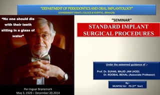
  • 1. Under the esteemed guidance of ;- Prof. Dr. SUHAIL MAJID JAN (HOD) Dr. ROOBAL BEHAL (Associate Professor) By:- MUMTAZ ALI PG (3nd Year) ‘’DEPARTMENTOF PERIODONTICS AND ORAL IMPLANTOLOGY’’ GOVNERNMENTDENATLCOLLEGE& HOSPITAL, SRINAGAR. “SEMINAR’’ STANDARD IMPLANT SURGICAL PROCEDURES “No one should die with their teeth sitting in a glass of water” Per-Ingvar Branemark May 3, 1929 – December 20,2014
  • 2. INTRODUCTION DEFINITION HISTORICAL BACKGROUND DENTAL IMPLANT COMPONENTS. STANDARD IMPLANT SURGICAL PROCEDURE General principles of implant surgery • Patient preparation • Implant site preparation • One stage versus two stage implant surgeries CONTENTS  Two stage “submerged” Implant placement • Flap designs, incisions and reflection • Implant site preparation • Flap closure and suturing • Post operative care • Second stage exposure surgery  One stage “non-submerged” implant placement • Flap designs, incisions and elevation • Implant site preparation • Flap closure and suturing • Postoperative care  Conclusion
  • 3. INTRODUCTION Dental implants ;- designed to provide a foundation for replacement of teeth that look, feel, and function like natural teeth. Partial and removable prosthesis may not bring satisfactory results. Goal of modern dentistry ;- restore normal contour, function, comfort, esthetics, speech and health of a patient. This leads to increased need and use of implant and implant supported prosthesis.
  • 4. DEFINITION Any object or material, such as an alloplastic substance or other tissue, which partially or completely inserted or grafted into body for therapeutic, diagnostic, prosthetic or experimental purposes. Can be defined as a substance that is placed into the jaw to support a crown or fixed or removable denture. - Charles M Weiss A prosthetic device or alloplastic material implanted into oral tissues beneath the mucosal or periosteal tissues and/or within the bone to provide retention and support for fixed or removal prosthesis. - Edward J Fredrickson IMPLANT DENTAL IMPLANT DENTAL IMPLANT
  • 5.  Archeological findings showed that the ancient Egyptian and South American civilizations already experimented with re-implanting lost teeth with hand-shaped ivory or wood substitutes.  In the 18th century lost teeth were sometimes replaced with extracted teeth of other human donors.  The implantation process was probably somewhat crude and the success rates extremely low due to the strong immune reaction of the receiving individual. HISTORY
  • 6. 2500 BC - Ancient Egyptians - gold ligature. 500 BC - Etruscan population - gold bands incorporating pontics. 500 BC - Phoenician population - gold wire. 300 AD - Phoenician population - Carved Ivory teeth. 600 AD - Mayan population - implantation of pieces of shell.
  • 7. 1911 - Greenfield –iridoplatinum basket soldered with 24 carat gold. 1943 –Dahl- subperiosteal type of implant Late 1970s and Early 1980s - Tatum - custom blade implants of Titanium alloy Early 1980s - Tatum - Titanium root form implant
  • 8. Modern Historical Developments The first Subperiosteal Implant was placed in 1948 by Gustav Dahl The Endosteal Blade Implant, introduced independently in 1967 by Leonard Linkow and Ralph and Harold Roberts After 1980s –hollow basket Core vent implant Niznick et al -Screw vent implant -Screw vent implant with Hydroxyapatite coating - Implant with titanium plasma spray
  • 9.  The quantum leap in Oral Implantology was achieved in 1952 in Sweden by PER INGVAR BRANEMARK (Father of Modern dental implantology)  He founded the phenomenon of Osseointegration.  Dr. Branemark's research shifted more towards the use of titanium appliances in human bone, including the use of titanium screws as bone anchors for lost teeth.  In 1982, the Toronto Conference on Osseointegration in Clinical Dentistry laid down the first parameters on what is to be considered successful implant treatment within the stringent confines of the scientific community.
  • 10. Classification Of Implant by Charles. A. Babbush There are five main types:
  • 11. 1.ENDOSTEAL IMPLANT  An implant which is placed into the alveolar bone and/ or basal bone of the mandible or maxilla  Transects only one corticle plate  Most commonly used Blade implant It consists of thin plates in the form of blade embedded into the bone Root form implant Designed to mimic the shape of the tooth For directional load distribution Ramus frame implant Horse shoe shaped stainless steel device Inserted from one retromolar pad to other
  • 12. 2. SUBPERIOSTEAL IMPLANT  Placed directly beneath the periosteum overlying the bony cortex  Do not penetrate into the jawbone.  Consists of non-Osseo integrated framework that rests on the surface of the jaw or beneath the mucoperiosteum.  Can be bilateral or unilateral
  • 13. 3. TRANSOSTEAL IMPLANT  Other names- staple bone implant Mandibular staple implant Transmandibular implant  Combines the subperiosteal and endosteal components  Penetrates both cortical plates  very similar to a nut and bolt arrangement  Used in mandibles only  penetrate the entire jaw to emerge opposite the entry site, usually at the bottom of the chin.
  • 14. 4. INTRAMUCOSAL IMPLANTS  Inserted into oral mucosa  Mucosa is used as attachment site for metal inserts
  • 15. •Described by Dr CHARLES WIESS •Complete encapsulation of implant with soft tissue •Soft tissue interface could resemble highly vascular periodontal fibers of natural dentition •Described by BRANEMARK •Direct contact between bone & surface of loaded implant •Bio active materials that stimulate formation of bone are used
  • 16.
  • 17. Cylindrical dental implants • In the form of cylinder • Depends on coating or surface conditioning to provide microscopic retension & bonding to bone • pushed or tapped into prepared bone site • Straight, tapered or conical Threaded dental implants • The surface is threaded, to increase surface area of implant • This results in distribution of forces over greater peri-implant bone volume Perforated dental implants • are made of inert micro porous membrane material (mixture of cellulose acetate) in intimate contact with & supported by layer of perforated metallic sheet material (pure titanium)
  • 18. Plateau dental implant • Plateau shaped implant with sloping shoulder Solid dental implant • They are of circular cross section without vent or hollow in the body Vented dental implant • It is hydroxyapetite coated cylinder with patented vertical groove connecting to apical vents designed to facilitate seating and allow bone in growth to prevent rotation Hollow dental implant Hollow design in apical portion Systematically arranged perforations along sides of implant Increased anchoring surface
  • 19.
  • 20.
  • 21. • Depending on the materials used: Metallic implants [titanium, titanium alloy, cobalt chromium molybdenum alloy] Non- metallic implants [ceramics, carbon] • According to loading • Immediate(<2weeks) • Early(2weeks -2mts) • Delayed (>3mts) • According to method of placement • Tapping system • Threading system
  • 22.  Based on the surface Machined surface Sand blasted Acid etched HA coating Plasma spray Bioactivesurface Oxidizedsurface Combination of one/more
  • 23. PARTS OF DENTAL IMPLANT
  • 24. Implant Body or Fixture: the component that is placed within the bone during first stage of surgery. Abutment Is the part of implant, which resembles a prepared tooth, and is designed to be screwed into the implant body via Abutment screw It is the primary component, which provides retention to the prosthesis Crown: replicate the original teeth to provide a biting surface and aesthetic appearance: Crown: Material Used: Porcelains (metal supported or metal free) or metal normally gold) Abutment: Materials Used: Titanium. Implant Body or Fixture: Materials Used: Titanium & titanium oxide --Crestal Module --Body --Apex
  • 25. OTHER IMPLANT COMPONENTS Healing Screw During the healing phase, this screw is normally placed in the superior surface of the body. functions -Facilitates the suturing of soft tissue over the edge of the implant. Healing Caps dome-shaped screws. Length ranges from 2-10mm. Project through the soft tissue into the oral cavity Function –prevent overgrowth of tissues around the implant during healing phase. Impression posts/coping: Is a small stem that facilitates the transfer of the intraoral location (of the implant or the abutment) to a similar position on the cast. They are screwed into implant body during impression making. Analogue or Implant Replica Analogues are used by laboratory technicians to replicate implants and their position in a patient’s mouth. The analogue,screwed onto the impression coping, is set into the plaster model during casting
  • 26. Patient preparation Implant site preparation One stage Vs two stage implant surgery GENERAL PRINCIPLES OF IMPLANT SURGERY
  • 27. 1. Explanation of risks and benefits to the patient. 2. Written / Informed consent 3. Local or General Anesthesia depending on patient’s needs. PATIENT PREPARATION
  • 28. 1. Implants must be sterile and made of a biocompatible material (e.g., titanium). 2. Implant site preparation should be performed under sterile conditions. 3. Implant site preparation should be completed with an atraumatic surgical technique that avoids overheating of the bone during preparation of the recipient site. 4. Implants should be placed with good initial stability. 5. Implants should be allowed to heal without loading or micro- movement (i.e., undisturbed healing period to allow for osseointegration) for 2 to 4 or 4 to 6 months, depending on the bone density, bone maturation, and implant stability. BASIC PRINCIPLES OF IMPLANT THERAPY TO ACHIEVE OSSEOINTEGRATION
  • 29. 1. Patient drape 2. Rinsing or swabbing the mouth with chlorhexidine gluconate for 1 to 2 minutes immediately before the procedure. 3. Atraumatic implant site preparation. 4. Avoid damage to bone or vital structures 5. Copious irrigation to avoid heating and debris removal. 6. The implant must be placed in healthy bone. 7. The surgical site should be kept aseptic. SURGICAL SITE PREPARATION
  • 30.  Good operating light  Good high volume suction  A dental chair which can be adjusted by foot controls  A surgical drilling unit which can deliver relatively high speeds (up to 3000 rpm) and low drilling speeds (down to about 10 rpm) with good control of torque  An irrigation system for keeping bone cool during the drilling process  The appropriate surgical instrumentation for the implant system being used.  Sterile drapes, gowns, gloves, suction tubing etc.  The appropriate number and design of implants planned plus an adequate stock to meet unexpected eventualities during surgery OPERATIVE REQUIRMENTS
  • 31.  The surgical stent  The complete radiographs including tomographs  A trained assistant  A third person to act as a get things in between to and from the sterile and non-sterile environment.  Light handles should be autoclaved or covered with sterile aluminum foil.  The instrument tray and any other surfaces which are to be used are covered in sterile drapes.
  • 32. ONE STAGE SURGICAL TECHNIQUE VS TWO STAGE SURGICAL TECHNIQUE
  • 33. In the one-stage approach, the implant or the abutment emerges through the mucoperiosteum/ gingival tissue at the time of implant placement. ONE STAGE TECHNIQUE ONE STAGE SURGICAL TECHNIQUE ADVANTAGES ONE STAGE TECHNIQUE  Easier Mucogingival management around the implant.  Patient management is simplified because a second stage exposure surgery is not necessary.
  • 34.  In the two-stage approach, the top of the implant and cover screw are completely covered with the flap closure.  Implants are allowed to heal, without loading or micro movement, for a period of time to allow for osseointegration.  The implant must be surgically exposed following an undisturbed healing period. TWO STAGE SURGICAL TECHNIQUE
  • 35.  In the second-stage (exposure) surgery, the implant is uncovered and a healing abutment is connected to allow emergence of the implant/abutment through the soft tissues, thus facilitating access to the implant from the oral cavity.  The restorative dentist then proceeds with the prosthodontic aspects of the implant therapy (impressions and fabrication of prosthesis) after soft tissue healing.  2-4 months – Dense cortical bone, good initial stability.  4-6 monts – Loose trabecular bone,Grafted sites, lesser implant stability.
  • 36.  Situations that require simultaneous bone augmentation procedures at the time of implant placement because membranes can be covered by primary flap closure, which will minimize postoperative exposure.  Prevents movement of the implant by the patient, who may inadvertently bite on the healing abutment during the healing period (one-stage protocol).  Mucogingival tissues can be augmented if desired at the second-stage surgery in a two-stage protocol. ADVANTAGES OF TWO STAGE TECHNIQUE
  • 37. The first stage ends by;-  Suturing  So the implant remains submerged and isolated from the oral cavity.  Mandible implants – 2 to 4 months  Maxillary implants – 4 to 6 months  Longer periods –  less dense bone  Less initial implant stability  Shorter periods –  More dense bone  Altered surface microtopography TWO STAGE “SUBMERGED” IMPLANT PLACEMENT
  • 38. In second stage The implant is uncovered and a healing abutment is connected to allow emergence of the implant through the soft tissue, thus facilitating access to the implant from the oral cavity.
  • 39. 1. Flap Design, Incisions, And Elevation  Vary slightly depending on the location and objective of the planned surgery.  Crestal  The incision is made from along the crest of the ridge, bisecting the existing zone of keratinized mucosa  Adv. Easy to manage, results in less bleeding, less edema, faster healing.  Suturing placed generally do not interfere with the healing.  Remote  The incision is made some distance from the planned osteotomy site.  Layer suturing is indicated to minimize the bone graft exposure. TWO STAGE “SUBMERGED” IMPLANT PLACEMENT.
  • 40.  A mucoperiosteal (full-thickness) flap is reflected up to or slightly beyond the level of the mucogingival junction, exposing the alveolar ridge of the implant surgical sites.  Elevated flaps may be sutured to the buccal mucosa or the opposing teeth to keep the surgical site open during the surgery.  The bone at the implant site(s) must be thoroughly debrided of all granulation tissue. 2. IMPLANT SITE PREPARATION
  • 41.  Once the flaps are reflected and the bone is prepared (i.e., all granulation tissue removed and knife-edge ridges flattened), the implant osteotomy site can be prepared.  A series of drills are used to prepare the osteotomy site precisely and incrementally for an implant.  A surgical guide or stent is inserted, checked for proper positioning, and used throughout the procedure to direct the proper implant placement.
  • 42. Sequence of drills used for standard- diameter (4.0-mm) implant site osteotomy preparation: Round,2mm twist, pilot,3mm twist, and countersink. Bone tap (not shown here) is an optional drill that is sometimes used in dense bone before implant placement. COUNTERSINK ROUND 2mm TWISTED PILOT 3mm TWISTED
  • 43. ROUND BUR/SPIRAL DRILL  A small round bur (or spiral drill) is used to mark the implant site(s). The surgical guide is removed, and the initial marks are checked for their appropriate buccal-lingual and mesial-distal location, as well as the positions relative to each other and adjacent teeth.  (1.5mm between implant and tooth)  (3mm between implants)  (1-1.5 mm buccally,lingually)  Slight modifications may be necessary to adjust spatial relationships and to avoid minor ridge defects. Any changes should be compared to the prosthetically-driven surgical guide positions.  Each marked site is then prepared to a depth of 1 to 2 mm with a round drill, breaking through the cortical bone and creating a starting point for the 2-mm twist drill.
  • 44. 2mm TWISTED DRILL A small twist drill, usually 2 mm in diameter and marked to indicate various lengths (i.e., corresponding to the implant sizes), is used next to establish the depth and align the long axis of the implant recipient site. Speed of approximately 800 to 1500 rpm, with copious irrigation to prevent overheating of the bone. Additionally, drills should be intermittently and repeatedly “pumped” or pulled out of the osteotomy site while drilling to expose them to the water coolant and to facilitate clearing bone debris from the cutting surfaces. In other words, clinicians should pump the drill (up and down) intermittently and avoid using a constant “push” of the drill in the apical direction only.
  • 45. PILOT DRILL A pilot drill with a noncutting 2-mm–diameter “guide” at the apical end and a cutting 3-mm–diameter (wider) midsection is used to enlarge the osteotomy site at the coronal end, thus facilitating the insertion of the subsequent drill in the sequence.
  • 46. TWIST DRILLS (TO ENLARGE THE OSTEOTOMY SITE TO TILL REQUIRED DIAMETER)
  • 47. The final drill in the osteotomy site preparation for a standard-diameter (4 mm) implant is the 3-mm twist drill. It is the last drill used to widen the site along the entire depth of the osteotomy from the previous diameter (2 mm) to final diameter (3 mm). It is critically important that the final diameter drilling be accomplished with a steady hand, without wobbling or changing direction so that the site is not overprepared. Finally, depending on bone density, the diameter of this final drill may be slightly increased or decreased to enhance implant support. The 3-mm Twist Drill 0.2-0.7 mm should be the difference between final osteotomy and fixture depending upon type of bone. - Carl Misch.
  • 48. GUIDE PINS When multiple implants are being placed next to one another, a guide pin should be placed in the prepared sites to check alignment, parallelism, and proper prosthetic spacing throughout the preparation process
  • 50. COUNTERSINK DRILL it is desirable to place the cover screw at or slightly below the crestal bone, countersink drilling is used to shape or flare the crestal aspect of the osteotomy site allowing the coronal flare of the implant head and cover screw to fit within the osteotomy site
  • 51. BONE TAP As the final step in preparing the osteotomy site in dense cortical bone, a tapping procedure may be necessary.  With self-tapping implants being almost universal, there is less need for a tapping procedure in most sites. However, in dense cortical bone or when placing longer implants into moderately dense bone, it is prudent to tap the bone (create threads in the osteotomy site) before implant placement to facilitate implant insertion and to reduce the risk of implant binding.
  • 52. It is better to allow the threaded implant to“cut” its own path into the osteotomy site. Bone tapping and implant insertion are both done at very slow speeds (e.g., 20 to 40 rpm). All other drills in the sequence are used at higher speeds (800 to 1500 rpm). It is important to create a recipient site that is very accurate in size and angulation.
  • 53.  In partially edentulous cases, limited jaw opening or proximity to adjacent teeth may prevent appropriate positioning of the drills in posterior edentulous areas.  In fact, implant therapy may be contraindicated in some patients because of a lack of inter occlusal clearance, lack of interdental space, or a lack of access for the instrumentation.  Therefore a combination of longer drills and shorter drills, with or without extensions, may be necessary.  Anticipating these needs before surgery facilitates the procedure and improves the results.
  • 54.  When wide-diameter drills are used for implant site preparation, it is advisable to reduce the drilling speed, according to the manufacturer's guidelines, to prevent overheating the bone.  Copious external irrigation is critical. In the case of wide diameter implants, a specific pilot drill is often indicated as a transition between each of the subsequent wider drills.
  • 55. Implant site preparation (osteotomy ) for a 4.0-mm diameter, 10 mm length screw-type, threaded (external hex) implant in a subcrestal position. A, Initial marking or preparation of the implant site with a round bur. B, Use of a 2-mm twist drill to establish depth and align the implant. C, Guide pin is placed in the osteotomy site to confirm position and angulation. D, Pilot drill is used to increase the diameter of the coronal aspect of the osteotomy site. SURGICAL STEPS
  • 56. E, Final drill used is the 3- mm twist drill to finish preparation of the osteotomy site. F, Countersink drill is used to widen the entrance of the recipient site and allow for the subcrestal placement of the implant collar and cover screw. G, Implant is inserted into the prepared osteotomy site with a handpiece or handheld driver (ratchet). H, Cover screw is placed and soft tissues are closed and sutured
  • 57. FLAP CLOSURE AND SUTURE  Once the implants are inserted and the cover screws secured, the surgical sites should be thoroughly irrigated with sterile saline to remove debris and clean the wound.  Proper closure of the flap over the implant(s) is essential.  One of the most important aspects of flap management is achieving good approximation and primary closure of the tissues in a tension free manner.  This is achieved by incising the periosteum (innermost layer of full- thickness flap), which is non-elastic.  Once the periosteum is released, the flap becomes very elastic and is able to be stretched over the implant(s) without tension.
  • 58.  One suturing technique that consistently provides the desired result is a combination of alternating horizontal mattress and interrupted sutures.  Horizontal mattress sutures evert the wound edges and approximate the inner, connective tissue surfaces of the flap to facilitate closure and wound healing.  Interrupted sutures help to bring the wound edges together, counterbalancing the eversion caused by the horizontal mattress sutures. FLAP CLOSURE AND SUTURE
  • 60.
  • 61. RATCHET/WRENCH: Fits on top of fixture mount & used to tighten fixture after placement.
  • 63.
  • 64.  Simple implant surgery in a healthy patient usually does not require antibiotic therapy.  However, patients can be premedicated with antibiotics (e.g., amoxicillin, 500 mg three times a day [tid]) starting 1 hour before the surgery and continuing for 1 week postoperatively if the surgery is extensive, if it requires bone augmentation, or if the patient is medically compromised. -CARL MISCH  Postoperative swelling is likely after flap surgery. POST-OPERATIVE CARE
  • 65.  This is particularly true when the periosteum has been incised (released).  As a preventive measure, patients should apply an ice pack to the area intermittently for 20 minutes (on and off) over the first 24 to 48 hours.  Chlorhexidine gluconate oral rinses can be prescribed to facilitate plaque control, especially in the days after surgery when oral hygiene is typically poorer. Adequate pain medication should be prescribed (e.g., ibuprofen, 600 to 800 mg tid).
  • 66.  Patients should be instructed to maintain a relatively soft diet after surgery.  Then, as soft tissue healing progresses, they can gradually return to a normal diet.  Patients should also refrain from tobacco and alcohol use at least 1 week before and several weeks after surgery.  Provisional restorations, whether fixed or removable, should be checked and adjusted so that impingement on the surgical area is avoided.
  • 67.  For implants placed using a two-stage “submerged” protocol, a second- stage exposure surgery is necessary after the prescribed healing period.  Thin soft tissue with an adequate amount of keratinized attached gingiva, along with good oral hygiene, ensures healthier peri- implant soft tissues and better clinical results SECOND STGAGE EXPOSURE SURGERY
  • 68. 1. To expose the submerged implant without damaging the surrounding bone. 2. To control the thickness of the soft tissue surrounding the implant. 3. To preserve or create attached keratinized tissue around the implant. 4. To facilitate oral hygiene. 5. To ensure proper abutment seating. 6. To preserve soft tissue aesthetics. OBJECTIVES OF SECOND STAGE SURGICAL TECHNIQUE
  • 69.  In areas with sufficient zones of keratinized tissue, the gingiva covering the head of the implant can be exposed with a circular or “punch” incision  Alternatively, a crestal incision through the middle of the keratinized tissue and full-thickness flap reflection can be used to expose implants.  This latter approach may be necessary when bone has grown over the implant and needs to be removed. SIMPLE CIRCULAR “PUNCH” /CRESTAL INCISION. PUNCH INCSION CRESTAL INCSION
  • 70. Clinical view of stage two, implant exposure surgery in a case with adequate keratinized tissue. A, Simple circular “punch” incision used to expose implant when sufficient keratinized tissue is present around the implant(s). B, Implant exposed. C, Healing abutment attached. D, Final restoration in place, achieving an esthetic result with a good zone of keratinized tissue.
  • 71. Clinical view of stage two implant exposure surgery in a case with inadequate keratinized tissue. A, Two endosseous implants were placed 4 months previously and are ready to be exposed. B, Two vertical incisions are connected by crestal incision. C, Buccal partial thickness flap is sutured to the periosteum apical to the emerging implants. D, Gingival tissue coronal to the cover screws is excised using the gingivectomy technique. E, Cover screws are removed, and heads of the implants are cleared. F, Abutments are placed. Visual inspection ensures intimate contact between the abutments and the implants.
  • 72. G, Healing at 2 to 3 weeks after second-stage surgery . H, Four months after the final restoration. Note the healthy band of keratinized attached gingiv a around the implants.
  • 73.  If a minimal zone of keratinized tissue exists at the implant site, a partial-thickness flap technique can be used to fulfill the objective of the second-stage surgery (exposing the implant) while increasing the width of keratinized tissue.  A partial-thickness flap is then raised in such a manner that a nonmobile, firm periosteum remains attached to the underlying bone. The flap, containing a narrow band of keratinized tissue, is then repositioned to the facial side of the emerging head of the implant and sutured to the periosteum with a fine needle and resorbable suture such as a 5.0 gut suture PARTIAL THICKNESS REPOSITIONED FLAP
  • 74.  A partial-thickness flap is apically displaced and sutured to the periosteum without exposing the alveolar bone.  A free gingival graft may be harvested from the palate and sutured to the periosteum on the labial surface of the implants to increase the zone of keratinized tissue.
  • 75. A, Partial-thickness flap is created from the lingual aspect of the crest toward the labial surf ace in order to preserve the keratinized tissue on the crest (over the implant). note: This tissue might be excised in a simple implant exposure. B, The split-thickness flap is repositioned to the labial surf ace. C, The flap is sutured to the periosteum at a more apical position preserving the amount of keratinized tissue (arrows). Finally , the remaining connective tissue over the cover screw (B) is excised with a sharp blade to expose the implant. Care should be taken to avoid removing keratinized tissue from the lingual aspect of the implant.
  • 76.  After the flap is repositioned and secured with periosteal sutures, the excess tissue coronal to the cover screw is excised, usually with a surgical blade.  When the excess tissue over the cover screw is removed or displaced, the outline of the cover screw is visible.  A sharp blade is used to eliminate all tissues coronal to the cover screw.  The cover screw is then removed, the head of the implant is thoroughly cleaned of any soft or hard tissue overgrowth, and the healing abutments or standard abutments are placed on the implant
  • 77.  remind the patient of the need for good oral hygiene around the implant and adjacent teeth.  rinse can be used to enhance oral hygiene for the initial few weeks after implant exposure.  oral hygiene procedures to avoid dislodging any repositioned or grafted soft tissues.  any direct pressure or movement directed toward the soft tissue from a provisional prosthesis can delay healing and should be avoided. POST OPERATIVE CARE
  • 78.  Impressions for the final prosthesis fabrication can begin about 2 to 6 weeks after implant exposure surgery, depending on healing and maturation of soft tissues.
  • 80. In the one-stage implant surgical approach, a second implant exposure surgery is not needed because the implant is exposed (per gingival) about 2 to 3 mm from the bone crest, and the flaps are adapted around the implant/abutment. In the standard (classic) implant protocol, the implants are left unloaded and undisturbed for a period similar to that for implants placed in the two-stage approach . (i.e., in areas with dense cortical bone and good initial implant support, the implants are left to heal undisturbed for a period of 2 to 4 months, whereas in areas of loose trabecular bone, grafted sites, and/or minimal implant support, they may be allowed to heal for periods of 4 to 6 months or more).
  • 81.  The flap design for the one-stage surgical approach is always a crestal incision bisecting the existing keratinized tissue.  Facial and lingual flaps in posterior areas should be carefully thinned before total reflection to minimize the soft tissue thickness (if needed or desired).  The soft tissue is not thinned in anterior or other esthetic areas of the mouth to maintain tissue height and to minimize metallic implant components from showing through tissue. FLAP DESIGN, INCISION, AND ELEVATION
  • 82.  The primary difference is that the coronal aspect of the implant or the healing abutment (two-stage implant) is placed about 2 to 3 mm above the bone crest and the soft tissues are approximated around the implant/implant abutment. IMPLANT SITE PREPARATION
  • 83.  The keratinized edges of the flap are sutured with single interrupted sutures around the implant.  Depending on the clinician's preference, the wound may be sutured with resorbable or nonresorbable sutures.  When keratinized tissue is abundant, scalloping around the implant(s) provides better flap adaptation.  However, if minimal keratinized tissue exists in an area, tissues should remain thick and soft tissue augmentation may be indicated. FLAP CLOSURE AND SUTURING
  • 84.  The postoperative care for one-stage surgical approach is similar to that for the two-stage surgical approach except that the cover screw or healing abutment is exposed to the oral cavity.  Patients are advised to avoid chewing in the area of the implant.  Prosthetic appliances should not be used if direct chewing forces can be transmitted to the implant, particularly in the early healing period (first 4 to 8 weeks). POST- OPERATIVE CARE
  • 85.  It is essential to understand and follow basic guidelines to achieve osseointegration predictably.  Fundamentals must be followed for implant placement and implant exposure surgery.  These fundamentals apply to all implant systems. CONCLUSION
  • 86. • Newman, Takei, Klokkevold, Carranza. Carranza’s Clinical Periodontology, 10th Edition and 11th Edition • Lindhe, Lang, Karring. Clinical Periodontology & Implant Dentistry, 5th Edition. • Carle E. Misch. Contemporary Implant Dentistry. 3rd edition. REFERENCES