SlideShare a Scribd company logo
1 of 18
Download to read offline
S K I N A ND S O FT
TI S SUE
I N FEC TIONS A N D
TREATMENT
By: Dr. VALERIA SIMONE MD
(SOUTHLAKE GENERAL SURGERY, TEXAS)
OVERVIEW
Skin and soft-tissue infections (SSTIs)
incorporate infections of the skin,
fascia, and muscle, hypodermic tissue,
wrap a wide scope of clinical overviews,
going from straightforward cellulitis to
quickly reformist necrotizing fasciitis.
Diagnosing the specific extent of the disease is critical for effective care of a patient with a
soft-tissue infection. Different types of Skin and soft-tissue infections (SSTI), mentioned
according to medical presentation and anatomic area, incorporate the following:
•Impetigo
•Folliculitis
•Furuncles
•Carbuncles
•Erysipelas
•Cellulitis
•Necrotizing fasciitis, also called hemolytic streptococcal gangrene, Meleney ulcer,
synergistic gangrene, and Fournier gangrene (when limited to the scrotum and perineal
area)
•Pyomyositis
What are the leading factors for Skin and soft-tissue
infections (SSTI)?
Factors to the formation of Skin and soft-tissue infections (SSTI) include:
•Breach in the epidermis
•Immunity Compromised Status – burns, malnutrition, diabetes mellitus, hypoproteinemia,
AIDS
•Chronic venous insufficiency (CVI)
•Dry and irritated skin
•Chronic neuropathy
•Chronic lymphatic insufficiency
Indications of
SSTI
Any kind of abscess, anyway small, should
be drained for complete treatment. Any
type of ulcer secured with dead and
necrotic tissue must be debrided for the
growth of healthy granulation tissue and
recuperating. Necrotizing fasciitis is
categorized under surgical treatment;
early surgical procedure enhances results
for these patients.
Preprocedural
SSTI Planning
The thumb rule of Skin and soft tissue
infections (SSTIs) is to control the source
of infection because it is the quickest
method of reducing bacterial infection.
Controlling the source of infection is
accomplished by methods of pus drainage
and debridement. The main reason for
debridement is to make an intense wound
milieu in order to trigger the body’s
wound healing mechanism and
accordingly initiate the healing process.
In accordance with surgical planning of an
SSTI, detailed examination, antibiotic
treatment, or both might be
demonstrated.
Different methods of debridement procedure
Debridement methods that might be utilized as options in contrast to surgical debridement
incorporate the accompanying:
•Autolytic debridement
•Ultrasonic debridement
•Chemical debridement
•Mechanical debridement
•Biological debridement
Autolytic
debridement
In this procedure, your body sheds the dead necrotic
tissue by the utilization of moisture. This cycle is helped
by the presence of compounds known as matrix
metalloproteinases (MMPs), which are delivered by
damaged tissue and disturb the proteins that
predicament the dead tissue in the body.
This cycle can be improved by applying dressings that
empower a decent moist environment in the injury.
Vigorously oozing wounds advantage using alginates,
cellulose dressings, and froths; these dressings
assimilate of abundance exudate and forestall
maceration of encompassing healthy tissue while yet
keeping up a moist environment that advances de-
sloughing. Dry wounds advantage using hydrogels and
hydrocolloids, which give moisture to the dead tissue to
encourage debridement.
Ultrasonic
debridement
It includes applying ultrasonic vibrations
to the injury bed through a fluid medium.
This causes cavitation (i.e., the creation
and demolition of little air pockets inside
the liquid encompassing the probe).
During cavitation, the air pockets sway fit
as a fiddle. They extend and quickly break
down, causing shockwave development,
and this collapse prompts the
disintegration of tissues. Ultrasonic
debridement causes necrotic tissue
disturbance, discontinuity, and emulsion.
Chemical
debridement
It is performed by utilizing certain
enzymatic synthetic substances on the
injury that cause lysis of the necrotic
tissue in the injury. Commercially
accessible collagenase enzyme granules
are sprinkled onto the injury every day
until the injury is away from necrotic
tissue. Customary dressings at that point
follow.
Mechanical
debridement
It is refined by utilizing the wet-to-dry
dressing technique. The injury is dressed
with a wet dressing (normally gauze
absorbed in saline) covered with a dry
dressing. The dressing is then left to dry
on the injury over the accompanying 24-
36 hours. Once the dry dressing is
removed, it strips the disciple necrotic
tissue away from the healthy tissue. This
is an exceptionally painful technique and
isn’t quite preferred.
Biological
debridement
Biological debridement is also known as worm
treatment; it includes presenting the injury to the
parasites of Lucilia sericata (the greenbottle fly).
These living beings digest the necrotic tissue and
bacteria in the injury yet save the underneath
healthy tissue. This strategy has not increased a
lot of favor among patients.
Debridement for
Necrotizing
Fasciitis at
Southlake
General Surgery
During the surgery of a limb, a tourniquet is
utilized in order to get a bloodless field. An
intense cut is made that goes through the
whole length of the injury from normal skin
proximally to normal skin distally. The incision
ought to stretch out to the muscle.
Necrotizing Fasciitis Zones
Delineation of zones of necrotizing fasciitis and relating degree of fascial extraction.
The deep belt is distinguished, and a finger is passed along it to test the degree of its
association. Healthy deep fascia can be distinguished by its flickering appearance and its
firm connection to the skin and subcutaneous tissue. In contrast to necrosed deep fascia, it
doesn’t disperse effectively from the skin and subcutaneous tissue.
Now, tissue samples are acquired and sent for aerobic and anaerobic culture and antibiotic
affectability and for histopathologic assessment to prepare for the diagnosis.
When the degree of deep fascial contribution has been set up, radical extraction of the deep
fascial is completed with scissors to uncover the underneath healthy muscle. The overlying
skin and subcutaneous tissue are then reviewed for suitability.
Tissues are necrosed and extracted. Tissues are reviewed cautiously for viability and
extracted when included. Viability can be checked by searching for dermal bleeding,
calcification and liquefaction of subcutaneous fat, and thrombosed veins. The included skin
and subcutaneous tissue are extracted.
The tourniquet is then emptied, and the tissue is analyzed for viability. Further debridement
is completed if vital. When debridement is finished, hemostasis is accomplished by methods
of electrocauterization, and the injury is washed altogether with saline.
The broad debridement needed in instances of necrotizing fasciitis brings about a huge raw
injury. A non-adherent dressing and a retentive dressing are used to properly dress the
wound and keep it in place after treatment. Within twenty-four hours, the dressing is
changed.
Complications of Necrotizing Fasciitis Procedures
Bleeding is the most well-known problem related to these methods. Bleeding after
debridement (particularly debridement for necrotizing fasciitis) may cause quick worsening
of a previously undermined patient. Meticulous hemostasis is henceforth compulsory. Much
of the time, a pressure dressing is adequate to control the bleeding. In a few cases, the
patient may go back to the surgery to control steady bleeding.
Let’s explore more: Skin and Soft Tissue Infections and Treatment - Southlake General
Surgery
Appointment
For more information on Skin and Soft
Tissue Infection diagnosis and treatment,
please contact our healthcare expert
at +1(817) 748-0200. For an online
appointment for consultation and
treatment. Click Here.
Follow us on Facebook and YouTube.
THANK YOU!
SOUTHLAKE GENERAL SURGERY
1545 E. Southlake Blvd, Suite 270 Southlake, TX 76092
EMAIL: info@southlakegeneralsurgery.com
VISIT US AT: www.southlakegeneralsurgery.com

More Related Content

More from MeghaSingh194

Choosing Top Hernia Surgeon In Texas.pdf
Choosing Top Hernia Surgeon In Texas.pdfChoosing Top Hernia Surgeon In Texas.pdf
Choosing Top Hernia Surgeon In Texas.pdfMeghaSingh194
 
Laparoscopic Hysterectomy - Recovery & Benefits.pdf
Laparoscopic Hysterectomy - Recovery & Benefits.pdfLaparoscopic Hysterectomy - Recovery & Benefits.pdf
Laparoscopic Hysterectomy - Recovery & Benefits.pdfMeghaSingh194
 
Hysteroscopy Explained - Procedure, Benefits, and Recovery.pdf
Hysteroscopy Explained - Procedure, Benefits, and Recovery.pdfHysteroscopy Explained - Procedure, Benefits, and Recovery.pdf
Hysteroscopy Explained - Procedure, Benefits, and Recovery.pdfMeghaSingh194
 
General Surgeons - Expertise and Care.pdf
General Surgeons - Expertise and Care.pdfGeneral Surgeons - Expertise and Care.pdf
General Surgeons - Expertise and Care.pdfMeghaSingh194
 
Understanding Small Bowel Resection.pdf
Understanding Small Bowel Resection.pdfUnderstanding Small Bowel Resection.pdf
Understanding Small Bowel Resection.pdfMeghaSingh194
 
Laparoscopic Surgery - Minimal Scars, Maximum Precision.pdf
Laparoscopic Surgery - Minimal Scars, Maximum Precision.pdfLaparoscopic Surgery - Minimal Scars, Maximum Precision.pdf
Laparoscopic Surgery - Minimal Scars, Maximum Precision.pdfMeghaSingh194
 
Surgical Options for Ruptured Gallbladder.pdf
Surgical Options for Ruptured Gallbladder.pdfSurgical Options for Ruptured Gallbladder.pdf
Surgical Options for Ruptured Gallbladder.pdfMeghaSingh194
 
Surgical Nutrition Insights - Dr Valeria Simone MD
Surgical Nutrition Insights - Dr Valeria Simone MDSurgical Nutrition Insights - Dr Valeria Simone MD
Surgical Nutrition Insights - Dr Valeria Simone MDMeghaSingh194
 
Diverticulitis Surgery - Procedure and Recovery Southlake.pdf
Diverticulitis Surgery - Procedure and Recovery Southlake.pdfDiverticulitis Surgery - Procedure and Recovery Southlake.pdf
Diverticulitis Surgery - Procedure and Recovery Southlake.pdfMeghaSingh194
 
Understanding Chronic Appendicitis - Unraveling the Mysteries.pdf
Understanding Chronic Appendicitis - Unraveling the Mysteries.pdfUnderstanding Chronic Appendicitis - Unraveling the Mysteries.pdf
Understanding Chronic Appendicitis - Unraveling the Mysteries.pdfMeghaSingh194
 
Acute Cholecystitis - Symptoms, Causes, & Treatment.pdf
Acute Cholecystitis - Symptoms, Causes, & Treatment.pdfAcute Cholecystitis - Symptoms, Causes, & Treatment.pdf
Acute Cholecystitis - Symptoms, Causes, & Treatment.pdfMeghaSingh194
 
Cellulitis - Causes, Treatments, and Recovery.pdf
Cellulitis - Causes, Treatments, and Recovery.pdfCellulitis - Causes, Treatments, and Recovery.pdf
Cellulitis - Causes, Treatments, and Recovery.pdfMeghaSingh194
 
Hemorrhoids - Essential Guide.pdf
Hemorrhoids - Essential Guide.pdfHemorrhoids - Essential Guide.pdf
Hemorrhoids - Essential Guide.pdfMeghaSingh194
 
Gallbladder Surgeons - Types of Surgery and Risks.pdf
Gallbladder Surgeons - Types of Surgery and Risks.pdfGallbladder Surgeons - Types of Surgery and Risks.pdf
Gallbladder Surgeons - Types of Surgery and Risks.pdfMeghaSingh194
 
Treatment for Gallstones-Symptoms, Causes, Risks, and Options.pdf
Treatment for Gallstones-Symptoms, Causes, Risks, and Options.pdfTreatment for Gallstones-Symptoms, Causes, Risks, and Options.pdf
Treatment for Gallstones-Symptoms, Causes, Risks, and Options.pdfMeghaSingh194
 
Surgical Nutrition – Enteral Feeding- How does it function and When to Use.pdf
Surgical Nutrition – Enteral Feeding- How does it function and When to Use.pdfSurgical Nutrition – Enteral Feeding- How does it function and When to Use.pdf
Surgical Nutrition – Enteral Feeding- How does it function and When to Use.pdfMeghaSingh194
 
Recovering After Gallbladder Surgery - What to Expect at Home.pdf
Recovering After Gallbladder Surgery - What to Expect at Home.pdfRecovering After Gallbladder Surgery - What to Expect at Home.pdf
Recovering After Gallbladder Surgery - What to Expect at Home.pdfMeghaSingh194
 
Parathyroid Diseases and Treatments Southlake.pdf
Parathyroid Diseases and Treatments Southlake.pdfParathyroid Diseases and Treatments Southlake.pdf
Parathyroid Diseases and Treatments Southlake.pdfMeghaSingh194
 
Understanding Bile Ducts Cancer (Cholangiocarcinoma).pdf
Understanding Bile Ducts Cancer (Cholangiocarcinoma).pdfUnderstanding Bile Ducts Cancer (Cholangiocarcinoma).pdf
Understanding Bile Ducts Cancer (Cholangiocarcinoma).pdfMeghaSingh194
 
Gallstone Pancreatitis – Symptoms and Treatment.pdf
Gallstone Pancreatitis – Symptoms and Treatment.pdfGallstone Pancreatitis – Symptoms and Treatment.pdf
Gallstone Pancreatitis – Symptoms and Treatment.pdfMeghaSingh194
 

More from MeghaSingh194 (20)

Choosing Top Hernia Surgeon In Texas.pdf
Choosing Top Hernia Surgeon In Texas.pdfChoosing Top Hernia Surgeon In Texas.pdf
Choosing Top Hernia Surgeon In Texas.pdf
 
Laparoscopic Hysterectomy - Recovery & Benefits.pdf
Laparoscopic Hysterectomy - Recovery & Benefits.pdfLaparoscopic Hysterectomy - Recovery & Benefits.pdf
Laparoscopic Hysterectomy - Recovery & Benefits.pdf
 
Hysteroscopy Explained - Procedure, Benefits, and Recovery.pdf
Hysteroscopy Explained - Procedure, Benefits, and Recovery.pdfHysteroscopy Explained - Procedure, Benefits, and Recovery.pdf
Hysteroscopy Explained - Procedure, Benefits, and Recovery.pdf
 
General Surgeons - Expertise and Care.pdf
General Surgeons - Expertise and Care.pdfGeneral Surgeons - Expertise and Care.pdf
General Surgeons - Expertise and Care.pdf
 
Understanding Small Bowel Resection.pdf
Understanding Small Bowel Resection.pdfUnderstanding Small Bowel Resection.pdf
Understanding Small Bowel Resection.pdf
 
Laparoscopic Surgery - Minimal Scars, Maximum Precision.pdf
Laparoscopic Surgery - Minimal Scars, Maximum Precision.pdfLaparoscopic Surgery - Minimal Scars, Maximum Precision.pdf
Laparoscopic Surgery - Minimal Scars, Maximum Precision.pdf
 
Surgical Options for Ruptured Gallbladder.pdf
Surgical Options for Ruptured Gallbladder.pdfSurgical Options for Ruptured Gallbladder.pdf
Surgical Options for Ruptured Gallbladder.pdf
 
Surgical Nutrition Insights - Dr Valeria Simone MD
Surgical Nutrition Insights - Dr Valeria Simone MDSurgical Nutrition Insights - Dr Valeria Simone MD
Surgical Nutrition Insights - Dr Valeria Simone MD
 
Diverticulitis Surgery - Procedure and Recovery Southlake.pdf
Diverticulitis Surgery - Procedure and Recovery Southlake.pdfDiverticulitis Surgery - Procedure and Recovery Southlake.pdf
Diverticulitis Surgery - Procedure and Recovery Southlake.pdf
 
Understanding Chronic Appendicitis - Unraveling the Mysteries.pdf
Understanding Chronic Appendicitis - Unraveling the Mysteries.pdfUnderstanding Chronic Appendicitis - Unraveling the Mysteries.pdf
Understanding Chronic Appendicitis - Unraveling the Mysteries.pdf
 
Acute Cholecystitis - Symptoms, Causes, & Treatment.pdf
Acute Cholecystitis - Symptoms, Causes, & Treatment.pdfAcute Cholecystitis - Symptoms, Causes, & Treatment.pdf
Acute Cholecystitis - Symptoms, Causes, & Treatment.pdf
 
Cellulitis - Causes, Treatments, and Recovery.pdf
Cellulitis - Causes, Treatments, and Recovery.pdfCellulitis - Causes, Treatments, and Recovery.pdf
Cellulitis - Causes, Treatments, and Recovery.pdf
 
Hemorrhoids - Essential Guide.pdf
Hemorrhoids - Essential Guide.pdfHemorrhoids - Essential Guide.pdf
Hemorrhoids - Essential Guide.pdf
 
Gallbladder Surgeons - Types of Surgery and Risks.pdf
Gallbladder Surgeons - Types of Surgery and Risks.pdfGallbladder Surgeons - Types of Surgery and Risks.pdf
Gallbladder Surgeons - Types of Surgery and Risks.pdf
 
Treatment for Gallstones-Symptoms, Causes, Risks, and Options.pdf
Treatment for Gallstones-Symptoms, Causes, Risks, and Options.pdfTreatment for Gallstones-Symptoms, Causes, Risks, and Options.pdf
Treatment for Gallstones-Symptoms, Causes, Risks, and Options.pdf
 
Surgical Nutrition – Enteral Feeding- How does it function and When to Use.pdf
Surgical Nutrition – Enteral Feeding- How does it function and When to Use.pdfSurgical Nutrition – Enteral Feeding- How does it function and When to Use.pdf
Surgical Nutrition – Enteral Feeding- How does it function and When to Use.pdf
 
Recovering After Gallbladder Surgery - What to Expect at Home.pdf
Recovering After Gallbladder Surgery - What to Expect at Home.pdfRecovering After Gallbladder Surgery - What to Expect at Home.pdf
Recovering After Gallbladder Surgery - What to Expect at Home.pdf
 
Parathyroid Diseases and Treatments Southlake.pdf
Parathyroid Diseases and Treatments Southlake.pdfParathyroid Diseases and Treatments Southlake.pdf
Parathyroid Diseases and Treatments Southlake.pdf
 
Understanding Bile Ducts Cancer (Cholangiocarcinoma).pdf
Understanding Bile Ducts Cancer (Cholangiocarcinoma).pdfUnderstanding Bile Ducts Cancer (Cholangiocarcinoma).pdf
Understanding Bile Ducts Cancer (Cholangiocarcinoma).pdf
 
Gallstone Pancreatitis – Symptoms and Treatment.pdf
Gallstone Pancreatitis – Symptoms and Treatment.pdfGallstone Pancreatitis – Symptoms and Treatment.pdf
Gallstone Pancreatitis – Symptoms and Treatment.pdf
 

Skin and Soft Tissue Infections and Treatment.pdf

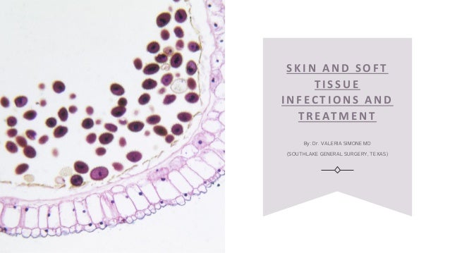
  • 1. S K I N A ND S O FT TI S SUE I N FEC TIONS A N D TREATMENT By: Dr. VALERIA SIMONE MD (SOUTHLAKE GENERAL SURGERY, TEXAS)
  • 2. OVERVIEW Skin and soft-tissue infections (SSTIs) incorporate infections of the skin, fascia, and muscle, hypodermic tissue, wrap a wide scope of clinical overviews, going from straightforward cellulitis to quickly reformist necrotizing fasciitis.
  • 3. Diagnosing the specific extent of the disease is critical for effective care of a patient with a soft-tissue infection. Different types of Skin and soft-tissue infections (SSTI), mentioned according to medical presentation and anatomic area, incorporate the following: •Impetigo •Folliculitis •Furuncles •Carbuncles •Erysipelas •Cellulitis •Necrotizing fasciitis, also called hemolytic streptococcal gangrene, Meleney ulcer, synergistic gangrene, and Fournier gangrene (when limited to the scrotum and perineal area) •Pyomyositis
  • 4. What are the leading factors for Skin and soft-tissue infections (SSTI)? Factors to the formation of Skin and soft-tissue infections (SSTI) include: •Breach in the epidermis •Immunity Compromised Status – burns, malnutrition, diabetes mellitus, hypoproteinemia, AIDS •Chronic venous insufficiency (CVI) •Dry and irritated skin •Chronic neuropathy •Chronic lymphatic insufficiency
  • 5. Indications of SSTI Any kind of abscess, anyway small, should be drained for complete treatment. Any type of ulcer secured with dead and necrotic tissue must be debrided for the growth of healthy granulation tissue and recuperating. Necrotizing fasciitis is categorized under surgical treatment; early surgical procedure enhances results for these patients.
  • 6. Preprocedural SSTI Planning The thumb rule of Skin and soft tissue infections (SSTIs) is to control the source of infection because it is the quickest method of reducing bacterial infection. Controlling the source of infection is accomplished by methods of pus drainage and debridement. The main reason for debridement is to make an intense wound milieu in order to trigger the body’s wound healing mechanism and accordingly initiate the healing process. In accordance with surgical planning of an SSTI, detailed examination, antibiotic treatment, or both might be demonstrated.
  • 7. Different methods of debridement procedure Debridement methods that might be utilized as options in contrast to surgical debridement incorporate the accompanying: •Autolytic debridement •Ultrasonic debridement •Chemical debridement •Mechanical debridement •Biological debridement
  • 8. Autolytic debridement In this procedure, your body sheds the dead necrotic tissue by the utilization of moisture. This cycle is helped by the presence of compounds known as matrix metalloproteinases (MMPs), which are delivered by damaged tissue and disturb the proteins that predicament the dead tissue in the body. This cycle can be improved by applying dressings that empower a decent moist environment in the injury. Vigorously oozing wounds advantage using alginates, cellulose dressings, and froths; these dressings assimilate of abundance exudate and forestall maceration of encompassing healthy tissue while yet keeping up a moist environment that advances de- sloughing. Dry wounds advantage using hydrogels and hydrocolloids, which give moisture to the dead tissue to encourage debridement.
  • 9. Ultrasonic debridement It includes applying ultrasonic vibrations to the injury bed through a fluid medium. This causes cavitation (i.e., the creation and demolition of little air pockets inside the liquid encompassing the probe). During cavitation, the air pockets sway fit as a fiddle. They extend and quickly break down, causing shockwave development, and this collapse prompts the disintegration of tissues. Ultrasonic debridement causes necrotic tissue disturbance, discontinuity, and emulsion.
  • 10. Chemical debridement It is performed by utilizing certain enzymatic synthetic substances on the injury that cause lysis of the necrotic tissue in the injury. Commercially accessible collagenase enzyme granules are sprinkled onto the injury every day until the injury is away from necrotic tissue. Customary dressings at that point follow.
  • 11. Mechanical debridement It is refined by utilizing the wet-to-dry dressing technique. The injury is dressed with a wet dressing (normally gauze absorbed in saline) covered with a dry dressing. The dressing is then left to dry on the injury over the accompanying 24- 36 hours. Once the dry dressing is removed, it strips the disciple necrotic tissue away from the healthy tissue. This is an exceptionally painful technique and isn’t quite preferred.
  • 12. Biological debridement Biological debridement is also known as worm treatment; it includes presenting the injury to the parasites of Lucilia sericata (the greenbottle fly). These living beings digest the necrotic tissue and bacteria in the injury yet save the underneath healthy tissue. This strategy has not increased a lot of favor among patients.
  • 13. Debridement for Necrotizing Fasciitis at Southlake General Surgery During the surgery of a limb, a tourniquet is utilized in order to get a bloodless field. An intense cut is made that goes through the whole length of the injury from normal skin proximally to normal skin distally. The incision ought to stretch out to the muscle.
  • 14. Necrotizing Fasciitis Zones Delineation of zones of necrotizing fasciitis and relating degree of fascial extraction. The deep belt is distinguished, and a finger is passed along it to test the degree of its association. Healthy deep fascia can be distinguished by its flickering appearance and its firm connection to the skin and subcutaneous tissue. In contrast to necrosed deep fascia, it doesn’t disperse effectively from the skin and subcutaneous tissue. Now, tissue samples are acquired and sent for aerobic and anaerobic culture and antibiotic affectability and for histopathologic assessment to prepare for the diagnosis. When the degree of deep fascial contribution has been set up, radical extraction of the deep fascial is completed with scissors to uncover the underneath healthy muscle. The overlying skin and subcutaneous tissue are then reviewed for suitability.
  • 15. Tissues are necrosed and extracted. Tissues are reviewed cautiously for viability and extracted when included. Viability can be checked by searching for dermal bleeding, calcification and liquefaction of subcutaneous fat, and thrombosed veins. The included skin and subcutaneous tissue are extracted. The tourniquet is then emptied, and the tissue is analyzed for viability. Further debridement is completed if vital. When debridement is finished, hemostasis is accomplished by methods of electrocauterization, and the injury is washed altogether with saline. The broad debridement needed in instances of necrotizing fasciitis brings about a huge raw injury. A non-adherent dressing and a retentive dressing are used to properly dress the wound and keep it in place after treatment. Within twenty-four hours, the dressing is changed.
  • 16. Complications of Necrotizing Fasciitis Procedures Bleeding is the most well-known problem related to these methods. Bleeding after debridement (particularly debridement for necrotizing fasciitis) may cause quick worsening of a previously undermined patient. Meticulous hemostasis is henceforth compulsory. Much of the time, a pressure dressing is adequate to control the bleeding. In a few cases, the patient may go back to the surgery to control steady bleeding. Let’s explore more: Skin and Soft Tissue Infections and Treatment - Southlake General Surgery
  • 17. Appointment For more information on Skin and Soft Tissue Infection diagnosis and treatment, please contact our healthcare expert at +1(817) 748-0200. For an online appointment for consultation and treatment. Click Here. Follow us on Facebook and YouTube.
  • 18. THANK YOU! SOUTHLAKE GENERAL SURGERY 1545 E. Southlake Blvd, Suite 270 Southlake, TX 76092 EMAIL: info@southlakegeneralsurgery.com VISIT US AT: www.southlakegeneralsurgery.com