Whiteman et al-1998-international_journal_of_cancer
Tierny et al
1. Cancer Therapy: Preclinical
Phase I Clinical Pharmacology Study of F14512, a
New Polyamine-Vectorized Anticancer Drug, in
Naturally Occurring Canine Lymphoma
Dominique Tierny1
, Fran¸cois Serres1
, Zacharie Segaoula1,2,3
, Ingrid Bemelmans1
,
Emmanuel Bouchaert1
, Aurelie Petain4
,Viviane Brel5
, Stephane Couffin6
,Thierry Marchal7
,
Laurent Nguyen4
, Xavier Thuru2,3
, Pierre Ferre4
, Nicolas Guilbaud5
, and Bruno Gomes5
Abstract
Purpose: F14512 is a new topoisomerase II inhibitor con-
taining a spermine moiety that facilitates selective uptake by
tumor cells and increases topoisomerase II poisoning. F14512
is currently in a phase I/II clinical trial in patients with acute
myeloid leukemia. The aim of this study was to investigate
F14512 potential in a new clinical indication. Because of the
many similarities between human and dog lymphomas, we
sought to determine the tolerance, efficacy, pharmacokinetic/
pharmacodynamic (PK/PD) relationship of F14512 in this
indication, and potential biomarkers that could be translated
into human trials.
Experimental Design: Twenty-three dogs with stage III–IV
naturally occurring lymphomas were enrolled in the phase I
dose-escalation trial, which consisted of three cycles of F14512
i.v. injections. Endpoints included safety and therapeutic efficacy.
Serial blood samples and tumor biopsies were obtained for
PK/PD and biomarker studies.
Results: Five dose levels were evaluated to determine the
recommended dose. F14512 was well tolerated, with the expected
dose-dependent hematologic toxicity. F14512 induced an early
decrease of tumoral lymph node cells, and a high response rate of
91% (21/23) with 10 complete responses, 11 partial responses, 1
stable disease, and 1 progressive disease. Phosphorylation of
histone H2AX was studied as a potential PD biomarker of F14512.
Conclusions: This trial demonstrated that F14512 can be safely
administered to dogs with lymphoma resulting in strong thera-
peutic efficacy. Additional evaluation of F14512 is needed to
compare its efficacy with standards of care in dogs, and to translate
biomarker and efficacy findings into clinical trials in humans. Clin
Cancer Res; 1–10. Ó2015 AACR.
Introduction
Non-Hodgkin lymphoma is the seventh most common human
systemic malignancy, with an estimated prevalence of 70,000
patients in the United States in 2013 (1). The addition of anti-
CD20 therapy to the multidrug chemotherapy regimen CHOP
(cyclophosphamide–adriamycin–vincristine–prednisolone) great-
ly improved the prognosis of diffuse large B-cell lymphoma
(DLBCL), but nearly one third of patients relapsed, underlining
the considerable possibility for therapeutic improvement (2).
Vectorization is an ingenious way to improve tumor selectivity
of known therapeutic agents, by conjugating them with a chem-
ical entity to target cancer cells more specifically. One possibility is
to exploit a selective transport system, such as the polyamine
transport system, which is overactive in many tumor cells (3). The
anticancer drug candidate F14512 is designed to target cancer cells
through the polyamine transport system. It contains a spermine
chain in place of the C4 glycosidic moiety of etoposide (4). The
positively charged spermine tail contributes to (i) favoring the
selective uptake of the drug by tumor cells via the polyamine
transport system, (ii) increasing DNA binding to reinforce topo-
isomerase II inhibition, (iii) enhancing the water solubility of the
drug. These properties translated into a favorable pharmacologic
profile: F14512 has demonstrated potent in vitro and in vivo
antitumor activities in preclinical studies, and shown to be super-
ior to etoposide, its parent compound (4–11).
With the support of this solid preclinical data, F14512 pro-
gressed to clinical development and a phase I clinical trial was
initiated in refractory/relapsing acute myeloid leukemia (AML).
Promising antileukemic activity was observed at different dose
levels (12) and F14512 is currently in a phase I/II trial in AML in
combination with cytarabine.
Preclinical data showed that lymphoma could be an indication
of interest for F14512 development (4). Dogs may be the most
relevant animal model to study new therapies for this indication.
Actually, naturally occurring lymphomas in dogs are closer to
their human counterparts than any xenograft mice models,
1
Oncovet Clinical Research, SIRIC ONCOLille, Avenue Paul Langevin,
Villeneuve d'Ascq, France. 2
Inserm, UMR-S1172, Jean Pierre Aubert
Research Centre, Lille, France. 3
Universite de Lille, Lille, France. 4
Insti-
tut de Recherche Pierre Fabre, Oncology Pharmacokinetics,Toulouse,
France. 5
Institut de Recherche Pierre Fabre, Experimental Oncology
Research Center, Toulouse, France. 6
Institut de Recherche Pierre
Fabre, Pharmacokinetics, Bel Air de Campans, Castres, France. 7
UPSP
2011-03-101, Interaction Cellules Environnement, Campus Veterinaire
de VetAgro-Sup, Marcy l'Etoile, France.
Note: Supplementary data for this article are available at Clinical Cancer
Research Online (http://clincancerres.aacrjournals.org/).
Corresponding Author: Bruno Gomes, Institut de Recherche Pierre Fabre,
Experimental Oncology Research Center, 3 Avenue Hubert Curien, Toulouse,
France. Current address: iTeos Therapeutics, Rue Auguste Piccard 48, B-6041,
Gosselies, Belgium. E-mail: gomesbruno@live.fr
doi: 10.1158/1078-0432.CCR-14-3174
Ó2015 American Association for Cancer Research.
Clinical
Cancer
Research
www.aacrjournals.org OF1
2. because of their growth over long periods of time in an intact
immune system, interindividual and intratumoral heterogene-
ity, development of recurrent or resistant diseases, and metas-
tasis (13). Clinical presentation, biologic behavior, tumor
genetics, and treatment response are very similar between
canine and human DLBCL (14, 15). Moreover, recent gene
profiling expression studies emphasized the molecular similar-
ities of DLBCL in both species (16, 17). Finally, the relevance of
dogs as a lymphoma model is supported by their use in clinical
trials (18, 19).
Etoposide has been extensively studied in dogs for pharmaco-
kinetics (PK) and toxicologic purposes (20, 21), but little data are
available concerning its potential antitumor efficacy in pet dogs
(22, 23). A retrospective study on 13 dogs with relapsing lym-
phoma treated with etoposide (24) showed that etoposide had a
minimal therapeutic effect. Etoposide phosphate administration
in dogs was associated with hematologic toxicity (20). An acute
pruritic cutaneous reaction was also observed, but it could have
been linked to the vehicle used in this study (24). As etoposide
had poor results in a pet dog lymphoma model, it was of great
interest to evaluate F14512, a vectorized and more soluble form of
etoposide, in this model to validate the proof of concept and the
benefits of this tumor-targeting agent.
The aims of this dose-escalation clinical trial were therefore (i)
to assess the potential clinical and hematologic tolerance and to
determine the MTD of F14512 in a population of dogs with
naturally occurring lymphoma, (ii) to describe the PK character-
istics of F14512 in these dogs, and (iii) to identify early signs of
efficacy of this treatment, with an evaluation of the remission rate
after three cycles of therapy. This was a translational research
study, as PK/pharmacodynamic (PD) relationships, biomarker,
and efficacy can be directly translated into ongoing and future
clinical trials of F14512.
Materials and Methods
Chemicals
F14512 was provided by Pierre Fabre Medicament. The design
and synthesis of F14512 have been patented (WO 2005/100363).
Cell culture
Namalwa (Burkitt's lymphoma—ATCC CRL-1432) cells were
grown in RPMI-1640 medium with 10% FCS, 2 mmol/L
L-glutamine, 100 mg/mL penicillin–streptomycin, and 1.25
mg/mL fungizone. Cells were incubated at 37
C in a humidified
atmosphere with 5% CO2 and maintained using standard cell
culture techniques.
Antiproliferative activity
Namalwa cells were seeded in 96-well plates, treated with
increasing concentrations of F14512 and incubated for 72 hours.
Cell viability was then evaluated by dosing the ATP released by
viable cells using the ATPlite assay (PerkinElmer). EC50 values
were determined with a curve-fitting analysis (a nonlinear regres-
sion model with a sigmoidal dose response, variable hill slope
coefficient), performed with the GraphPad Software algorithm.
Cell-cycle analysis
Namalwa cells were seeded in 96-well plates and incubated
with 6 different concentrations ranging from 0.125- to 5-fold the
IC50 value (determined in the ATPlite assay) for 24, 48, and 72
hours. Cells were labeled with the Kit Coulter DNA-Prep Reagents
(Beckman Couter) and analyzed with a Guava PCA-96 flow
cytometer (Merck Millipore). The percentage of cells in each
cell-cycle phase was calculated using M Cycle software.
DNA double-strand breaks detection
Namalwa cells were seeded in 12-well plates and exposed to
increasing concentrations of F14512 for 4, 16, and 24 hours. To
detect DNA double-strand breaks (DSB), cells were fixed and
permeabilized with DNA-prep LPr reagent (Beckman Coulter)
and stained with an Alexa Fluor 647 anti–H2AX-phosphorylated
(ser139) antibody (Biolegend). For the DNA content analysis,
cells were stained with DNA-prep STAIN reagent. Phospho-
H2AX–positive cells were analyzed on a LSR-II cytometer (BD
Biosciences).
Annexin V–PE/Nexin-7AAD staining experiments
Namalwa cells were seeded in 96-well plates and treated with
increasing concentrations of tested compound for 16 hours. The
cells were then trypsinized and stained with the Guava PCA-96
Nexin Kit (Guava Technologies). This kit contained Annexin V
combined with phycoerythrin and 7-amino actinomycin D (7-
AAD) that bound to the intracellular nucleic acid when the
membrane integrity was impaired. Thereafter, samples were ana-
lyzed with a Guava PCA-96 flow cytometer.
Caspases 3/7 activation measurements
Namalwa cells were seeded in 96-well plates, treated with
different concentrations of F14512 and incubated for 16 hours.
Caspases 3/7 enzyme activities were measured using the Caspase-
Glo 3/7 assay (Promega Corp.), in accordance with the manu-
facturer's instructions.
Fine-needle aspirates
Fine-needle aspirates were performed by a clinical trial and
veterinary care assistant using 21-gauge needles. Cells were col-
lected from three different lymph nodes on which an average of
three punctures had been performed. Aspirates were then flushed
in a 4 mL vial containing PBS (adapted from refs. 25 and 26). The
needle was redirected several times during aspiration to avoid cell
Translational Relevance
Despite the approval over the past 10 years of several
targeted therapeutic drugs, it is important to continue to test
cytotoxic agents that still play a major role in high-grade
lymphoma therapy. Drugs that are specifically vectorized to
cancer cells, such as the new polyamine-vectorized drug
F14512, should offer reinforced activity to tackle tumor cells
while sparing normal cells, resulting in an improved thera-
peutic index and/or reducing unwanted toxicities. Although
traditional preclinical mice models often fail to accurately
predict the antitumor activity and toxicity of new therapies,
comparative oncology has revealed that spontaneous lympho-
mas in dogs share clinical, biologic, genetic, and therapeutic
similarities with their human counterparts. This study showed
the therapeutic relevance of the vectorized drug F14512 in a
canine lymphoma model and has potential translational
relevance for both the ongoing clinical development of
F14512 and the treatment of lymphomas in humans.
Tierny et al.
Clin Cancer Res; 2015 Clinical Cancer ResearchOF2
3. collection from the same area. Subsequent sample collections
were carried out on the same lymph nodes used for the initial
diagnosis and evaluation (as described by Williams and collea-
gues; ref. 27). A cell count was performed twice on each sample
using Nexcelom Cellometer Auto T4 (Ozyme Biosciences) and
Covalab cell chambers.
Ex vivo P-H2AX cytometry analysis
Phospho-H2AX levels were quantified using the flow-cytome-
try technique adapted from Huang and colleagues (28). Experi-
ments were performed on fine-needle aspirate samples. Cells were
fixed in a 2% formalin solution and permeabilized in a 70% cold
ethanol solution then washed and rehydrated in PBS, 4% FCS and
0.1% Triton X-100. Nuclear H2AX staining was performed using a
rabbit anti canine-gamma-H2AX antibody (NB100-384; Novus
biologicals) and a secondary antibody (Cy5-goat anti-rabbit IgG
A10931; Invitrogen Life Technologies).
Dog selection
This study was conducted by Oncovet Clinical Research (OCR)
as part of a collaborative research project between OCR and Pierre
Fabre Medicament. As the drug provider, Pierre Fabre Medica-
ment also provided technical and scientific assistance. The trial
was available for all dogs that (i) had new or previously diagnosed
multicentric lymphoma; (ii) had a measurable disease at study
entry (allowing diagnosis and staging) with no restriction on the
stage of the disease; (iii) had not responded to standard therapies
(including chemotherapy and/or glucocorticoids) or whose own-
ers had declined standard therapies; (iv) had no anticancer
treatment in the month before entry to the study; (v) had no
significant biochemical abnormality or cytopenia, which preclud-
ed the use of cytotoxic drugs; (vi) had no concurrent serious
systemic disorder incompatible with this study; (vii) had a life
expectancy of at least 9 weeks, according to veterinary opinion.
Initial staging was performed on all dogs using a standardized
protocol, with a histologic confirmation of diagnosis. The dogs
were staged at the time of diagnosis based on the World Health
Organization (WHO) classification: five-stage criteria for canine
lymphoma and lymph node size were assessed using published
recommendations (29). Staging tests included a complete blood
count, chemistry panel, two-view chest X-rays, an abdominal
ultrasound, and a bone marrow aspirate.
This study was approved by the OCR Ethical Committee. Dogs
were only enrolled after the obtention of a signed informed
consent from the owner and were able to withdraw from the
study at any time.
Pathology
Biopsy specimens from enlarged lymph nodes were fixed in
10% neutral-buffered formalin for 48 hours and embedded in
paraffin wax. Four micrometer-thick sections were stained with
hematoxylin and eosin. Immunophenotyping was performed
using antibodies targeting human antigens but cross-reacting with
the equivalent canine antigens. An antibody targeting CD-3 was
used as a pan-T marker (monoclonal mouse anti-human F7.2.38;
Dako) and an antibody targeting CD-20 was used as a pan-B
marker (rabbit anti-human RB-9013-P; Thermoscientific). One
null cell neoplasm (negative for both CD20 and CD3) was also
evaluated for PAX5 (clone 24; Cell Mark), and BLA36 (clone A27-
42; Biogenex) expression (two antigens expressed by B-cell neo-
plasms). The original diagnosis for each case was made by one
pathologist (I. Bemelmans). A second pathologist worked inde-
pendently as an expert reviewer (T. Marchal). The classification of
the 23 cases was based on cellular morphology and immuno-
phenotypes according to WHO criteria.
Assessment of hematologic and coagulation status
Blood was collected using jugular or cephalic venipuncture.
Blood samples were placed into EDTA vacutainer glass tubes and
heparinized blood tubes. Analyses were performed immediately.
The baseline assessment included the measurement of the chem-
istry profile, including an ionized calcium assessment (Catalyst
biochemistry analyzer, IDEXX Laboratories Inc.), a complete
blood count, including platelet concentration (Procyte Hematol-
ogy analyzer, IDEXX Laboratories Inc.), and D-dimers concentra-
tion using a turbidometric immunoassay (Nyco Card Reader II,
NYCOMED).
Treatment schedule
All the dogs involved in the study followed the same protocol
over a period of 6 weeks. This consisted of a 3-hour F14512 i.v.
infusion once daily for 3 consecutive days, repeated every 2 weeks
(days 1–3, days 15–17, and days 22–24). The first dose level of
0.05 mg/kg and the schedule of administration were chosen
following previous preclinical studies performed in beagle dogs
and to be consistent with ongoing and future clinical trials in
humans.
Side effect assessment and medical intervention
Toxicities were graded according to the Veterinary Cooperative
Oncology Group criteria for adverse events (30). The toxicity
grade assigned to each dog was based on the nadir neutrophil
or platelet count, the highest documented biochemical enzyme
noted and the highest grade of gastrointestinal or constitutional
symptom/toxicity noted. A total of 16 interim visits were planned
in the protocol (on days 1, 3, 4, 7, 9, 11, 15, 17, 18, 22, 25, 29, 31,
32, 36, and 38). At each follow-up visit, owners were questioned
about signs of adverse clinical effects, daily water intake, appetite,
urination, vomiting, stool consistency/frequency, energy level,
mood, and exercise tolerance. A complete blood count was
performed at each visit. All dogs were monitored for a mini-
mum of 8 weeks. Dose-limiting toxicity (DLT) was defined as
prolonged (48 hours) asymptomatic grade 4 neutropenia,
febrile neutropenia, grade 4 anorexia, grade 4 vomiting, grade
4 diarrhea, or death. In all cases, the maximal toxicity grade
recorded during all follow-up examinations was used for the
determination of the MTD. Gastrointestinal adverse events
were treated with symptomatic treatments. A prophylactic
broad spectrum antibiotherapy was administrated in the case
of severe asymptomatic neutropenia (grades 3 and 4). Dogs
with febrile neutropenia were hospitalized and treated with
intravenous fluids and antibiotics.
Response assessment and follow-up
The response to the drug was evaluated at each treatment
session by an oncologist in accordance with previously published
criteria (31). The remission status was assessed on the basis of
physical examination and mandatory lymph node cytology of the
remaining enlarged lymph nodes. Two weeks after the end of the
protocol (day 45), all dogs underwent complete end-staging,
similar to initial staging. If requested, a complementary CHOP-
based chemotherapy protocol was proposed to the dog's owner.
F14512 Dose-Escalation Trial in Spontaneous Canine Lymphoma
www.aacrjournals.org Clin Cancer Res; 2015 OF3
4. F14512 analysis in plasma
The PK of F14512 and its active metabolite F16490 were
evaluated throughout the treatment. Six blood samples were
drawn on day 1 of cycle 1 (before dosing and 1.5, 3, 3.5, 5, and
6 hours after the start of the 3-hour infusion). Several samples
were also obtained during and after cycles 2 and 3. Plasma
concentrations of F14512 and F16490 were quantified using a
validated LC/MS-MS method with a lower limit of quantification
(LLOQ) of 0.25 ng/mL.
Statistical analysis
Data are expressed either as mean Æ SD or as percentages. The
primary endpoint of this study was to determine the clinical
response rate (stable disease, complete or partial remission) on
day 45. The selected level of significance was set at P 0.05.
Results
F14512 effects in lymphoma cell lines
We previously found that F14512 (see structure in Supplemen-
tary Fig. S1A) displayed strong efficacy in a human lymphoma cell
line, both in vitro and in vivo in a mouse model (4, 8). In this study,
wefurtheranalyzed the effect ofF14512 on the Burkitt's lymphoma
cell line Namalwa. This drug induced a dose-dependent growth
inhibition after a 72-hour treatment (Supplementary Fig.S1B) with
a calculated EC50 value of 46 nmol/L (95% confidence interval;
35–61 nmol/L). We observed that Namalwa cells treated with
F14512 were blocked in the G2–M phase in a dose-dependent
manner (55% and 62% after a 48-hour and 72-hour treatment,
respectively, at 2 EC50; Supplementary Fig. S1C). The induction of
apoptosis was first evaluated via the conventional annexin V–PE/
7-AAD staining procedure. F14512 showed no effect on Namalwa
cells after a 16-hour treatment at doses up to 10 mmol/L (Supple-
mentary Fig. S1D). These results were confirmed by measuring
caspases 3 and 7 activities after exposure to the drug for 16 hours.
F14512 only induced a dose-dependent increase of caspases-3/7
activity at high doses (above 3 mmol/L; Supplementary Fig. S1E),
underlining that F14512 did not behave as a proapoptotic agent.
A key step of the mechanism of action of F14512 is the
topoisomerase II-dependent generation of DNA damages. Inhi-
bition of topoisomerase II leads to the generation of cleavable
complexes and DSB of DNA that are characterized by phosphor-
ylation of histone H2AX on Ser-139. P-H2AX is used as a reporter
of DNA damage.
We then evaluated the impact of F14512 on DNA damage by
measuring the phosphorylation of histone H2AX on Ser-139 in
Namalwa cells. F14512 induced DNA-damage in a dose- and
time-dependent manner (Supplementary Fig. S2). P-H2AX
increased as early as 4 hours after F14512 incubation and the
percentage of stained cells stabilized after a 16-hour incubation
period (55% and 67% of P-H2AX–positive cells at the dose
corresponding to the antiproliferative EC50 and 2 mmol/L, respec-
tively). The presence of Ser-139–phosphorylated H2AX was also
explored as an in vivo PD biomarker of F14512.
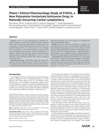
P-H2AX is a PD biomarker of F14512 in dogs
As a proof of concept of F14512-targeting tumors in dogs with
naturally occurring lymphoma, we looked at P-H2AX induction
in tumoral lymph node fine-needle aspirates. Four lymphoma-
bearing dogs received a single F14512 i.v. injection at a low dose
of 0.05 mg/kg, and serial fine-needle aspirates were performed. A
P-H2AX induction was observed by flow cytometry as early as 2
hours (not shown) after the end of the F14512 infusion and
increased at 4 hours (Fig. 1). P-H2AX induction was heteroge-
neous among the 4 patients but an increase was observed in all
dogs. These PD data further supported the clinical evaluation and
dose-escalation trial of F14512 in lymphoma-bearing dogs. These
4 dogs were subsequently included in the first cohort of the trial.
Epidemiologic characteristics and staging of dogs with
spontaneous lymphoma
Twenty-three dogs with naturally occurring lymphomas were
enrolled in the dose-escalation trial consisting of three cycles of
F14512 i.v. injection between November 2013 and March 2014.
The epidemiologic characteristics of these patients are summa-
rized in Table 1. There were 14 female and 9 male dogs. The mean
age of all 23 dogs was 8.0 Æ 2.6 years. The mean weight of all dogs
was 29.5 Æ 17 kg. Seventeen different canine breeds known as
being predisposed to lymphoma were represented (32). The
majority of cases (15/23) were classified as DLBCLs, 12 as cen-
troblastic (DLBCL-CB) and 3 as immunoblastic (DLBCL-IB).
Three other B-cell lymphoma cases were identified as late-stage
marginal zone lymphomas in transition into DLBCL-IB. The other
subtypes were: two peripheral T-cell lymphomas (2/23), includ-
ing one pleomorphic small lymphoma and one pleomorphic
mixed lymphoma (according to the updated Kiel Malignant
Lymphoma Classification); and a T-cell lymphoblastic lympho-
ma (1/23). The lymphoma subtype could not be determined in
two cases due to the lack of biopsy material, but the diagnosis of
high-grade lymphoma was based on the cytologic examination.
Two dogs had previously received long-term corticosteroid mono-
therapy before the study, with partial responses and rapid
relapses. Four dogs had previously received various CHOP-based
chemotherapy treatments with a complete response and relapse
before inclusion.
Pharmacokinetics
A total of five cohorts were successively initiated, with an
initial dosage of 0.050, 0.060, 0.070, 0.085, and 0.075 mg/kg
(cohorts 1 to 5). Although the cohort size per dose level was
small and the dose range explored was narrow, F14512 and
F16490 plasma AUC increased overall with the dose level (Fig.
2A). Figure 2B and C represents plasma concentrations versus
time of F14512 and its metabolite F16490 on day 1 for all dogs
treated at 0.075 mg/kg (recommended dose). F14512 plasma
concentrations were in the range of the IC50 value estimated in
the Namalwa model (46 nmol/L ¼ 29 ng/mL) for approxi-
mately 2 to 3 hours in most dogs. Interestingly, the patient with
the lowest plasma concentration was the only one that did not
have a clinical response to treatment at this dose level. Else-
where, the AUC of the active metabolite F16490 and of F14512
was proportional and, on average, the F16490 AUC represented
23% of the F14512 AUC.
Pharmacodynamics
Lymph node tumor cells and blood cell numeration were
monitored early during this clinical trial to determine whether
F14512 therapy was associated with any biologic effect. Serial
fine-needle aspirates were performed in tumoral lymph nodes in
the hours following the first injection of F14512 to look for PD
markers of F14512. The total cell number in these serial aspirates
was evaluated and normalized to the aspirate volume. A rapid and
Tierny et al.
Clin Cancer Res; 2015 Clinical Cancer ResearchOF4
5. dramatic decrease in the number of cells was observed (Fig. 3A) as
early as 2 hours after the beginning of the F14512 infusion. By the
end of the first cycle of the F14512 therapy, the number of cells
was dramatically reduced. Such a potent effect on tumoral cells
made the analysis of P-H2AX induction impossible in all patients.
Indeed, F14512 induced a strong decrease in viable cells, gener-
ating a lot of necrotic cells and debris (Supplementary Fig. S3),
and thus rendering any reliable P-H2AX evaluation using flow
cytometry impossible. This significant decrease in total lymph
node cell numbers was noticed at all dose levels (not shown) and
was clearly a PD marker of the F14512 efficacy.
Blood cell counts could be both a marker of efficacy and
toxicity. As expected, a decrease in the white blood cell count,
including neutrophils, lymphocytes, and monocytes, was
observed after each cycle of F14512 therapy, with a nadir on day
nine. A dose–effect relationship was observed between the dose of
F14512 and the number of neutrophils (Fig. 3B). This decrease
was reversible and baseline levels were recovered by the beginning
of the next cycle. The evaluation of the decrease in white blood
cells as well as the duration of the decrease correlated with the
dose of F14512 and allowed to determine DLTs.
Toxicities
All 23 dogs were evaluated for tolerance. Signs of toxicity
included neutropenia, anemia, thrombocytopenia, and digestive
disorders (diarrhea and vomiting). Toxicities are reported
in Table 2. Gastrointestinal adverse events (diarrhea for 6 dogs
and vomiting for 2 dogs) were mild in 6 dogs (grade 1) and
moderate in 1 dog (grade 2). They were not dose related (no
gastrointestinal toxicity observed with the highest dosage) and the
side effects resolved rapidly with the use of symptomatic treat-
ments. Severe adverse events (grade 3 and above) were limited to
neutropenia, thrombocytopenia, and anemia, and were all revers-
ible. Grade 4 neutropenia was observed at all dosages, but was
short (less than 48 hours) and clinically well tolerated for the first
three dose levels. Cohorts 1 and 2 were extended to confirm the
short duration and reversibility of the neutropenia at these dose
levels. Grade 4 neutropenia lasting more than 48 hours was
observed in 2 dogs from cohort 4, and was considered as a DLT.
A fifth cohort with an intermediate dosage (0.075 mg/kg) was,
therefore, constituted and determined as the recommended dose.
There were no treatment-related deaths, but 1 dog died due to the
progress of the disease on day 27.
Figure 1.
P-H2AX is a PD biomarker of F14512 in dogs. In vivo analysis of P-H2AX staining in fine-needle aspirates of tumor lymph nodes from 4 dogs (A–D) treated
with 0.05 mg/kg F14512. Isotype control (blank), before treatment (shaded) and 4 hours after the end of F14512 infusion (black).
F14512 Dose-Escalation Trial in Spontaneous Canine Lymphoma
www.aacrjournals.org Clin Cancer Res; 2015 OF5
6. Clinical outcome
Stable or progressive disease was observed in 2 dogs, both
having been treated before entering the study (Table 3). One of
these dogs, treated in cohort 1, died on day 27 with progressive
disease, whereas the other dog, treated in cohort 5, died on day
128 (after showing a stable disease on day 45). Ten dogs achieved
a complete regression, whereas 11 dogs experienced a partial
remission (Table 3). Pretreatment and inclusion in one of the
first three cohorts were associated with a tendency toward a lower
chance of achieving a complete remission, although the difference
was not statistically significant. Twenty dogs received additional
treatments following F14512 (consisting of various chemother-
apy protocols).
Discussion
Naturally occurring canine tumors represent a useful and
valuable model for deciphering many aspects of human cancers
(33). As part of the broader field of comparative oncology,
translational drug development studies in dogs with spontaneous
cancers have been used to define doses and schedules for thera-
peutic agents through rigorous PK–PD endpoints, often involving
serial biopsies of tumor tissue and the collection of biologic
materials before and after exposure to novel therapeutics (34,
35). Comparative oncology has been focusing on the study of
homologies, differences, and translational relevance of various
cancers, including lymphomas (36), osteosarcomas (37, 38), soft
tissue sarcomas (39), urinary bladder cancer (40), mammary
cancers (41), and others.
In this dose-escalating study, we have shown that the response
rate (complete and partial responses) of F14512 in treated canine
patients with lymphoma is 91%. Direct comparison of median
survival time or time to relapse with published data with other
chemotherapy agents is difficult because of the heterogeneity of
our population. Moreover, for obvious ethical reasons, some
patients were treated with another type of chemotherapy after
the F14512 study. Nevertheless, we can compare the clinical
response rate with F14512 with the reported response rate with
doxorubicin, which is commonly recognized as the most efficient
single agent for the treatment of canine high-grade non-Hodgkin
lymphoma. In a previous study (42), the response rate of dogs
treated with doxorubicin as a first line agent was 74%, which can
be compared with the result observed in our study (91%).
Therefore, our results are promising and emphasize the potential
of F14512, which could be investigated further either alone or in
combination with other agents. Because the treatment schedule
only included three cycles of F14512 injections, additional cycles
could be considered in future studies to potentially increase
clinical efficacy. As the main toxicity of F14512 is febrile neutro-
penia, a combination with conventional chemotherapy should be
chosen rationally to avoid major toxicities. In human lympho-
mas, the CHOP-based chemotherapy is associated with anti-
CD20 antibodies. It should be noted that some laboratories are
now working on therapeutic canine anti-CD20 antibodies (43).
Apart from rituximab, no other targeted therapy has emerged and
been developed so far for DLBCL, and the canine patient as a
cancer model may accelerate research. Ibrutinib, an irreversible
BTK inhibitor, benefited from its evaluation in canine lymphomas
(44) and is now proven to be efficacious in humans (45): The
canine lymphoma model enabled the authors to demonstrate the
full-target occupancy at various dosages, and to achieve some
objective clinical responses (44). Second-generation BTK inhibi-
tors are now being evaluated in dogs (46) in proof-of-concept
studies along with their development pathway, whereas clinical
Table 1. Epidemiologic and clinical characteristics at diagnosis of the whole study population composed of 23 dogs with na€ve and relapsing lymphoma
Epidemiologic characteristics Population studied
Sex
Male 39.1% (9/23)
Female 60.9% (14/23)
Age, y, mean Æ SD (range) 8.0 Æ 2.6 (4.0–15.0)
Body weight, kg, mean Æ SD (range) 29.5 Æ 17 (4.3–54.0)
Clinical characteristics
Breed
Rottweiler 13.0% (3/23)
Cavalier King Charles spaniel, Bernese mountain dog, Jack Russel terrier, Labrador Retriever 8.7% (2/23 each 4 breeds)
Yorkshire terrier, Cocker spaniel, West highland white terrier, Golden Retriever, Bull terrier, French
Bouledogue, Dogue de Bordeaux, Weimaraner, Mastiff, Greyhound, German Shepherd, cross-breed
4.3% (1/23 each 12 breeds)
Clinical stage
Stage 3 17.4% (4/23)
Stage 4 82.6% (19/23)
Clinical substage
a 52.1% (12/23)
b 47.9% (11/23)
Hypercalcemia 8.7% (2/23)
Pretreatment
Corticosteroid 8.7% (2/23)
Chemotherapy 17.4% (4/23)
Lymphoma subtype
B cell
Diffuse large B-cell lymphoma 65.2% (15/23)
Marginal zone lymphoma 13.0% (3/23)
T cell
Peripheral T cell 8.7% (2/23)
T-cell lymphoblastic 4.3% (1/23)
Unclassified 8.7% (2/23)
Tierny et al.
Clin Cancer Res; 2015 Clinical Cancer ResearchOF6
7. studies of combination of ibrutinib with polychemotherapy are
ongoing in DLBCL in humans. Thus, we believe that a combina-
tion of F14512 with targeted therapies such as anti-CD20 mAb or
BTK inhibitors should be considered in the future.
As a vectorized form of etoposide, F14512 was shown to be
more potent than etoposide in vitro (4) and to be superior to
etoposide in terms of efficacy and therapeutic window in xeno-
graft mice models (8, 9). F14512 also revealed a convincing
antitumor activity in naturally occurring lymphomas in dogs,
with 22 of 23 patients displaying a clinical response (either stable
disease, partial, or complete remission). These clinical results in
dogs clearly appear to be superior to those described for etoposide
(24). Etoposide was also shown to be poorly active in a retro-
spective study on 13 animals, with only 2 dogs displaying a
Figure 2.
F14512 PK data in dogs. A, descriptive statistics of F14512 and F14512 main
metabolite (F16490; AUClast by dose level). B and C, F14512 and
F16490 plasma concentrations versus time for all dogs treated at the
recommended dose level of 0.075 mg/kg, after the first infusion (cycle 1,
day 1). F14512 infusions started at t ¼ 0 hours and lasted between
3.25 and 3.63 hours ($).
Figure 3.
Tumoral and hematologic PD markers of F14512. A, F14512 induces a strong
and early decrease of tumoral lymph node cell number in dogs. Fine-needle
aspirates were performed in tumoral lymph nodes all along the first cycle
of F14512 administration. F14512 infusions were initiated at t ¼ 0 hours and
lasted 3 hours at day 1 (0–3 hours), day 2 (24–27 hours), and day 3
(48–51 hours). The graph shows the total cell count per milliliter of fine-needle
aspirates, from all dogs treated at all dose levels (results, mean þ SD; data
were analyzed using ANOVA followed by PLSD Fisher post hoc; Ã
, P 0.05;
ÃÃ
, P 0.01; and ÃÃÃ
, P 0.001, n ¼ 23 dogs. B, F14512 induces a dose-
dependent decrease in circulating neutrophils in treated dogs. The graph
shows the dose–effect relationship between the dose level of F14512 and the
number of neutrophils (median absolute neutrophil count vs. time by dose
level in mg/kg). F14512 was administered on days 1, 2, and 3.
F14512 Dose-Escalation Trial in Spontaneous Canine Lymphoma
www.aacrjournals.org Clin Cancer Res; 2015 OF7
8. clinical response. Moreover, as F14512 was shown to be a poor
substrate of the efflux protein PgP (unpublished data), it would be
of great interest to look for F14512 efficacy in relapsing and/or
refractory chemoresistant dog lymphomas who have a poor prog-
nosis (47). The canine multidrug resistance associated protein has
been molecularly identified (48). In thepresent study, 4 dogs hada
previous polychemotherapy course. Even if their PgP/Mdr status
was not known, a clinical response was observed in 3 of them.
Therefore, this F14512 phase I study warrants further evaluation of
F14512 in relapsing and/or chemoresistant lymphoma cases.
Because of the unequal distribution of dogs depending on the
lymphomasubtype, it was not possible to see any relation between
the clinical response and the lymphoma subtype.
In this study, we looked at F14512-induced DNA damages and
demonstrated that P-H2AX signaling (49) could be used as a PD
biomarker. We took the opportunity to design a preliminary study
in 4 dogs treated with a single injection of low-dose F14512 and
unraveled an early in vivo P-H2AX induction as a PD response.
Unfortunately, we could not correlate P-H2AX induction with the
F14512 AUC or the clinical response owing to the rapid onset of
lymphoma cell death induced by F14512. We were not able to
recover enough living cells from all treated dogs to perform a
relevant flow-cytometry study of P-H2AX. We did not expect such
a tumoral cell death (as early as a couple of hours after initiating
F14512 infusion). A preferential and rapid uptake of F14512 by
tumor cells, with an added cytotoxic effect brought by F16490
metabolite, may explain this strong tumoral cytotoxicity. We
previously demonstrated the preferential uptake of F14512 by
tumor cells (4, 5), and as a consequence plasma levels of F14512
and F16490 may only partially reflect the concentrations reached
in tumors. F16490 alone displays cytotoxic activity with an EC50
of 74 nmol/L in the Namalwa cell line. This rapid cell death
induction was observed in all dogs, even in the 2 dogs that did not
display a clinical response later. This latter observation confirms
the strong cytotoxicity of F14512 in dog lymphoma and supports
further evaluation either in combination and/or with the addition
of other cycles of treatment to reinforce and extend its efficacy.
Careful monitoring of patients may be required in human clinical
trials for the early detection of tumor lysis syndrome that may
occur.
An interesting point about the study we performed on dogs is
that there is an ongoing clinical assessment of F14512 in humans,
so we are able to compare and translate the clinical data and
findings from both species, even if the clinical tumor indications
are not the same. The first-in-man multicenter phase I trial on
F14512 as a single drug was conducted in adult patients with
relapsed or refractory AML (12, 50). Patients received a daily i.v.
infusion for 5 consecutive days every 2 to 6 weeks depending on
the leukemia response as well as the recovery of sufficient hema-
topoiesis and the resolution of toxicities. The main toxicity was
myelosuppression, which was dose dependent and reversible. The
MTD reached was 44 mg/m2
and the recommended dose was
determined to be 39 mg/m2
. Antileukemic activity was observed
at different dose levels with 10% complete responses, 8% com-
plete responses with incomplete recovery and 3 patients who
experienced hematologic improvements. As with humans, we
Table 2. Toxicity observed in the whole study population composed of 23 dogs with na€ve and relapsing lymphoma
Hematologic toxicity grade (number of dogs)
Gastrointestinal toxicity
grade (number of dogs)
Cohort (dose) Number of dogs Hemoglobin HCT Platelet Neutrophils Diarrhea Vomiting
Cohort 1 (0.05 mg/m2
) 6 Grade 2 (1) Grade 2 (1) Grade 3 (1) Grade 4 (3)a
Grade 1 (1) Grade 1 (1)
Grade 1 (4) Grade 1 (4) Grade 2 (3) Grade 3 (1)
Cohort 2 (0.06 mg/m2
) 4 Grade 4 (1) Grade 4 (1) Grade 2 (2) Grade 4 (2)a
Grade 1 (2) Grade 1 (1)
Grade 2 (1) Grade 3 (1) Grade 1 (1) Grade 3 (1)
Grade 1 (1) Grade 2 (1)
Cohort 3 (0.07 mg/m2
) 3 Grade 3 (1) Grade 3 (1) Grade 2 (1) Grade 4 (1)a
Grade 2 (1) 0
Grade 1 (2) Grade 1 (2) Grade 3 (1)
Cohort 4 (0.085 mg/m2
) 4 Grade 2 (1) Grade 2 (2) 0 Grade 4 (2)b
0 0
Grade 1 (3) Grade 1 (2) Grade 4 (1)a
Grade 2 (1)
Cohort 5 (0.075 mg/m2
) 6 Grade 2 (3) Grade 2 (4) Grade 4 (1) Grade 4 (1)b
Grade 1 (2) 0
Grade 1 (3) Grade 1 (2) Grade 3 (1) Grade 4 (2)a
Grade 2 (2) Grade 3 (1)
a
Asymptomatic neutropenia lasting less than 48 hours.
b
Febrile neutropenia lasting more than 48 hours.
Table 3. Clinical response of the whole study population composed of 23 dogs with na€ve and relapsing lymphoma
Clinical response at day 45
Population Progressive disease Stable disease Partial response Complete response
Whole population 4.4% (1/23) 4.4% (1/23) 47.8% (11/23) 43.5% (10/23)
Pretreatment
Yes (n ¼ 6) 16.7% (1/6) 16.7% (1/6) 66.6% (4/6) 0
No (n ¼ 17) 0 0 58.8% (10/17) 41.2% (7/17)
Cohort
Cohort 1: 0.050 mg/kg (n ¼ 6) 16.7% (1/6) 0 50.0% (3/6) 33.3% (2/6)
Cohort 2: 0.060 mg/kg (n ¼ 4) 0 0 75.0% (3/4) 25.0% (1/4)
Cohort 3: 0.070 mg/kg (n ¼ 3) 0 0 66.6% (2/3) 33.3% (1/3)
Cohort 4: 0.085 mg/kg (n ¼ 4) 0 0 50.0% (2/4) 50.0% (2/4)
Cohort 5: 0.075 mg/kg (n ¼ 6) 0 16.7% (1/6) 16.7% (1/6) 66.6% (4/6)
Tierny et al.
Clin Cancer Res; 2015 Clinical Cancer ResearchOF8
9. also observed clinical responses in dogs at all dose levels, but
relapses were more frequently noticed at the first dose levels (even
if not statistically significant). When we looked at PD markers of
F14512 in dogs, the tumor lymph node cell counts and the
circulating blood cells counts (Fig. 3) revealed a favorable ther-
apeutic window: A strong decrease of tumor cells was observed at
all doses whereas the hematologic adverse events were increased
only at higher doses. Interestingly, the tolerance profile was very
similar between humans and dogs, with the hematotoxicity being
the major adverse event observed. A phase II trial of F14512 in
combination with cytosine arabinoside is currently ongoing in
patients with AML.
In conclusion, our results provide a strong evidence of the
clinical efficacy of the new vectorized drug F14512 in a pet dog
model of lymphoma. The translational value of canine lympho-
ma warrants further studies of F14512 and strongly supports its
clinical development.
Disclosure of Potential Conflicts of Interest
No potential conflicts of interest were disclosed.
Authors' Contributions
Conception and design: D. Tierny, F. Serres, A. Petain, N. Guilbaud, B. Gomes
Development of methodology: Z. Segaoula, I. Bemelmans, E. Bouchaert
Acquisition of data (provided animals, acquired and managed patients,
provided facilities, etc.): D. Tierny, F. Serres, V. Brel, T. Marchal
Analysis and interpretation of data (e.g., statistical analysis, biostatistics,
computational analysis): D. Tierny, Z. Segaoula, A. Petain, V. Brel, S. Couffin,
L. Nguyen, X. Thuru, B. Gomes
Writing, review, and/or revision of the manuscript: D. Tierny, F. Serres, V. Brel,
S. Couffin, P. Ferre, N. Guilbaud, B. Gomes
Administrative, technical, or material support (i.e., reporting or organizing
data, constructing databases): Z. Segaoula, I. Bemelmans, E. Bouchaert
Study supervision: D. Tierny, B. Gomes
Acknowledgments
The authors thank Marie Bosredon for her kind assistance in the F14512
batch management and the flow core facility of BICeL-IFR114 for the technical
assistance.
Grant Support
This work was supported by grant from BPI-France, through the CaninCa
project.
The costs of publication of this article were defrayed in part by the
payment of page charges. This article must therefore be hereby marked
advertisement in accordance with 18 U.S.C. Section 1734 solely to indicate
this fact.
Received December 8, 2014; revised June 13, 2015; accepted July 4, 2015;
published OnlineFirst July 13, 2015.
References
1. Siegel R, Naishadham D, Jemal A. Cancer statistics, 2013. CA Cancer J Clin
2013;63:11–30.
2. Younes A, Thieblemont C, Morschhauser F, Flinn I, Friedberg JW, Amorim
S, et al. Combination of ibrutinib with rituximab, cyclophosphamide,
doxorubicin, vincristine, and prednisone (R-CHOP) for treatment-naive
patients with CD20-positive B-cell non-Hodgkin lymphoma: a non-ran-
domised, phase 1b study. Lancet Oncol 2014;15:1019–26.
3. Casero RA Jr, Marton LJ. Targeting polyamine metabolism and function in
cancer and other hyperproliferative diseases. Nat Rev Drug Discov
2007;6:373–90.
4. Barret JM, Kruczynski A, Vispe S, Annereau JP, Brel V, Guminski Y, et al.
F14512, a potent antitumor agent targeting topoisomerase II vectored into
cancer cells via the polyamine transport system.Cancer Res 2008;68:
9845–53.
5. Annereau JP, Brel V, Dumontet C, Guminski Y, Imbert T, Broussas M,
et al. A fluorescent biomarker of the polyamine transport system to
select patients with AML for F14512 treatment. Leuk Res 2010;34:
1383–9.
6. Brel V, Annereau JP, Vispe S, Kruczynski A, Bailly C, Guilbaud N. Cyto-
toxicity and cell death mechanisms induced by the polyamine-vectorized
anti-cancer drug F14512 targeting topoisomerase II. Biochem Pharmacol
2011;82:1843–52.
7. Gentry AC, Pitts SL, Jablonsky MJ, Bailly C, Graves DE, Osheroff N.
Interactions between the etoposide derivative F14512 and human type II
topoisomerases: implications for the C4 spermine moiety in promoting
enzyme-mediated DNA cleavage. Biochemistry 2011;50:3240–9.
8. Kruczynski A, Vandenberghe I, Pillon A, Pesnel S, Goetsch L, Barret JM, et al.
Preclinical activity of F14512, designed to target tumors expressing an
active polyamine transport system. Invest New Drugs 2011;29:9–21.
9. Kruczynski A, Pillon A, Creancier L, Vandenberghe I, Gomes B, Brel V, et al.
F14512, a polyamine-vectorized anti-cancer drug, currently in clinical trials
exhibits a marked preclinical anti-leukemic activity. Leukemia 2013;27:
2139–48.
10. Mouawad F, Gros A, Rysman B, Bal-Mahieu C, Bertheau C, Horn S, et al.
The antitumor drug F14512 enhances cisplatin and ionizing radiation
effects in head and neck squamous carcinoma cell lines. Oral Oncol
2014;50:113–9.
11. Leblond P, Boulet E, Bal-Mahieu C, Pillon A, Kruczynski A, Guilbaud N,
et al. Activity of the polyamine-vectorized anti-cancer drug F14512 against
pediatric glioma and neuroblastoma cell lines. Invest New Drugs 2014;
32:883–92.
12. De Botton S, Berthon C, Bulabois CE, Prebet T, Vey N, Chevallier P ,
et al. F14512 a novel polyamine-vectorized anti-cancer drug targeting
topoisomerase II in adults patients with acute myeloid leukemia (AML):
results from a phase I study. EHA 17th Congress. 14–17 June 2012;
Amsterdam.
13. Marconato L, Gelain ME, Comazzi S. The dog as a possible animal model
for human non-Hodgkin lymphoma: a review. Hematol Oncol 2013;31:
1–9.
14. Ponce F, Marchal T, Magnol JP, Turinelli V, Ledieu D, Bonnefont C, et al. A
morphological study of 608 cases of canine malignant lymphoma in
France with a focus on comparative similarities between canine and human
lymphoma morphology. Vet Pathol 2010;47:414–33.
15. Ranieri G, Gadaleta CD, Patruno R, Zizzo N, Daidone MG, Hansson MG,
et al. A model of study for human cancer: spontaneous occurring tumors in
dogs. Biological features and translation for new anticancer therapies. Crit
Rev Oncol Hematol 2013;88:187–97.
16. Richards KL, Motsinger-Reif AA, Chen HW, Fedoriw Y, Fan C, Nielsen DM,
et al. Gene profiling of canine B-cell lymphoma reveals germinal center and
postgerminal center subtypes with different survival times, modeling
human DLBCL. Cancer Res 2013;73:5029–39.
17. Mudaliar MA, Haggart RD, Miele G, Sellar G, Tan KA, Goodlad JR, et al.
Comparative gene expression profiling identifies common molecular
signatures of NF-kB activation in canine and human diffuse large B-cell
lymphoma (DLBCL). PLoS ONE 2013;8:e72591.
18. Marconato L, Frayssinet P, Rouquet N, Comazzi S, Leone VF, Laganga P,
et al. Randomized, placebo-controlled, double-blinded chemoimmu-
notherapy clinicaltrial in apet dog model of diffuse large B-cell lymphoma.
Clin Cancer Res 2014;20:668–77.
19. Ito D, Frantz AM, Modiano JF. Canine lymphoma as a comparative model
for human non-Hodgkin lymphoma: recent progress and applications. Vet
Immunol Immunopathol 2014;159:192–201.
20. Igwemezie LN, Kaul S, Barbhaiya RH. Assessment of toxicokinetics and
toxicodynamics following intravenous administration of etoposide phos-
phate in beagle dogs. Pharm Res 1995;12:117–23.
21. Flory AB, Rassnick KM, Balkman CE, Kiselow MA, Autio K, Beaulieu BB,
et al. Oral bioavailability of etoposide after administration of a single dose
to tumor-bearing dogs. Am J Vet Res 2008;69:1316–22.
F14512 Dose-Escalation Trial in Spontaneous Canine Lymphoma
www.aacrjournals.org Clin Cancer Res; 2015 OF9
10. 22. Lana S, U'ren L, Plaza S, Elmslie R, Gustafson D, Morley P, et al. Continuous
low-dose oral chemotherapy for adjuvant therapy of splenic hemangio-
sarcoma in dogs. J Vet Intern Med 2007;21:764–9.
23. Willmann M, M€ullauer L, Schwendenwein I, Wolfesberger B, Kleiter M,
Pagitz M, et al. Chemotherapy in canine acute megakaryoblastic leukemia:
a case report and review of the literature. In Vivo 2009;23:911–8.
24. Hohenhaus AE, Matus RE. Etoposide (VP-16). Retrospective analysis of
treatment in 13 dogs with lymphoma. J Vet Intern Med 1990;4:239–41.
25. Fernandes NC, Guerra JM, Ressio RA, Wasques DG, Etlinger-Colonelli D,
Lorente S, et al. Liquid-based cytology and cell block immunocytochem-
istry in veterinary medicine: comparison with standard cytology for the
evaluation of canine lymphoid samples. Vet Comp Oncol 2015 Feb 9.
[Epub ahead of print].
26. Joetzke AE, Eberle N, Nolte I, Mischke R, Simon D. Flow cytometric
evaluation of peripheral blood and bone marrow and fine-needle aspirate
samples from multiple sites in dogs with multicentric lymphoma. Am J Vet
Res 2012;73:884–93.
27. Williams LE, Broussard MT, Johnson JL, Neel J. Comparison of results of
clinicians' assessments, cytologic examination of fine-needle lymph node
aspirates, and flow cytometry for determination of remission status of
lymphoma in dogs. J Am Vet Med Assoc 2005;226:562–6.
28. Huang X, Darzynkiewicz Z. Cytometric assessment of histone H2AX
phosphorylation: a reporter of DNA damage. Methods Mol Biol 2006;
314:73–80.
29. Valli VE, San Myint M, Barthel A, Bienzle D, Caswell J, Colbatzky F, et al.
Classification of canine malignant lymphomas according to the World
Health Organization criteria. Vet Pathol 2011;48:198–211.
30. Veterinary Co-operative Oncology Group (VCOG). Veterinary Cooperative
Oncology Group - Common Terminology Criteria for Adverse Events
(VCOG-CTCAE) following chemotherapy or biological antineo-plastic
therapy in dogs and cats v1.0. Vet Comp Oncol 2004;2:195–213.
31. Vail DM, Michels GM, Khanna C, Selting KA, London CA. Veterinary
Cooperative Oncology Group. Response evaluation criteria for periph-
eral nodal lymphoma in dogs (v1.0)—a Veterinary Cooperative Oncol-
ogy Group (VCOG) consensus document. Vet Comp Oncol 2010;8:
28–37.
32. Modiano JF, Breen M, Burnett RC, Parker HG, Inusah S, Thomas R, et al.
Distinct B-cell and T-cell lymphoproliferative disease prevalence among
dog breeds indicates heritable risk. Cancer Res 2005;65:5654–61.
33. Paoloni M, Khanna C. Translation of new cancer treatments from pet dogs
to humans. Nat Rev Cancer 2008;8:147–56.
34. London CA, Hannah AL, Zadovoskaya R, Chien MB, Kollias-Baker C,
Rosenberg M, et al. Phase I dose-escalating study of SU11654, a small
molecule receptor tyrosine kinase inhibitor, in dogs with spontaneous
malignancies. Clin Cancer Res 2003;9:2755–68.
35. Pryer NK, Lee LB, Zadovaskaya R, Yu X, Sukbuntherng J, Cherrington JM,
et al. Proof of target for SU11654: inhibition of KIT phosphorylation in
canine mast cell tumors. Clin Cancer Res 2003;9:5729–34.
36. Richards KL, Suter SE. Man's best friend: what can pet dogs teach us about
non-Hodgkin's lymphoma? Immunol Rev 2015;263:173–91.
37. Khanna C, Fan TM, Gorlick R, Helman LJ, Kleinerman ES, Adamson PC,
et al. Toward a drug development path that targets metastatic progression
in osteosarcoma. Clin Cancer Res 2014;20:4200–9.
38. Fenger JM, London CA, Kisseberth WC. Canine osteosarcoma: a naturally
occurring disease to inform pediatric oncology. ILAR J 2014;55:69–85.
39. Milovancev M, Hauck M, Keller C, Stranahan LW, Mansoor A, Malarkey DE.
Comparative pathology of canine soft tissue sarcomas: possible models of
human non-rhabdomyosarcoma soft tissue sarcomas. J Comp Pathol
2015;152:22–7.
40. Knapp DW, Ramos-Vara JA, Moore GE, Dhawan D, Bonney PL, Young KE.
Urinary bladder cancer in dogs, a naturally occurring model for cancer
biology and drug development. ILAR J 2014;55:100–18.
41. Liu D, Xiong H, Ellis AE, Northrup NC, Rodriguez CO Jr, O'Regan RM, et al.
Molecular homology and difference between spontaneous canine mam-
mary cancer and human breast cancer. Cancer Res 2014;74:5045–56.
42. Simon D, Moreno SN, Hirschberger J, Moritz A, Kohn B, Neuman S, et al.
Efficacy of a continuous, multiagent chemotherapeutic protocol versus a
short-term single-agent protocol in dogs with lymphoma. J Am Vet Med
Assoc 2008;232:879–885.
43. Ito D, Brewer S, Modiano JF, Beall MJ. Development of a novel anti-canine
CD20 monoclonal antibody with diagnostic and therapeutic potential.
Leuk Lymphoma 2015;56:219–25.
44. Honigberg LA, Smith AM, Sirisawad M, Verner E, Loury D, Chang B, et al.
The Bruton tyrosine kinase inhibitor PCI-32765 blocks B-cell activation
and is efficacious in models of autoimmune disease and B-cell malignancy.
Proc Natl Acad Sci U S A 2010;107:13075–80.
45. Aalipour A, Advani RH. Bruton tyrosine kinase inhibitors: a promising
novel targeted treatment for B-cell lymphomas. Br J Haematol 2013;163:
436–43.
46. Gardner HL, Harrington BK, Izumi R, Hamdy A, Kapstein A, Van Lith B,
et al. ACP-196: a second generation Btk inhibitor demonstrates biologic
activity in a canine model of B-cell non-Hodgkin lymphoma. AACR 2014
Annual Meeting. 5–9 April. 2014; San Diego. Poster #1744.
47. LeBlanc AK, Mauldin GE, Milner RJ, LaDue TA, Mauldin GN, Bartges JW.
Efficacy and toxicity of BOPP and LOPP chemotherapy for the treatment of
relapsed canine lymphoma. Vet Comp Oncol 2006;4:21–32.
48. Ma L, Pratt SE, Cao J, Dantzig AH, Moore RE, Slapak CA. Identification and
characterization of the canine multidrug resistance-associated protein. Mol
Cancer Ther 2002;1:1335–42.
49. Matthaios D, Hountis P, Karakitsos P, Bouros D, Kakolyris S. H2AX a
promising biomarker for lung cancer: a review. Cancer Invest 2013;31:
582–99.
50. De Botton S, Quesnel B, Audoly L, Bailly C, Brandely-Talbo M, Provendier
O, et al. Tacklingleukemia: phase I study ofF14512 in relapsed orrefractory
AML patients. 12th International Congress on Targeted Anticancer Ther-
apies, 2014; Washington.
Clin Cancer Res; 2015 Clinical Cancer ResearchOF10
Tierny et al.