High Profile Call Girls Jaipur Vani 8445551418 Independent Escort Service Jaipur
inmunofenotipo para leucemias linfociticas en perros.pdf
1. Immunophenotype Predicts Survival Time in Dogs with Chronic
Lymphocytic Leukemia
S. Comazzi, M.E. Gelain, V. Martini, F. Riondato, B. Miniscalco, L. Marconato,
D. Stefanello, and M. Mortarino
Background: Chronic lymphocytic leukemia (CLL) is a hematologic disorder in dogs, but studies on prognostic factors and
clinical outcome are lacking. In people, several prognostic factors have been identified and currently are used to manage pa-
tients and determine therapy.
Objectives: The aim of the study was to determine if the immunophenotype of neoplastic cells predicts survival in canine
CLL.
Design: Retrospective study.
Animals: Forty-three dogs with CLL.
Procedures: Records of dogs with a final diagnosis of CLL were reviewed. For each included dog, a CBC, blood smear for
microscopic reevaluation, and immunophenotyping data had to be available. Data on signalment, history, clinical findings,
therapy, follow-up, as well as date and cause of death were retrieved.
Results: Seventeen dogs had B-CLL (CD211), 19 had T-CLL (CD31 CD81), and 7 had atypical CLL (3 CD3 CD81, 2
CD31 CD4 CD8 , 1 CD31 CD41 CD81, and 1 CD31 CD211). Among the variables considered, only immunophenotype
was associated with survival. Dogs with T-CLL had approximately 3-fold and 19-fold higher probability of surviving than dogs
with B-CLL and atypical CLL, respectively. Old dogs with B-CLL survived significantly longer than did young dogs, and
anemic dogs with T-CLL survived a significantly shorter time than dogs without anemia.
Conclusions: Although preliminary, results suggested that immunophenotype is useful to predict survival in dogs with CLL.
Young age and anemia are associated with shorter survival in dogs with B-CLL and T-CLL, respectively.
Key words: Blood; Canine; Lymphoproliferative disorders; Oncology; Prognosis.
Chronic lymphocytic leukemia (CLL) is a hemato-
logic disorder occurring in middle-aged to old dogs,
although studies on its actual prevalence still are lacking.
Also, no extensive description of clinical behavior and
prognostic factors is available. Hematologic features of
CLL1–4
and data on clinical outcome have been reported
on a limited number of patients.5,6
In human medicine, 2
widely accepted staging systems (the so-called modified
Rai and Binet staging systems) have been developed and
routinely applied in the clinical setting.7,8
Several prog-
nostic factors, including lymphoadenomegaly, spleno-
megaly or hepatomegaly, anemia, thrombocytopenia,
severe lymphocytosis, rapid lymphocyte doubling
time, 410% prolymphocytes or selected chromosomal
anomalies, have been linked to a worse prognosis, thus
requiring a more aggressive therapeutic approach.
In people, CLL primarily is a B-cell neoplasm caused
by the expansion of a subpopulation of cells with CD5
positivity. However, some cytogenetic similarities have
been reported between humans and dogs.9
In people, B-CLL tends to be an indolent disease
and, even if cure is not possible, at the moment, median
survival times often are 4150 months. On the
other hand, some studies report that T-CLL often fol-
lows a more aggressive course.10
In dogs, T-CLL is the
most common disease but, to our knowledge, few studies
focusing on immunophenotype-related prognosis
have been carried out. According to some authors,
T-CLL with large granular lymphocyte (LGL) morpho-
logy does not harbor a worse prognosis than B-CLL.2
A study on neoplastic lymphocytosis has been
published,11
and found no differences in survival based
on immunophenotype. An important finding of that
study was that dogs with B-cell neoplasms characterized
by small circulating cells or with T-cell lympho-
cytosis with o30,000 cells/mL had long survival times. A
limitation of that study was the lack of differentiation
between CLL and lymphoma with blood infiltration, be-
ing attributable to the unavailability of consensus
definitions.
Chemotherapy is not always indicated, because CLL
has an indolent course in most cases.12
In agreement with
human oncology, chemotherapy may be indicated in the
case of progressive disease with cytopenia, high lympho-
cyte count, lymphoadenomegaly and splenomegaly,
fever, or infections. To date, the efficacy of different ther-
apeutic approaches and outcomes have not been
explored in dogs.
The aim of this retrospective study was to determine if
immunophenotype predicts survival in dogs with CLL.
Additional factors associated with long survival also
were evaluated.
From the Dipartimento di Patologia Animale, Igiene e Sanità Pub-
blica Veterinaria, Università degli Studi di Milano, Milan, Italy
(Comazzi, Gelain, Martini, Mortarino); the Dipartimento di Pat-
ologia Animale, Università di Torino, Turin, Italy (Riondato,
Miniscalco); the Animal Oncology and Imaging Center, Hünenberg,
Switzerland (Marconato); and the Dipartimento di Scienze Cliniche
Veterinarie, Università degli Studi di Milano, Milan, Italy (Stefanello).
Corresponding author: Stefano Comazzi, DVM, PhD, Dip-
ECVCP, Dipartimento di Patologia Animale, Igiene e Sanità
Pubblica Veterinaria, Università degli Studi di Milano, Via Celoria
10, 20133 Milan, Italy; e-mail: stefano.comazzi@unimi.it.
Submitted February 23, 2010; Revised July 20, 2010; Accepted
September 29, 2010.
Copyright r 2010 by the American College of Veterinary Internal
Medicine
10.1111/j.1939-1676.2010.0640.x
J Vet Intern Med 2011;25:100–106
2. Materials and Methods
Eligibility
The databases of the authors’ institutions were reviewed to
identify dogs with CLL, blood samples of which were referred for
immunophenotyping (September 2006 to January 2010).
To be included in the study, flow cytometry of peripheral blood
showing lymphocyte counts 46109
/L12
had to be available.
Additionally, immunophenotyping data had to be suggestive of a
homogeneous expansion of lymphocytes.11
Exclusion criteria included:
(1) morphology suggestive of immature or blast cells in 430%
of cells (based on medium to large cells; round or indented,
medium to large nucleus with poorly condensed chromatin;
presence of nucleoli or some combination of these),
(2) CD34 positivity,
(3) moderate or severe lymphadenomegaly, splenomegaly, or both
with nodal, splenic, or both having cytological features compat-
ible with lymphoma. Mild lymphadenopathy or splenomegaly
was not considered an exclusion criteria, except for those cases
showing cytological features suggestive of specific lymphoma
subtypes, and
(4) positive serologic titer for Ehrlichia or Leishmania or any other
identifiable cause of lymphocytosis (eg, hypoadrenocorticism,
postvaccinal lymphocytosis, stress lymphocytosis).
Dogs that were severely symptomatic or had specific clinical signs
attributable to multiorgan infiltration were reevaluated by means of
clinical reassessment, cytology of presumptive lesions, and imaging
to exclude lymphoma. Any clinical sign attributable to anemia or
thrombocytopenia was not considered an exclusion criterion.
For each included case, the following data had to be available: a
CBC, at least 1 high quality blood smear for microscopic reevalua-
tion, a complete medical record including signalment, history,
clinical findings, therapy, follow-up as well as date and presumptive
cause of death (if applicable).
Hematology
CBC was performed on EDTA blood samples with an automated
laser analyzer. Automated differentials were validated by micro-
scopic evaluation of blood smears stained with May Grünwald-
Giemsa. Blood smears were accurately evaluated, paying particular
attention to the percentage of cells with prolymphocytic morphology
(ie, medium cells with slightly basophilic cytoplasm and centrally
placed, round, or slightly indented nucleus with minimal coarse chro-
matin and a large, round bluish nucleolus) or with LGL appearance
(medium cells with abundant clear cytoplasm and fine cytoplasmic
granules, round nucleus with clumped chromatin, and an inconspic-
uous nucleolus). Anemia was defined by PCV o37% (i.e., lower limit
of the reference range). Thrombocytopenia was confirmed if both
platelet count was o100,000/mL and blood smear showed low
platelet estimation and no evidence of platelet clumping.13
Immunophenotype
Flow cytometric immunophenotype was performed as reported
previously14
with a flow cytometer.a
The following monoclonal
antibodies were used: CD45-PEb
(clone YKIX716.13, Serotec),
CD3-FITC (clone CA17.2A12, Serotec, T cells), CD4-FITC
(clone YKIX302.9, Serotec, T-helper and neutrophils), CD8-PE
(clone YCATE55.9, Serotec, T-cytotoxic/suppressor), CD21-PE
(clone CA21D6 Serotec, B cells), CD34-PE (clone 1H6 Pharmingen,
precursor cells).c
Analysis was conducted by specific software
(Cell Quest). Based on flow cytometric results, cases were grouped
as follows: B-CLL (CD451 CD211), T-CLL (CD31 CD81), and
atypical CLL (other phenotypes).
Statistical Analysis
Data were recorded in a spreadsheet and summarized by descrip-
tive statistics with standard statistical software.d
Hematological features of different immunophenotype groups
were analyzed by means of the Kruskal-Wallis test for independent
samples. To test correlation between different hematologic
variables, the Spearman test was performed. Frequency of anemia
(PCV o37%), thrombocytopenia, and extreme lymphocytosis
(lymphocytes 450109
/L) were compared within different
phenotypic groups by the Pearson w2
-test.
To assess whether immunophenotype influenced survival, curves
were generated by the Kaplan-Meier method and compared with
the log-rank test. Survival time was calculated from the date of
diagnosis of CLL to death. Cases were censored if lost to follow-up,
if they died or were euthanized because of unrelated causes, or
if they were alive at the end of the study.
The following factors were investigated with multivariate Cox’s
proportional hazard regression analysis to assess association with
long survival: immunophenotype, age, PCV, total lymphocyte
count. Age, PCV, and total lymphocyte count were analyzed as
continuous noncategorical variables. Analysis was performed in 2
steps: initially, all cases were included in the analysis, thereafter only
B-CLL and T-CLL were evaluated to define possible variables
predicting survival within different phenotypic groups. The
Cox proportional hazards regression model assumption was tested
by plotting the ‘‘log negative log of survival’’ among cases.
Finally, curves were generated by the Kaplan-Meier method and
compared with the log-rank test to verify whether therapy affected
survival times. These data were not included in the multivariate
analysis because they were not considered independent from some
prognostic features (eg, anemia, severe lymphocytosis, organ
failure, clinical signs) and because therapy was not started immedi-
ately after diagnosis in all cases. Therapeutic approaches were
arbitrarily grouped into 3 categories: (A) dogs receiving neither anti-
neoplastic chemotherapy nor corticosteroids, (B) dogs receiving
only corticosteroids, and (C) dogs receiving cytotoxic antineoplas-
tic chemotherapy and corticosteroids.
P-values o .05 were considered significant.
Results
Of 272 cases with homogeneous lymphocytosis, 153
were excluded because of clinical and cytological aspects
suggestive of lymphoma, whereas 53 were excluded be-
cause of CD34 positivity or morphological appearance of
precursor blast cells.
Two cases were excluded because low-grade lym-
phoma and CLL could not be differentiated based on
clinical signs (moderate lymphoadenomegaly and
splenomegaly) and cytological features (medium or small
lymphocytes).
Ten cases with moderate to marked lymphadenome-
galy were excluded because cytological smears were
absent or of poor quality, thereby precluding the possi-
bility of reevaluation. Seven cases were excluded because
of absence of clinical and follow-up data.
Four cases showing a homogeneous expansion of lym-
phocytes were excluded because of serologic positivity to
Ehrlichia (n 5 3) or Leishmania (n 5 1).
101
Immunophenotypes and Survival in Canine CLL
3. Eight cases showing moderate (PCV o 30%) anemia
and 7 cases with thrombocytopenia were included because
of the absence of masses or lymph node enlargement. Mild
to moderate splenomegaly and lymphadenomegaly were
observed in 7 and 11 cases, respectively; these dogs were
included based on the presence of a prevalent homoge-
neous population of small mature lymphocytes.
Forty-three dogs met the inclusion criteria: there were
17 B-CLL (CD211), 19 T-CLL (CD31 CD81), and
7 atypical CLL, including 3 cases with CD3 CD81
immunophenotype 2 double-negative T-CLL (CD31
CD4 CD8 ), 1 double-positive T-CLL (CD31 CD41
CD81), and 1 biphenotypic CLL (CD31 CD211).
Median age was 10 years (range, 2–16 years).
Hematology
Results of the main hematological features are shown
in Table 1. Twenty-one (48.8%) dogs were anemic (9/17;
9/19; 3/7 in B-, T-, and atypical CLL, respectively); ane-
mia generally was mild (PCV 4 31%). Leukocytosis
with lymphocytosis was uniformly present and in 22
(51.2%) of 43 cases (6/17; 12/19; 4/7 in B-, T-, and atyp-
ical CLL, respectively) lymphocyte count was 450109
/L
(extreme lymphocytosis). Ten (23.3%) dogs were throm-
bocytopenic. The percentage of prolymphocytes was
o10% in all samples except for 1 B-CLL, 1 T-CLL, and
1 atypical CLL, reaching 20%. LGLs were found in
14/19 T-CLL and in 4 atypical CLL (2 CD3 CD81, 1
double-negative, and 1 biphenotypic). No statistical
differences were found among B-, T-, and atypical CLL
for any variable examined, with the exception of platelet
count, which was higher in T-CLL (P 5 .039). Pearson’s
w2
-test showed no significant difference between fre-
quency of anemia, thrombocytopenia, and extreme
lymphocytosis among different phenotypic groups.
Erythrocyte number, PCV, and hemoglobin concen-
tration were directly correlated with platelet count (P 5
.012 r2
5 0.384; P 5 .002 r2
5 0.470; P 5 .025 r2
5 .344,
respectively) and inversely correlated with lymphocyte
count (P o .001 r2
5 0.539; P 5 .003 r2
5 0.437; P 5
.001 r2
5 0.470, respectively).
Clinical Outcome
B-CLL. Among the 17 cases with B-CLL, 11 (64.7%)
dogs died during the study period. In 1 case only, death
was unrelated to CLL and occurred 150 days after diag-
nosis. In 8 cases, cause of death was attributable to pro-
gressive CLL, with deterioration of clinical condition,
development of anemia, thrombocytopenia, progressive
increase of lymphocytes, or some combination of these. In
2 cases, a B-cell high-grade lymphoma developed, being
confirmed by cytology and immunophenotyping. Death
occurred after 571 and 292 days, respectively, most likely
attributable to hepatic and renal failure because of neo-
plastic infiltration. Among the dogs that died of CLL, 8
received chemotherapy (chlorambucil and prednisone in 7
cases, doxorubicin and prednisone in 1 case) whereas 2
dogs received no treatment. Six (35.3%) dogs were alive at
the end of study, with a median follow-up time of 323 days
(range, 99–797 days). These dogs showed stable disease
(confirmed by serial CBC follow-ups) without clinical
signs. Regarding treatment, 4 dogs received chlorambucil
and prednisone, whereas 2 dogs were not treated.
T-CLL. Among the 19 cases with T-CLL, 7 (36.8%)
died during the study period whereas 3 dogs were cen-
sored because they were lost to follow-up 150, 240, and
300 days after diagnosis. Among the dogs that died, 1
developed a high-grade T-lymphoma and died 270 days
after diagnosis, whereas death was because of progressive
disease in the other 6. With respect to treatment, 3 dogs
received no therapy, 2 received chlorambucil and pre-
dnisone, 1 was treated with melphalan and prednisone,
and 1 received prednisone only.
Nine (47.3%) dogs were alive at the end of study, with
a median follow-up time of 380 days (range, 41–1,101
days). With respect to treatment, 5 dogs received chemo-
therapy (chlorambucil and prednisone), 1 received
prednisone only, and 3 dogs were not treated.
Atypical CLL. Six of the 7 dogs with atypical CLL died
during the study period because of progressive disease.
Three dogs received no treatment, 2 were treated with che-
motherapy (chlorambucil and prednisone in 1 case and L-
asparaginase in 1 case), and 1 dog received prednisone alone.
One dog was alive at the end of the study, 356 days
after diagnosis and without treatment. This dog had an
incidental finding of lymphocytosis, with the cells show-
ing positivity to CD3 and CD21.
Survival Analysis
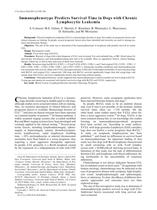
The Kaplan-Meier survival curve is shown in Figure 1.
Based on log-rank test, immunophenotype was signifi-
Table 1. Main hematological features (median) and minimum-maximum intervals (in parenthesis) for each
phenotypic group of CLL.
Group No.
Erythrocyte
(106
/mL)
Hemoglobin
(g/dL)
PCV
(L/L)
Platelets
(103
/mL)
Leukocytes
(103
/mL)
Lymphocytes
(103
/mL)
Neutrophils
(103
/mL)
B-CLL 17 5.50 12.90 0.35 225 48.74 35.98 8.25
(2.57–7.70) (6.00–16.80) (0.20–0.47) (24–576) (13–288) (8–201) (0.74–75.0)
T-CLL 19 5.58 13.20 0.38 405 85.43 75.61 7.45
(2.07–7.84) (5.20–16.80) (0.19–0.51) (50–757) (19–419) (12–380) (3.6–33.0)
Atypical 7 5.66 14.10 0.40 219 73.06 61.30 13.25
CLL (3.1–6.61) (8.40–14.30) (0.30–0.42) (60–280) (27–836) (19–819) (7.0–88.4)
No statistical differences were detected between the groups except for platelets number (P 5 .039).
CLL, chronic lymphocytic leukemia.
102 Comazzi et al
4. cantly associated with survival (P o .001) . Median over-
all survival time was 22 days for atypical CLL, 480 days
for B-CLL, and 930 days for T-CLL. Therapeutic
approach was not associated with survival in either
group (P 5 .71).
Results of Cox’s multivariate analysis are shown in
Table 2. Only immunophenotype was significantly
associated with survival. In particular, dogs with atypi-
cal CLL had approximately 19-fold higher likelihood of
dying compared with dogs with T-CLL, whereas dogs
with B-CLL had 43-fold higher likelihood of dying
compared with dogs with T-CLL. Degree of anemia and
lymphocytosis at diagnosis were not associated with
survival. With regard to B-CLL, young age at diagnosis
was associated with a poor prognosis (P 5 .014, hazard
ratio 5 0.65, CI 0.47–0.92). With regard to T-CLL,
low PCV was associated with shorter survival (P 5 .023,
hazard ratio 5 0.72, CI 0.54–0.96).
Discussion
The distinction between CLL and lymphoma with
blood involvement is challenging. For our study
purposes, we arbitrarily applied the following exclusion
criteria: dogs with circulating immature or large lym-
phoid cells (nuclei 2 RBC in diameter), with moderately
to severely enlarged lymph nodes or with mild lymph
node enlargement with cytology suggestive of a specific
lymphoma subtype.15
Furthermore, dogs with severe
clinical signs were accurately reevaluated to eliminate
occult lymphoma. Use of these criteria allowed exclusion
most cases of stage V lymphoma. Nevertheless, a limited
number of stage V small cell lymphoma (lymphocytic
subtype), cases may have been included in our cases.
However, this subtype occurs quite rarely in dogs,
accounting for o1% of all lymphoma cases.16
According to the REAL/WHO classification system,
the nomenclature is linked to the tissue with the greatest
volume of tumor, meaning that if the neoplasm is mostly
in the bone marrow, it is called leukemia. On the other
hand, if the neoplasm is mostly in the peripheral tissues,
it is called lymphoma. In practice, CLL and low-grade
lymphocytic lymphoma represent different manifesta-
tions of the same disease.17,18
CD34-positive cases were excluded. Although not all
acute lymphoid leukemias are reported to be CD34 pos-
itive,2
cases in which immature or blast cells were the
prevalent population also were excluded, thereby elimi-
nating potentially CD34-negative acute leukemias from
the cases.
CLL is frequent in dogs, but no studies focusing on
prognosis have been published. Some case series have
been reported describing hematological features and
immunophenotype.2–4
CLL rarely has been associated
with severe cytopenias, with no differences between
phenotypes. T-CLL with CD31 CD81 phenotype has
been reported as the most frequent form, representing
73% of CLL, mainly showing LGL morphology (54%).2
In the present work, T-CLL represented only 44% of the
cases, but this finding may have been biased by the inclu-
sion criteria, as some cases were excluded because of lack
of complete clinical data or lymph node cytology for
reevaluation. In agreement with previous studies2,3
our
results confirm that there are no statistical differences
within phenotypic groups with regard to hematological
variables, with the exception of a higher platelet count in
T-CLL. Prolymphocytes rarely are found in canine CLL,
without differences within the phenotypic groups. In
people, prolymphocyte percentage 410% (but o55%)
is considered a poor prognostic factor.19
In the present
study, dogs with high prolymphocyte percentages did not
have a worse prognosis.
According to our results, LGL morphology was a
frequent feature of T-CLL and atypical CLL. However,
atypical CLL were of T lineage, except for 1 case that
coexpressed CD3 and CD21. LGL morphology also
has been reported to be frequent in T-CLL by other
authors,2,3
but no correlation has been found with bio-
logical behavior, although extensive survival studies are
lacking. In our case series, the presence of LGL was not
associated with a specific survival time, and this may be
Fig 1. Kaplan-Meier curves for survival times in dogs with CLL
with different phenotypes. Dotted line indicates atypical CLL; solid
line indicates B-CLL; broken line indicates T-CLL. The difference is
significant, P o .001. CLL, chronic lymphocytic leukemia.
Table 2. Results of Cox’s multivariate analysis for
analyzed variables in all CLL cases.
Variables
No. of
Cases
Hazard
Ratio 95% CI P-Value
Phenotype
B-CLL 17 3.43 1.10–10.68 .034
Atypical-CLL 7 19.17 4.57–80.35 o.001
T-CLL 19 Reference — —
Age 43 0.91 0.78–1.05 .204
PCV 43 0.96 0.90–1.02 .262
Lympho-
cytes (mmc)
43 1.00 0.99–1.01 .695
CI, confidence interval; CLL, chronic lymphocytic leukemia.
P o .05.
103
Immunophenotypes and Survival in Canine CLL
5. attributable to the phenotype, as T-CLL had a longer
survival time in comparison with atypical CLL.
CLL generally is a disease of old animals,20
with a me-
dian age at diagnosis of 10.5 years.1
Our results support
this finding, with a median age at diagnosis of 10 years.
However, 6 dogs (2 with B-CLL, 2 with T-CLL, and 2
with atypical CLL) were younger than 8 years. Among
these 6 dogs, 4 harbored a poor prognosis, surviving o40
days. The 2 dogs with T-CLL showed survival times that
were similar to those of the older dogs. For B-CLL,
young age was statistically associated with shorter sur-
vival, suggesting that leukemia in these dogs may have a
more aggressive behavior. In dogs with B-CLL, young
age therefore may be considered as a possible negative
prognostic factor. Conversely, T-CLL may have a more
homogeneous clinical behavior with respect to age,
whereas the severity of anemia was correlated to a worse
prognosis in T-CLL only.
To the best of our knowledge, there is only 1 published
study11
identifying prognostic factors in dogs with neo-
plastic lymphocytosis. The authors found no difference
in survival with regard to phenotype, but the inclusion
criteria used were very different from those used in the
present study, because no attempt was made to distin-
guish between CLL and stage V lymphoma. The authors
found that small size of cells in B-neoplasms and a lym-
phocyte count o30,000 cells/mL in T-cell neoplasms were
predictive of longer survival time.
Our study shows that T-CLL generally is associated
with very long survival times, although anemia must be
considered as a negative prognostic factor. On the other
hand, atypical CLL (mainly of T lineage) has a very ag-
gressive course and is associated with a short survival
time. The finding of 1 case with biphenotypic CLL that
lived more than 1 year without evident clinical signs re-
mains to be elucidated and the different outcome might
be related to the particular cell type involved in this very
infrequent leukemia.
Short survival times are quite unusual for CLL. CD34
negativity and lack of severe cytopenia allowed excluding
acute lymphoid leukemia. All dogs were accurately eval-
uated (including imaging and cytology) to rule out
presence of masses or visceral involvement. However,
despite our effort to differentiate CLL from stage V lym-
phoma, we cannot be certain that we did not include dogs
with occult small cell lymphoma in this series. Regardless
of the possible misclassification, our results indicate that
a more aggressive course is expected in dogs with he-
matological and clinical signs of CLL showing atypical
phenotypes.
B-CLL may be considered an intermediate phenotype,
with old dogs surviving quite long, and young dogs show-
ing a more aggressive course. These results partially dis-
agree with those of the study of Williams et al,12
which
may be attributable to the different inclusion
criteria. Although no consensus exists on how to differ-
entiate CLL from stage V lymphoma, to the authors’
experience, the distinction between these 2 entities is
relatively easy in most cases, with the exception of
small cell lymphomas. Clinical features and an accurate
evaluation of lymph node cytology may be of use in most
cases to make this distinction. In the present study, we
made an effort to differentiate between stage V lym-
phoma and CLL based on clinical and cytological
presentation.18
Williams and colleagues found that lymphocytosis be-
cause of small cells (according to an arbitrarily fixed cut-
off) were linked to very long survival. In the present
study, the low number of samples did not allow us to dis-
tinguish leukemias based on cellular size. Furthermore,
we found that anemia, but not lymphocytosis, was cor-
related with survival in T-CLL. Both of these findings
disagree with the previously cited study, but the low
number of cases may account for such differences. Pro-
spective studies including more dogs and by similar
inclusion criteria are warranted.
In our study, 2 dogs with B-CLL and 1 with T-CLL
developed high-grade aggressive lymphoma. This partic-
ular condition is similar to the well-known Richter’s
syndrome reported in people, occurring in about 5% of
patients with CLL.21
In most cases, the aggressive lym-
phoma evolves from the original leukemic cell clone.
Conversely, lymphoma may represent a second malig-
nancy.22
Several molecular predictors of Richter’s
transformation have been identified in people.22
To our
knowledge, this evolution has been reported previously
only in 2 of 22 dogs with CLL,1
but immunophenotype
data were not provided. Additional studies will be useful
to clarify analogies between the canine and human
forms.
In human CLL, mutational status of the variable re-
gion of immunoglobulin heavy chain genes, cytogenetic
aberrations, expression level of ZAP-70, CD38, and mol-
ecules regulating the process of angiogenesis have been
evaluated as possible molecular markers having prog-
nostic value.23
Recently, telomere length has been
proposed as an independent predictor of outcome in hu-
man CLL, including overall survival and Richter’s
syndrome transformation.24
Taken together, the above
findings encourage research efforts aimed at the evalua-
tion of the potential utility of these molecular markers
for canine CLL.
Another possible unfavorable evolution of CLL is pro-
gressive active disease, being characterized by massive
lymphoadenopathy and splenomegaly, increased number
of lymphocytes, progressive anemia, and thrombocyto-
penia. Death generally occurs secondary to organ failure.
Indeed, all dogs that were alive at the end of the study
showed stable disease with absent or minimal clinical
signs; stable erythrocytes, lymphocytes, and platelets; no
evidence of massive splenomegaly or lymphadenopathy;
and no evidence of organ failure.
With regard to treatment, chronic immunosuppres-
sion with chlorambucil and prednisone is indicated in
cases with severe hematological abnormalities, bulky
or symptomatic lymphadenopathy, or organomegaly.5,20
Treatment also may be valuable in cases with clinical ev-
idence of disease, including lethargy or anorexia. On the
other hand, asymptomatic dogs are followed and receive
treatment only if disease progression occurs.
Even in the face of treatment, CLL remains incur-
able with standard therapies. Affected dogs eventually
104 Comazzi et al
6. relapse, become refractory to treatment, or undergo dis-
ease transformation. According to the results obtained
here, no benefit in overall survival has been documented
when considering treated and untreated dogs. Because of
the retrospective nature of this study, however, treatment
varied among dogs and different subgroups of CLL,
thereby possibly preventing differences from being iden-
tified. Additionally, whether treatment was indicated or
not was left at the discretion of the referring clinicians,
who were not necessarily oncologists, and incorrect
decisions may have been made, including providing un-
necessary treatments or not treating dogs that actually
may have benefited from chemotherapy. Future prospec-
tive trials should be carried out to better elucidate the
role of chemotherapy in the treatment of specific subsets
of canine CLL and to illustrate which populations of
dogs will require chemotherapy.
In conclusion, this is the first study focused on prog-
nostic factors associated with survival in canine CLL.
Based on our results, determination of flow cytometric
immunophenotype is strongly recommended as an accu-
rate approach to canine lymphocytosis, not only because
it allows the clinician to identify clonal versus nonclonal
expansion in most cases, but also because it permits one
to distinguish T-CLL (showing the classic indolent
behavior and long survival time), atypical CLL (charac-
terized by very aggressive behavior and short survival),
and B-CLL with long survival time, especially in older
dogs. Conversely, young animals have shorter survival
times. Moreover, we described development of high-
grade aggressive lymphoma as a possible negative evolu-
tion of canine CLL, similarly to human Richter’s
syndrome. Additional studies are needed to confirm our
results and to identify possible molecular biomarkers
associated with different survival times or disease
evolution.
Footnotes
a
FACSCalibur, Becton Dickinson, San Jose, CA
b
Serotec, Oxford, UK
c
Pharmingen, BD Bioscience, San Jose, CA
d
SPSS Statistics 17.0, SPSS Company, Wacker Drive, Chicago, IL
Acknowledgments
The authors thank Dr G. Romanelli and the private
practitioners who provided follow-up data. Supported
PUR 2008 (Dr Comazzi), University of Milan, PRIN
2008 (Dr Comazzi).
References
1. Leifer CE, Matus RE. Chronic lymphocytic leukemia in the
dog: 22 cases (1974–1984). J Am Vet Med Assoc 1986;189:214–217.
2. Vernau W, Moore PF. An immunophenotypic study of
canine leukemias and preliminary assessment of clonality by
polymerase chain reaction. Vet Immunol Immunopathol 1999;69:
145–164.
3. Tasca S, Carli E, Caldin M, et al. LS Hematologic abnor-
malities and flow cytometric immunophenotyping results in dogs
with hematopoietic neoplasia: 210 cases (2002–2006). Vet Clin
Pathol 2009;38:2–12.
4. Adam F, Villiers E, Watson S, et al. Clinical pathological
and epidemiological assessment of morphologically and immuno-
logically confirmed canine leukaemia. Vet Comp Oncol 2009;7:
181–195.
5. Harvey JW, Terrell TG, Hyde DM, et al. Well-differentiated
lymphocytic leukemia in a dog: Long-term survival without ther-
apy. Vet Pathol 1981;18:37–47.
6. Fujino Y, Sawamura S, Kurakawa N, et al. Treatment of
chronic lymphocytic leukaemia in three dogs with melphalan and
prednisolone. J Small Anim Pract 2004;45:298–303.
7. Rai KR, Sawitsky A. A review of the prognostic role of cyto-
genetic, phenotypic, morphologic, and immune function
characteristics in chronic lymphocytic leukemia. Blood Cells 1987;
12:327–338.
8. Binet JL, Auquier A, Dighiero G, et al. A new prognostic
classification of chronic lymphocytic leukemia derived from a multi-
variate survival analysis. Cancer 1981;48:198–206.
9. Breen M, Modiano JF. Evolutionarily conserved cytogenetic
changes in hematological malignancies of dogs and humans—man
and his best friend share more than companionship. Chromosome
Res 2008;16:145–154.
10. Hoyer JD, Ross CW, Li CY, et al. True T-cell chronic
lymphocytic leukemia: A morphologic and immunophenotypic
study of 25 cases. Blood 1995;86:1163–1169.
11. Workman HC, Vernau W. Chronic lymphocytic leukemia in
dogs and cats: The veterinary perspective. Vet Clin North Am Small
Anim Pract 2003;33:1379–1399.
12. Williams MJ, Avery AC, Lana SE, et al. Canine lympho
proliferative disease characterized by lymphocytosis: Immuno-
phenotypic markers of prognosis. J Vet Intern Med 2008;22:596–
601.
13. Botsch V, Küchenhoff H, Hartmann K, et al. Retrospective
study of 871 dogs with thrombocytopenia. Vet Rec 2009;164:647–
651.
14. Comazzi S, Gelain ME, Spagnolo V, et al. Flow cytometric
patterns in blood from dogs with non-neoplastic and neoplastic he-
matologic diseases using double labeling for CD18 and CD45. Vet
Clin Pathol 2006;35:47–54.
15. Fournel-Fleury C, Magnol JP, Bricaire P, et al. Cytohisto-
logical and immunological classification of canine malignant
lymphomas: Comparison with human non-Hodgkin’s lymphomas.
J Comp Pathol 1997;117:35–59.
16. Ponce F, Marchal JP, Turinelli V, et al. A morphological
study of 608 cases of canine malignant lymphoma in France with a
focus on comparative similarities between canine and human lym-
phoma morphology. Vet Pathol 2010;47:414–433.
17. Jacobs RM, Messick JB, Valli VE. Tumors of the hemolym-
phatic system. In: Meuten DJ., ed. Tumors in Domestic Animals,
4th ed. Ames, IA: Iowa State Press; 2002:177–180.
18. Vail DM, Young KM. Haematopoietic tumors. In: Withrow
SJ, Vail DM, eds. Withrow and Mac Ewen’s Small Animal Clinical
Oncology. St Louis, MO: Saunders; 2007:722–725.
19. Vallespı́ T, Montserrat E, Sanz MA. Chronic lymphocytic leu-
kaemia: Prognostic value of lymphocyte morphological subtypes. A
multivariate survival analysis in 146 patients. Br J Haematol
1991;77:478–485.
20. Helfand SC, Modiano JF. Chronic lymphocytic leukemia.
In: Feldman BF, Zinkl JG, Jain NC, eds. Schalm’s Veterinary He-
matology, 5th ed. Baltimore, MD: Lippincott Williams Wilkins;
2000:638–641.
21. Tsimberidou AM, Keating MJ. Richter syndrome:
Biology, incidence, and therapeutic strategies. Cancer 2005;103:
216–228.
105
Immunophenotypes and Survival in Canine CLL
7. 22. Rossi D, Gaidano G. Richter syndrome: Molecular insights
and clinical perspectives. Hematol Oncol 2009;27:1–10.
23. Vroblová V, Smolej L, Vrbacký F, et al. Biological prognos-
tic markers in chronic lymphocytic leukemia. Acta Medica (Hradec
Kralove) 2009;52:3–8.
24. Rossi D, Lobetti Bodoni C, Genuardi E, et al. Telomere
length is an independent predictor of survival, treatment require-
ment and Richter’s syndrome transformation in chronic
lymphocytic leukemia. Leukemia 2009;23:1062–1072.
106 Comazzi et al