1. Proceedings of the 1
st
ITOW
Newport, Rhode Island, USA
October 25-26, 2013
A STATISTICAL SHAPE MODEL OF THE THUMB CARPOMETACARPAL JOINT
M.T.Y.Schneider1
, J. Crisco2
, A. C. Weiss2
, A. L. Ladd3
, P. Nielsen1,4
, T. Besier1,4
, J. Zhang1
1) Auckland Bioengineering Institute, The University of Auckland, Auckland, New Zealand
2) Department of Orthopaedics, Brown University, RI, USA
3) Department of Orthopaedic Surgery, Stanford, Stanford University, CA, USA
4) Department of Engineering Science, The University of Auckland, Auckland, New Zealand
INTRODUCTION. The first carpometacarpal (CMC) joint performs a range of dexterous movements, all of
which are influenced by the morphology and articulation between the first metacarpal and trapezium bones.
However, the CMC joint is highly susceptible to osteoarthritis, which is more prevalent with age and 5-7 times
more prevalent in women than in men. Here we present a statistical shape model of the CMC joint to investigate
age and sex differences in CMC joint morphology.
METHODS. A training set of 50 CMC joints were manually segmented from CT images of the hand with a
resolution of 0.4x0.4x0.625mm (age range: 18 yrs to 67 yrs; 24 females and 26 males). A template mesh
consisting of parametric surface elements was created for a single segmentation and then fitted to the entire
training set (n=50), resulting in a set of correspondent meshes of the metacarpal and trapezium. Principal
Component Analysis was performed on these meshes to determine significant modes of shape variation. We
then performed linear regression and one-way ANOVA on the mode scores of the meshes against age and sex.
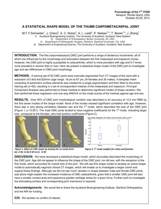
RESULTS. Over 95% of CMC joint morphological variation was described to an accuracy of ~0.2 mm RMS by
the first seven modes of the shape model. None of the modes showed significant correlation with age. However,
there was a very strong correlation between sex and the 1
st
mode, which described the size of the CMC joint
(Figure 1, p <0.001). The male CMC joints tended to have negative coefficients for the 1
st
mode, indicating larger
size, compared to the females, who had positive coefficients (Figure 2).
Figure 1. Effect on CMC shape by varying the 1st mode from: Figure 2. 1
st
mode weights for males and females
(a) -2 SD, to (b) 0 SD to (c) +2 SD
DISCUSSION. We have developed a statistical shape model, which accurately described the morphology of
the CMC joint. Age did not appear to influence the shape of the CMC joint, nor did sex, with the exception of the
first mode, which accounted for overall size of the joint. We will use this shape model to develop an active shape
model to automatically segment clinical CT images, which will enable us to investigate a larger cohort and
support these findings. Although we did not see much variation in shape between male and female CMC joints,
size alone might explain the increased incidence of CMC osteoarthritis, given that a smaller CMC joint will also
have a smaller contact area and experience greater cartilage stress for a given force. Further work to investigate
the articulating surface and corresponding joint mechanics is required.
Acknowledgements. We would like to thank the Auckland Bioengineering Institute, Stanford Orthopaedics,
and the NIH for funding.
COI. We declare no conflict of interest.
A B C