exhuma plot and synopsis from the exhuma movie.pptx
Microscopy Factsheet.pdf
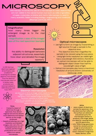
1. Microscopy
Microscopy involves the use of a microscope to view objects that
are not visible to the naked human eye such as cells organelles,
viruses, molecules, proteins and more. Microscopy is used in
many scientific fields including biology, engineering and medicine.
(Shampo and Kyle, 1997)
Magnification
how many times bigger the
enlarged image is to the real
sample
Magnification = size of the image/
size of the real specimen (sample)
Resolution
the ability to distinguish between
adjacent structures, essentially
how clear and detailed the image
appears.
use light to form an image by transmitting a
light source through a sample to the
objective lens.
The objective lens then magnifies the
sample to be viewed using the eye piece.
Using light limits resolution as visible light
has a wavelength 500-600nm, therefore
an optical microscope will not be able to
distinguish object closer than half the
wavelength value of light.
Optical microscopes also have a maximum
resolution of around 200nm. (University of
Edinburgh, 2018)
Optical microscopes
In 1931 German
electrical engineer
Ernst Ruska was
credited for
inventing the earliest
electron microscope.
Thus the electron
microscope went on
to being mass-
produced and
commercialised in
1939 (Shampo and
Kyle, 1997)
transmit a beam of
electrons through a
specimen, thus denser
regions/ structures absorb
more electrons and appear
darker on the image
produced.
Despite producing very high
resolution and detailed
images, only very thin, non-
living specimens can be
viewed, and the image
produced is not in colour
unlike in an optical
microscope.
TEMs
use electrons to form an image, which results in a much
higher resolution than optical microscopes.
This gives a more detailed image as they have a
maximum resolution of around 0.2nm.
Electron microscopes can be further categorised into
Transmission electron microscopes (TEMs) or Scanning
electron microscopes (SEMs).
Electron microscopes
scan a beam of electrons
across the specimen, thus
the electrons that reflect off
the surface are detected
and picked up by the
microscope’s sensor to
produce a 3D image. SEMs
are ideal for viewing thicker
and 3D specimens.
Like TEMs living specimens
cannot be viewed and the
image is not produced in
colour plus they produce a
lower resolution image than
TEMs. (Ilitchev, 2019)
SEMs