The document outlines the qualifications and educational background of Elham Ali Ahmed Ali, including degrees in nursing and diplomas in therapeutic nutrition, medical quality, and infection control. It then provides an outline for a presentation on heart attacks, defining them, listing symptoms and risk factors, describing types and complications, and discussing diagnosis and treatment approaches. Key points covered include definitions of STEMI and NSTEMI heart attacks based on ECG patterns and damage levels, as well as risk factors such as high cholesterol, smoking, diabetes, and family history. Complications discussed relate to heart and psychological damage.
2. 2
| P a g e
Prepared by...
Elham Ali Ahmed Ali
Holds a Bachelor's degree
In Nursing,
Ain Shams University,
Holds a diploma in therapeutic nutrition
Approved by the Arab Studies Center
And
Holds a diploma in a medical quality
Approved by the Arab Studies Center
And
Holds a diploma in an infection control
Approved by the Arab Studies Center and accredited by
the foreign consulate
And
Holds a Mini master’s in Medical administration
Approved by the Arab Studies Center and accredited by
the foreign consulate.
3. 3
| P a g e
Out lines
subject
Introduction
Definition
Symptoms and signs
Incidence
Etiology
Types of heart attack
Types of heart attack related to ECG , Radiography
and Bio marks
Types of heart attack related to causes
The risk factors
Complications
Complication related to damage in heart during
heart attack
Complication related to psychological condition
Complication related to stop medication
Diagnosis
Treatments
Immediate treatment
Medications for heart attacks
Surgical treatment
Prevention
Rehabilitation
Nurse care plan
Reference
5. 5
| P a g e
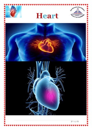
Introduction
A heart attack occurs when
the flow of blood to the heart
is blocked.
The blockage is most often
a buildup of fat, cholesterol and other substances,
which form a plaque in the arteries that feed the heart
(coronary arteries( .
a plaque can rupture and form a clot .
Coronary artery is carry blood, oxygen, and nutrients
to the heart .
When coronary artery become blocked .
The heart doesn’t get enough blood supply.
The interrupted blood flow can damage or destroy part
of the heart muscle.
A heart attack, also called a myocardial infarction, can
be fatal, but treatment has improved dramatically over
the years.
6. 6
| P a g e
Definition
A heart attack , or myocardial infarction ( MI ) :-
- Is permanent damage to the heart muscle .
"myo" means muscle ,
"cardial" refer to the heart
"infraction" means death of tissue due to lack of blood
supply .
Symptoms and signs
7. 7
| P a g e
chest pain, and pain that travels to one or both arms
- pain in the center of the chest.
- This chest discomfort may be described as a
pressure or tightness rather than a sharp pain.
- Some people describe feeling pain in one or both
arms or their back, neck, or jaw.
nausea
shortness of breath
anxiety
lightheadedness
breaking out in a cold sweat
sudden and extreme fatigue
rapid heartbeat
sweatiness (without exercising)
dizziness or faintness
leg swelling
Women are more likely to have different
symptoms.
8. 8
| P a g e
Women are more likely to experience symptoms
such as:
“atypical” chest pain — not the classic sensation of
chest pressure
shortness of breath
nausea
vomiting
back pain
jaw pain
Incidence
9. 9
| P a g e
According to the latest WHO data published in 2018
Coronary Heart Disease Deaths in Egypt reached
163,171 or 29.38% of total deaths.
The age adjusted Death Rate is 271.69 per 100,000
of population ranks Egypt #15 in the world .
Heart Attack Statistics for 2021
One in five heart attacks is silent.
CVDs represent 40% of total deaths in Egypt.
With the coronavirus pandemic, people’s chances of
dying from heart attacks have doubled.
The leading cause of death in 2020 in the US was
heart disease.
Annually, 805,000 people in the US have a heart
attack.
Coronary heart disease affects 1 in 13 White men.
High blood pressure causes 47% of coronary heart
diseases.
26% of women die within a year of a heart attack.
10. 10
| P a g e
People who have suffered heart failure live ten years
less than those who haven’t.
7% of the hospital visits related to snow shoveling
are due to heart problems, mainly heart attacks
Cardiovascular diseases (CVD) are amongst the top
10 leading causes of death and disability in the
world.
Etiology
A heart attack occurs when one or more of a
coronary arteries becomes blocked
might be a complete or partial blockage of the coronary
artery.
11. 11
| P a g e
A complete blockage means you've had an ST
elevation myocardial infarction (STEMI) .
A partial blockage means you've had a non-ST
elevation myocardial infarction (NSTEMI).
A heart attack occurs when a coronary
arteries becomes a spasm .
a spasm of a coronary artery that shuts down blood flow
to part of the heart muscle.
12. 12
| P a g e
A heart attack occurs as result of complications
of procedure or operation .
during the diagnostic cardiac catheterization
(3–5 percent of people) .
Blood clot or damage to the blood vessel that the
catheter is put into .
Leading to Sudden blockage of a coronary artery.
13. 13
| P a g e
Allergy reaction from dye leading to spasm to
coronary artery .
A heart attack occurs as result of complications
during or post operation
Post operation of Coronary artery bypass graft
Heart attack
14. 14
| P a g e
The main reason why people undergo coronary
artery bypass surgery is to reduce their risk of heart
attack and stroke.
But in some cases, stroke and heart attack are a
serious complication of the surgery.
According to a 2014 study, neurological dysfunction
after coronary bypass surgery may include stroke in
up to five percent of patients.
A heart attack can occur if lifestyles changes have
not been shifted to include lowering of artery-
clogging cholesterol.
Blood clot
Coronary artery bypass surgery is designed to
bypass an existing blood clot to allow oxygen and
blood flow to the heart.
15. 15
| P a g e
However, if a person’s lifestyle habits remain the
same, a blood clot may form in the newly
constructed path to the heart.
Infection
Site infections occur at the location of the surgical
puncture .
Infections that occur near the bloodstream or in
blood vessels .
A heart attack occurs as result of angioplasty
and stent
After angioplasty, the treated coronary artery can
become narrowed or blocked again, often within 6
months of angioplasty.
16. 16
| P a g e
( This is called restenosis )
When a stent (small mesh tube) isn't used during
angioplasty, 4 out of 10 people have restenosis.
The growth of scar tissue in and around a stent also
can cause restenosis.
When a stent is used, 2 out of 10 people have
restenosis
Stents coated with medicine reduce the growth of
scar tissue around the stent and lower the chance of
restenosis even more.
When these stents are used, about 1 in 10 people has
restenosis.
Infection with COVID-19 also may damage your
heart in ways that result in a heart attack.
Types of heart attack
Types of heart attack related to ECG ,
Radiography and Bio marks
The three types of heart attacks are:
ST segment elevation myocardial infarction
(STEMI( .
17. 17
| P a g e
non-ST segment elevation myocardial infarction
(NSTEMI( .
coronary spasm, or unstable angina .
ST segment elevation myocardial infarction
(STEMI( :-
18. 18
| P a g e
A STEMI occurs when a coronary artery becomes
completely blocked and a large portion of the muscle
stops receiving blood .
It’s a serious heart attack that can cause significant
damage.
“ST segment” refers to the pattern that appears on an
electrocardiogram, which is a display of your
heartbeat.
Only a STEMI will show elevated segments.
STEMI: The classic or major heart attack
When most people think of a heart attack, they often
think of a STEMI.
NSTEMI heart attacks
Unlike in a STEMI, the affected coronary artery is
only partially blocked in a NSTEMI .
A NSTEMI sometimes show depression in the ST
segment on the electrocardiogram , sometimes don't
show any change in the ST segment on the
electrocardiogram .
19. 19
| P a g e
A coronary angiography will show the degree to
which the artery is blocked.
A blood test will also show elevated troponin protein
levels .
Normal range: below 0.04 ng/ml
Probable heart attack: above 0.40 ng/ml
While there may be less heart damage ,
an NSTEMI is still a serious condition.
Coronary spasm, or unstable angina .
CAS, silent heart attack, or heart attack without
blockage
The coronary artery spasm is also known as a
coronary spasm, unstable angina, or silent heart
attack.
The symptoms, which can be the same as a STEMI
heart attack, may be mistaken for muscle pain,
indigestion, and more.
20. 20
| P a g e
It occurs when one of the heart’s arteries tightens so
much that blood flow stops or becomes drastically
reduced.
Only imaging and blood test results can tell your
doctor if you’ve had a silent heart attack.
There is no permanent damage during a coronary
artery spasm.
While silent heart attacks aren’t as serious, they do
increase your risk of another heart attack or one
that may be more serious.
The risk factors :-
Types of heart attack related to causes :-
Five types of heart attack
Type 1
Spontaneous Myocardial Infarction
Atherosclerotic plaque rupture or intraluminal thrombus
in one or more of the coronary arteries
NOTE
Both STEMI and NSTEMI heart attacks can cause enough
damage to be considered major heart attacks.
21. 21
| P a g e
Type 2
Myocardial Infarction Secondary to an Ischemic
Imbalance
Condition other than CAD contributes to an imbalance
between myocardial oxygen supply
Type 3
Cardiac Death Due to Myocardial Infarction
-
Suffer cardiac death with symptoms suggestive of
myocardial ischemia without elevated biomarkers
Type 4
Myocardial Infarction Associated With Revascularization
Procedure
a: Related to PCI
b: Related to Stent Thrombosis
Type 5
22. 22
| P a g e
Myocardial Infarction Related to CABG Procedure
The risk factors
high levels of LDL (“bad”) cholesterol .
- Cholesterol is a waxy, fat-like substance made
by the liver or found in certain foods.
- Extra cholesterol can build up in artery walls,
causing them to become narrow and decrease
the blood flow to the heart, brain, and other
parts of the body.
23. 23
| P a g e
high blood pressure .
- High blood pressure occurs when the pressure
of the blood in your arteries and other blood
vessels is too high and can cause the arteries to
stiffen.
diabetes.
- Not producing enough of a hormone secreted by
your pancreas (insulin) or not responding to
insulin properly causes your body's blood sugar
levels to rise, increasing your risk of a heart
attack.
obesity .
- Obesity is linked with high blood cholesterol
levels, high triglyceride levels, high blood
pressure and diabetes.
- Losing just 10% of your body weight can lower
this risk.
Metabolic syndrome.
24. 24
| P a g e
- This syndrome occurs when you have obesity,
high blood pressure and high blood sugar.
- Having metabolic syndrome makes you twice as
likely to develop heart disease than if you don't
have it .
alcohol consumption
- Drinking too much alcohol can raise your blood
pressure and produce an irregular heartbeat.
Substance use disorders,
- including marijuana and cocaine use, may also
be factors.
- Younger people who had heart attacks were
more likely to report overusing these substances
smoking .
- because it increases the blood pressure and risk
for clots by reducing oxygen cells within the
bloodstream.
- This means that the heart works harder to
pump blood and has fewer healthy oxygen cells
to maintain optimal performance.
advanced age .
25. 25
| P a g e
- increases after age 65.
- This is due to age-related changes that can
occur in the heart, including high blood
pressure (hypertension) and hardening of the
arteries (arteriosclerosis( .
- Older adults may also be at a higher risk for
cognitive issues and reduced functional
movements , so , decreasing physical activity .
physical activity is help strengthen the heart
muscle and protect it from future damage.
gender :-
- For example,
- until age 55
- men are at a higher risk of heart attack.
- After menopause, though, women tend to have
similar risks as men.
- Also, men tend to have problems in the heart’s
larger arteries, while women often experience
blockage in the smaller arteries of the heart.
Heredity
- Children of parents with heart disease are more
likely to develop heart disease themselves.
Lack of physical activity.
- Being inactive contributes to high blood
cholesterol levels and obesity.
26. 26
| P a g e
- People who exercise regularly have better
heart health, including lower blood pressure .
Stress.
- You might respond to stress in ways that can
increase your risk of a heart attack.
Birth control pills
- A European study has raised new concerns
about the safety of women's long-term use of
birth control pills, as it indicated an increased
risk of heart attacks or strokes.
- The researchers told a meeting of the American
Heart Association that women who use oral
contraceptives are more likely than their
counterparts who do not take such pills of
deposits on the walls of the arteries.
- "The main concern is that if a woman has high
levels of plaque, she may develop a clot of one
of these plaques and have a stroke, heart attack
or sudden death from heart disease," Dr. Ernst
Ritzschell of the University of Ghent in
Belgium, who led the research, told reporters.
Illicit drug use.
- Using stimulant drugs, such as cocaine or
amphetamines,
27. 27
| P a g e
- can trigger a spasm of your coronary arteries
that can cause a heart attack.
A history of preeclampsia.
- This condition causes high blood pressure
during pregnancy and increases the lifetime
risk of heart disease.
An autoimmune condition.
- Having a condition such as rheumatoid
arthritis or lupus can increase your risk of a
heart attack.
Coronary artery spasm risk factors
The factors above also put you at risk of coronary spasm.
But having other conditions can increase your risk of
coronary artery spasms as well.
These conditions include:
Migraines .
excess thyroid hormone .
chronic allergy conditions .
excessive alcohol consumption .
low magnesium levels .
28. 28
| P a g e
taking drugs for chemotherapy .
Complications
29. 29
| P a g e
Complication related to damage in heart during
heart attack :-
Abnormal heart rhythms (arrhythmias).
- Electrical "short circuits" can develop, resulting
in abnormal heart rhythms, some of which can
be serious, and may lead to death.
Heart failure.
- A heart attack might damage so much heart
tissue that the remaining heart muscle can't
pump enough blood out of your heart.
- Heart failure can be temporary, or it can be a
chronic condition resulting from extensive and
permanent damage to your heart.
Sudden cardiac arrest.
30. 30
| P a g e
- Without warning, your heart stops due to an
electrical disturbance that causes an
abnormal heart rhythm (arrhythmia).
- Heart attacks increase the risk of sudden
cardiac arrest, which can cause death without
immediate treatment.
Complication related to psychological condition:-
31. 31
| P a g e
feel very fatigued its normal
feel weak and mentally exhausted.
It’s common to have mental health complication
after a heart attack. These can last between
2 and 6 months.
Some mental health-related symptoms include:
- anger
- irritability
- fear
- insomnia and daytime fatigue
- sadness
- feelings of guilt and hopelessness
- loss of interest in hobbies
complication related to stopped the medication :-
a stent closes, you’ll need surgery to open the artery
up again.
32. 32
| P a g e
Repeated Heart attack
Diagnosis
Electrocardiogram
33. 33
| P a g e
An electrocardiogram (ECG) is an important test in
suspected heart attacks.
It should be done within 10 minutes of being
admitted to hospital.
An ECG is important because:
it helps confirm the diagnosis of a heart attack
it helps determine what type of heart attack you
have had, which will help determine the most
effective treatment
The leads affected determine the site of The
infarct
Inferior II, III, aVF ƒ
Anteroseptal V1 -V4 ƒ
Anterolateral V4 -V6, I, aVL ƒ
Posterior Tall wide R and depression ST in V1 and
V2
A myocardial infarction occurs when blood flow to
the heart muscle stops
or is suddenly decreased long enough to cause cell
death Infarcted cells
34. 34
| P a g e
are without function and cannot respond to
electrical stimulus or provide any mechanical
function
EKG Changes:
ST-segment elevation,
T-wave inversion,
abnormal Q waves
Abnormal Q Waves
An abnormal Q wave
indicates the presence of dead myocardial tissue and
subsequently a loss of electrical activity
Pathological Q waves
35. 35
| P a g e
represent transmural MI and are most commonly
seen with STEMI
Sometimes occurs within hours of onset of chest pain
More commonly appears 1-3 days after the event
Most Post MI Q waves are permanent
Pathologic Q Waves
1/3 or greater than the amplitude of an R wave R Wave 5
mm Q Wave 2.5 mm And/or greater than 40 ms (0.04
secs)
Thrombus partially or intermittently occludes
the coronary artery
Diagnostic Findings:
ST-segment depression
or T-wave inversion
36. 36
| P a g e
Normal Cardiac Markers
Myocardial ischemia on ECG
T wave peaking or inversion
watch out for pseudo-normal (look at pervious ECGs)
37. 37
| P a g e
ST elevation
(1mm) indicating transmural (subepi) injury (hrs - days)
ST depression: Subendo (not transmural) injury
Significant Q wave
1square (0.04) wider and/or deeper than 25% of the R
wave
38. 38
| P a g e
Chest X-ray
A chest X-ray can be useful if diagnosis of a heart
attack is uncertain
and there are other possible causes of your
symptoms, such as a pocket of air trapped between
the layers of your lungs (pneumothorax.)
used to check whether complications have happened
because of the heart attack, such as a build-up of
fluid inside your lungs (pulmonary oedema).
39. 39
| P a g e
Echocardiogram
An echocardiogram is a type of scan that uses sound
waves to build a picture of the inside of your heart.
This can be useful to identify exactly which areas of
the heart have been damaged and how this damage
has affected your heart's function.
Coronary angiography
Coronary angiography can help determine whether
there is a blockage or narrowing in the coronary
arteries and, if so, to locate the exact place of the
blockage or narrowing.
The test involves inserting a thin tube (catheter),
into one of the blood vessels in your groin or arm.
The catheter is guided into your coronary arteries
using X-rays.
Blood test
Cardiac Enzymes
A.K.A. ACP (Acute Cardiac Profile)
40. 40
| P a g e
Normal Ranges:
CPK: 39 – 308
CK-MB: 0 – 3.60
Troponin I : 0 – 0.099
Ordered for patients c/o chest pain and suspected AMI
CE’s are drawn in sets of three 6 to 8 hours apart
Sometimes initial results are negative
CK-MB & Troponins are released within hours of a
cardiac event
CK-MB or CPK-MB Creatine Phosphokinase Rise
within 4-6 hours after an AMI
Peak @ 18 – 24 hours (6x > normal)
Return to normal within 3 – 4 days
Troponin I Rise within 3 hours of an AMI
Preferred cardiac enzyme in diagnosis of an AMI
ABGs
41. 41
| P a g e
An arterial blood gas (ABG) test measures oxygen
and carbon dioxide levels in your blood.
It also measures your body’s acid-base (pH) level,
which is usually in balance when you’re healthy.
Urea and Electrolytes
which measures the levels of sodium, potassium and
other important chemicals in your blood such as
magnesium and calcium.
These chemicals are important for the overall
function of your heart and also help
assess kidney function. Imbalances in the blood can
be linked to medication that you may be taking.
coagulation studies (PT, a PTT, clotting times).
his measures how quickly your blood clots. This is
important if you take blood thinning medication
Total cholesterol.
This is the amount of your blood's cholesterol
content. A high level can increase your risk of heart
disease.
42. 42
| P a g e
Ideally, your total cholesterol
should be below 200 milligrams per deciliter
(mg/dL) or 5.2 millimoles per liter (mmol/L).
Low-density lipoprotein (LDL) cholesterol.
Too much LDL cholesterol in your blood causes
plaque to buildup in your arteries, which reduces
blood flow.
These plaque deposits sometimes rupture and lead
to major heart and blood vessel problems.
Your LDL cholesterol level should be less than 130
mg/dL (3.4
High-density lipoprotein (HDL) cholesterol.
it helps carry away LDL ("bad") cholesterol, keeping
arteries open and your blood flowing more freely.
If you're a man, your HDL cholesterol level should
be over 40 mg/dL (1.0 mmol/L). Women should aim
for an HDL over 50 mg/dL (1.3 mmol/L).
Natriuretic peptides
43. 43
| P a g e
these show the level of a hormone in your blood
which if elevated can be a sign of heart failure
Brain natriuretic peptide, also called B-type
natriuretic peptide (BNP), is a protein that your
heart and blood vessels make.
BNP helps your body eliminate fluids, relaxes blood
vessels and moves sodium into your urine.
When your heart is damaged, your body secretes
high levels of BNP into your bloodstream to try to
ease the strain on your heart.
One of the most important uses of BNP is to try to
determine whether shortness of breath is due to
heart failure
44. 44
| P a g e
Treatments
Immediate treatment
If the doctor suspects a heart attack, may be treated
immediately with:
aspirin , ticagrelor (Brilinta) or clopidogrel
(Plavix) to prevent blood clotting .
nitroglycerin to relieve chest pain and improve
blood flow .
oxygen therapy .
After the doctor confirms the heart attack,
they will prescribe medications.
They may recommend surgery, if needed.
45. 45
| P a g e
Medications for heart attacks
These drugs may include:
clot busters to dissolve clots that are blocking
arteries
ACE inhibitors to help reduce the heart’s
workload and control blood pressure
blood thinners to prevent blood clots
statins
to help lower LDL cholesterol
HMG-CoA Reductase Inhibitors
“Statins”
Decreases the rate of cholesterol production Liver
needs HMG-CoA reductase to make cholesterol
When less cholesterol is produced liver needs to
“recycle” LDL from the blood circulation
46. 46
| P a g e
the first way to block cholesterol synthesis is by
interrupting the conversion of HMG CoA to
mevalonate (so that mevalonate cannot generate
cholesterol).
In order for HMG CoA to become mevalonate,
the reaction must be catalyzed by the enzyme HMG
CoA reductase.
If this enzyme is blocked, mevalonate cannot be
generated and cholesterol cannot be synthesized
Receptor-mediated endocytosis of LDL, whereby the
liver cells, or hepatocytes, attach cholesterol via LDL
receptors on their membranes and remove it from
circulation
47. 47
| P a g e
beta-blockers,
which decrease the heart rate and cardiac output
Beta-Adrenergic Blockers Negative Inotropic and
Chronotropic Effects
Reduces myocardial contractility and heart rate
resulting in decreased demand for oxygen
Calcium Blockers
Calcium Channel Blockers Non-dihydropyridines
Negative Inotropic and Chronotropic Effects
Reduces myocardial contractility and heart rate
resulting in decreased demand for oxygen
Also work to decrease workload of the heart by
coronary arterioles
Nitroglycerin
Promotes decrease O2 demand by
dilating veins
- which decreases venous return to the heart thus
decreasing ventricular filling
- (decreases preload)
48. 48
| P a g e
- Decrease in wall tension decreases O2 demand
Morphine Sulfate
Decreases pain and anxiety
decreasing heart rate and oxygen consumption
Reduces cardiac preload and afterload
decreasing workload of the heart Relaxes
bronchioles
increasing oxygenation
Antiplatelet Agents
Aspirin (acetylsalicylic acid):
Low dose, long- term
use irreversibly blocks formation of thromboxane A2
in platelets,
producing an inhibitory effect on platelet
aggregation
Plavix (clopidogrel):
Inhibits 1st and 2nd Phases
ADP-induced effects of platelet aggregation
49. 49
| P a g e
Glycoprotein IIb/IIIa inhibitors
The glycoprotein IIb/IIIa inhibitors such as
abciximab (Reopro) and eptifibatide (Integrilin)
prevent aggregation of platelets by inhibiting the
glycoprotein receptors on the platelets.
They are the most potent anti-platelet agents,
approximately 9 times
more potent than aspirin, and three times more
potent than the thienopyridines.
The glycoprotein II b/III a inhibitors are also the
most expensive anti-platelet agents.
They usually are given along with aspirin and
heparin.
They are quick acting; their maximal anti-platelet
effects are achieved within minutes of infusion.
These inhibitors have become important in the
treatment of patients with heart attacks, patients
with unstable angina, and patients undergoing
PTCA with or without stenting.
Numerous studies have shown that
glycoprotein II b/III a inhibitors:
Decrease the size of the blood clot blocking the
coronary arteries, thus improving blood flow,
50. 50
| P a g e
limiting damage to heart muscle, and improving
survival among patients with heart attacks
Decrease the incidence of heart attacks and improve
survival among patients with unstable angina
Prevent the formation of blood clots inside
coronary stents and in coronary arteries
unblocked by PTCA, thus decreasing the incidence
of heart attacks and improving survival,
specifically, when given intravenously at the time
of PTCA and stenting and followed by oral aspirin
and clopidogrel
Surgical treatment for major heart attack
Grafting:
A blocked artery may also be treated with coronary
artery bypass grafting, sometimes referred to as
bypass surgery.
In this procedure, a blood vessel is taken from
elsewhere in the body and attached, or grafted, onto
the blocked artery.
With this, blood flow can be rerouted around the
blockage.
51. 51
| P a g e
Stent:
A stent is a tiny, flexible, wire mesh tube that is
placed at the site of the blockage .
This opens up your blocked artery for normal blood
flow and increases blood flow to the heart muscle.
The plaque is pressed against the wall of the artery
and the stent allows blood to pass through it .
A stent is used to reduce the chances of a heart
attack .
The stent is left in place permanently to improve
your condition.
Stents reduce your overall risk of experiencing
narrowing of that same artery.
52. 52
| P a g e
Prevention
Lifestyle changes
A heart-healthy lifestyle can complement a medical
treatment plan for heart disease.
Exercise
exercise help increase the amount of oxygen circulating in
your body and also strengthen the heart’s ability to pump
it through the bloodstream to the rest of your body.
Spend at least 150 minutes (2.5 hours) per week doing
moderate-intensity exercise, such as
- brisk walking
- swimming
- bicycling
- jogging or running
53. 53
| P a g e
As an added bonus, regular aerobic exercise also helps
reduce:
- high blood pressure
- stress
- cholesterol
Diet
Follow a heart-healthy diet
A low-fat diet
- Avoid trans fats and saturated fats whenever
possible.
- These fats directly contribute to plaque
formation in the arteries.
- When your arteries become clogged, blood can
no longer flow to the heart, resulting in a heart
attack
low-calorie diet
- Eating too many calories and having
overweight can also strain your heart.
limit sodium
54. 54
| P a g e
- Reducing your daily sodium intake to decrease
blood pressure and the overall strain on your
heart.
while focusing on potassium-rich sources of fruits and
vegetables, whole grains, lean proteins (such as fish),
beans, lentils, nuts and plant oils ( Such as olive oil )
Avoid processed foods, and beverages with added
sugars.
avoid canned versions
One helpful eating plan is called the dietary
approaches to stop hypertension, or DASH.
The Mediterranean diet is similar to DASH in that they
both emphasize plant-based foods.
Stop smoking
Stop alcohol consumption
Take your medications consistently.
NOTE
Research suggests that a plant-based diet may decrease
inflammation and oxidative stress, which contributes
to heart failure, also decrease heart disease severity .
55. 55
| P a g e
Sleep
Get 7 to 9 hours of sleep each night.
Reduce stress.
Get regular checkups and blood work done.
Rehabilitation
the doctor may recommend cardiac rehabilitation
(rehab).
Cardiac rehab is a medically supervised program
that helps improve the health and well-being of
people who have heart problems.
Cardiac rehab includes exercise training, education
on heart healthy living, and counseling to reduce
stress and help you return to an active life.
the doctor can tell you where to find a cardiac rehab
program near your home.
56. 56
| P a g e
Nurse care plan
Nursing diagnosis:
Pain, Acute
May be related to
Tissue ischemia (coronary artery occlusion)
Possibly evidenced by
Subjective
o Reports of chest pain with/without radiation
Objective
o Restlessness, changes in level of consciousness
o Changes in pulse, BP
Demonstrates the Levine’s sign
Observed evidence of pain
Facial mask; sleep disturbance (eyes lack luster,
beaten look, fixed or scattered movement, grimace)
Expressive behavior (e.g., restlessness, moaning,
crying, vigilance, irritability, sighing)
Distraction behavior (e.g., pacing, seeking out other
people and/or activities, repetitive activities)
57. 57
| P a g e
Change in muscle tone (may span from listless
[flaccid] to rigid)
Diaphoresis; change in blood pressure/heart
rate/respiratory rate; pupillary dilation
Desired outcomes/evaluation criteria—patient
will:
Report pain is relieved/ controlled.
Follow prescribed pharmacological regimen.
Verbalize non-pharmacologic methods that provide
relief.
Demonstrate use of relaxation skills and diversional
activities, as indicated, for individual situation
58. 58
| P a g e
Nursing Interventions Rationale
Pain Management
Independent
Monitor/document
characteristics of pain, noting
verbal reports, nonverbal cues
(e.g., moaning, crying,
restlessness, diaphoresis,
clutching chest, rapid
breathing), and hemodynamic
response (BP/heart rate
changes).
Variation of appearance and
behavior of patients in pain
may present a challenge in
assessment. Most patients with
an acute MI appear ill,
distracted, and focused on pain.
Verbal history and deeper
investigation of precipitating
factors should be postponed
until pain is relieved.
Respirations may be increased
as a result of pain and
associated anxiety; release of
stress-induced catecholamines
increases heart rate and BP.
Obtain full description of pain
from patient including location,
intensity (0–10), duration,
characteristics(dull/crushing),
and radiation.
Assist patient to quantify pain
by comparing it to other
experiences.
Pain is a subjective experience
and must be described by
patient. Provides baseline for
comparison to aid in
determining effectiveness of
therapy,
resolution/progression of
problem.
Review history of previous
angina, anginal equivalent, or
MI pain.
Discuss family history if
pertinent.
May differentiate current pain
from preexisting patterns, as
well as identify complications
such as extension of infarction,
pulmonary embolus, or
pericarditis.
Instruct patient to report pain
immediately.
Delay in reporting pain hinders
pain relief/may require
increased dosage of medication
to achieve relief. In addition,
59. 59
| P a g e
severe pain may induce shock
by stimulating the sympathetic
nervous system, thereby
creating further damage and
interfering with diagnostics
and relief of pain.
Provide quiet environment, calm
activities, and comfort measures
(e.g., dry/wrinkle-free linens,
backrub).
Approach patient calmly and
confidently.
Decreases external stimuli,
which may aggravate anxiety
and cardiac strain, limit coping
abilities and adjustment to
current situation.
Assist/instruct in relaxation
techniques, e.g., deep/slow
breathing, distraction behaviors,
visualization, guided imagery.
Helpful in decreasing
perception of/ response to pain.
Provides a sense of having
some control over the situation,
increase in positive attitude.
Check vital signs before and after
narcotic medication.
Hypotension/respiratory
depression can occur as a result
of narcotic administration.
These problems may increase
myocardial damage in presence
of ventricular insufficiency.
Collaborative
Administer supplemental oxygen
by means of nasal cannula or
face mask, as indicated.
Increases amount of oxygen
available for myocardial
uptake and thereby may relieve
discomfort associated with
tissue ischemia.
Administer medications as
indicated:
Antianginals,
e.g., nitroglycerin (Nitro-Bid,
Nitrostat, Nitro-Dur),
Nitrates are useful for pain
control by coronary
vasodilating effects, which
increase coronary blood flow
and myocardial perfusion.
60. 60
| P a g e
isosorbide denitrate (Isordil),
mononitrate (Imdur)
Peripheral vasodilation effects
reduce the volume of blood
returning to the heart
(preload), thereby decreasing
myocardial workload and
oxygen demand.
Beta-blockers, e.g., atenolol
(Tenormin), pindolol(Visken),
propranolol (Inderal), nadolol
(Corgard), metoprolol
(Lopressor)
Important second-line agents
for pain control through effect
of blocking sympathetic
stimulation, thereby reducing
heart rate, systolic BP, and
myocardial oxygen demand.
May be given alone or with
nitrates. Note: beta-blockers
may be contraindicated if
myocardial contractility is
severely impaired, because
negative inotropic properties
can further reduce contractility.
Analgesics, e.g., morphine,
meperidine (Demerol)
Although intravenous (IV)
morphine is the usual drug of
choice, other injectable
narcotics may be used in acute-
phase/recurrent chest pain
unrelieved by nitroglycerin to
reduce severe pain, provide
sedation, and decrease
myocardial workload. IM
injections should be avoided, if
possible, because they can alter
the CPK diagnostic indicator
61. 61
| P a g e
and are not well absorbed in
underperfused tissue.
Nursing diagnosis:
Cardiac Output, risk for decreased
Risk factors may include
Changes in rate, rhythm, electrical conduction
Reduced preload/increased SVR
Infarcted/dyskinetic muscle, structural defects, e.g.,
ventricular aneurysm,
septal defects
Possibly evidenced by
62. 62
| P a g e
Subjective
o Palpitations
o Fatigue
o Difficulty of breathing
o Anxiety
Objective
o Dysrhythmias; tachycardia; bradycardia
o ECG changes
o Distended jugular vein; edema; weight gain;
increased/decreased central venous pressure
(CVP); increased/decreased pulmonary artery
wedge pressure (PAWP); murmurs
o Dyspnea; clammy skin; skin color changes
[cyanosis, pallor]; prolonged capillary refill
o Crackles; cough
o Restlessness
Desired outcomes/evaluation criteria—patient
will:
Cardiac Pump Effectiveness (NOC)
Maintain hemodynamic stability, e.g., BP, cardiac
output within normal range, adequate urinary
output, and decreased frequency/absence of
dysrhythmias.
Report decreased episodes of dyspnea, angina.
63. 63
| P a g e
Demonstrate an increase in activity tolerance.
Display hemodynamic stability (e.g., blood pressure,
cardiac output, renal perfusion/urinary output,
peripheral pulses).
Verbalize knowledge of the disease process,
individual risk factors, and treatment plan.
Participate in activities that reduce the workload of
the heart
Identify signs of cardiac decompensation, alter
activities, and seek help appropriately.
Nursing Interventions Rationale
Cardiac Care: Acute
Independent
Auscultate BP. Compare both
arms and obtain lying,
sitting, and standing
pressures when able
Hypotension may occur related to
ventricular dysfunction, hypo
perfusion of the myocardium, and
vagal stimulation. However,
hypertension is also a common
phenomenon, possibly related to
pain, anxiety, catecholamine
release, and/or preexisting
vascular problems. Orthostatic
(postural) hypotension may be
associated with complications of
infarct, e.g., HF.
64. 64
| P a g e
Evaluate quality and
equality of pulses, as
indicated
Decreased cardiac output results
in diminished weak/thready
pulses. Irregularities suggest
dysrhythmias, which may require
further evaluation/monitoring.
Auscultate heart sounds:
Note development of S3, S4;
S3 is usually associated with HF,
but it may also be noted with the
mitral insufficiency
(regurgitation) and left
ventricular overload that can
accompany severe infarction. S4
may be associated with
myocardial ischemia, ventricular
stiffening, and pulmonary or
systemic hypertension.
Presence of murmurs/rubs Indicates disturbances of normal
blood flow within the heart, e.g.,
incompetent valve, septal defect,
or vibration of papillary
muscle/chordae tendineae
(complication of MI). Presence of
rub with an infarction is also
associated with inflammation,
e.g., pericardial effusion and
pericarditis.
Auscultate breath sounds. Crackles reflecting pulmonary
congestion may develop because
of depressed myocardial function.
Cardiac Care: Acute
Independent
Monitor heart rate and
rhythm. Document
Heart rate and rhythm respond to
medication, activity, and
developing complications.
Dysrhythmias (especially
65. 65
| P a g e
dysrhythmias via telemetry. premature ventricular
contractions or progressive heart
blocks) can compromise cardiac
function or increase ischemic
damage. Acute or chronic atrial
flutter/fibrillation may be seen
with coronary artery or valvular
involvement and may or may not
be pathological.
Note response to activity and
promote rest appropriately.
(Refer to ND: Activity
intolerance.)
Overexertion increases oxygen
consumption/demand and can
compromise myocardial function.
Provide small/easily
digested meals. Limit
caffeine intake, e.g., coffee,
chocolate, cola
Large meals may increase
myocardial workload and cause
vagal stimulation, resulting in
bradycardia/ectopic beats.
Caffeine is a direct cardiac
stimulant that can increase heart
rate.
Note: New guidelines suggest no
need to restrict caffeine in regular
coffee drinkers
Have emergency
equipment/medications
available
Sudden coronary occlusion, lethal
dysrhythmias, extension of
infarct, and unrelenting pain are
situations that may precipitate
cardiac arrest, requiring
immediate life-saving
therapies/transfer to CCU.
Collaborative
Administer supplemental
oxygen, as indicated
Increases amount of oxygen
available for myocardial uptake,
reducing ischemia and resultant
cellular irritation/dysrhythmias.
Measure cardiac output and
other functional parameters
as appropriate.
Cardiac index, preload/afterload,
contractility, and cardiac work
can be measured noninvasively
66. 66
| P a g e
with thoracic electrical
bioimpedance (TEB) technique.
Useful in evaluating response to
therapeutic interventions and
identifying need for more
aggressive/emergency care.
Maintain IV/Hep-Lock
access as indicated.
Patent line is important for
administration of emergency
drugs in presence of persistent
lethal dysrhythmias or chest pain.
Review serial ECGs Provides information regarding
progression/resolution of
infarction, status of ventricular
function, electrolyte balance, and
effects of drug therapies.
Review chest x-ray. May reflect pulmonary edema
related to ventricular dysfunction.
Monitor laboratory data,
e.g., cardiac enzymes, ABGs,
electrolytes.
Enzymes monitor
resolution/extension of infarction.
Presence of hypoxia indicates need
for supplemental oxygen.
Electrolyte imbalance, e.g.,
hypokalemia/hyperkalemia,
adversely affects cardiac
rhythm/contractility.
Cardiac Care: Acute
Collaborative
Administer antidysrhythmic
drugs as indicated.
(Refer to CP:
Dysrhythmias.)
Dysrhythmias are usually treated
symptomatically, except for PVCs,
which are often treated
prophylactically.
Early inclusion of ACE inhibitor
therapy (especially in presence of
large anterior MI, ventricular
aneurysm, or HF) enhances
67. 67
| P a g e
ventricular output, increases
survival, and may slow
progression of HF.
Note: Use of routine lidocaine is
no longer recommended.
Assist with
insertion/maintain
pacemaker, when used.
Pacing may be a temporary
support measure during acute
phase or may be needed
permanently if infarction severely
damages conduction system,
impairing systolic function.
Evaluation is based on
echocardiography or radionuclide
ventriculography.
68. 68
| P a g e
Nursing diagnosis:
Tissue Perfusion, ineffective
Risk factors may include
Reduction/interruption of blood flow, e.g.,
vasoconstriction, hypovolemia/shunting, and
thromboembolic formation
Decreased hemoglobin concentration in blood;
enzyme poisoning
Altered affinity of hemoglobin for oxygen; impaired
transport of oxygen
Hypoventilation
Possibly evidenced by
Subjective
o Cardiopulmonary
o Chest pain
o Dyspnea
o Sense of “impending doom”
o Gastrointestinal
o Nausea
69. 69
| P a g e
o Abdominal pain or tenderness
o Peripheral
o Claudication
Objective
o Altered blood pressure outside of acceptable
parameters
o Oliguria; anuria; hematuria
o Elevation in BUN/creatine ratio
o Restlessness
o Extremity weakness; paralysis
o Changes in pupillary reactions
o Difficulty in swallowing
o Cardiopulmonary
o Arrhythmias
o Capillary refill >3 sec
o Chest retraction; nasal flaring
o Bronchospasms
o Abnormal arterial blood gases
o Hypoactive/absent bowel sounds
o Abdominal distention
o Paleness of the skin
o Edema
70. 70
| P a g e
o Delayed healing
o Positive Homans’ sign
Desired outcomes/evaluation criteria—patient
will:
Cardiac Pump Effectiveness (NOC)
Demonstrate adequate perfusion as individually
appropriate, e.g., skin warm and dry, peripheral
pulses present/strong, vital signs within patient’s
normal range, patient alert/oriented, balanced I&O,
absence of edema, free of pain/discomfort.
Verbalize understanding of condition, therapy
regimen, side effects of medications, and when to
contact healthcare provider
Demonstrate behaviors/lifestyle changes to improve
circulation (e.g., cessation of smoking, relaxation
techniques, exercise/ dietary program)
71. 71
| P a g e
Nursing Interventions Rationale
Hemodynamic Regulation
Independent
Investigate sudden changes
or continued alterations in
mentation, e.g., anxiety,
confusion, lethargy, stupor.
Cerebral perfusion is directly
related to cardiac output and is
also influenced by
electrolyte/acid-base variations,
hypoxia, and systemic emboli.
Inspect for pallor, cyanosis,
mottling, cool/clammy skin.
Note strength of peripheral
pulse.
Systemic vasoconstriction
resulting from diminished cardiac
output may be evidenced by
decreased skin perfusion and
diminished pulses.
Monitor respirations, note
work of breathing.
Cardiac pump failure and/or
ischemic pain may precipitate
respiratory distress; however,
sudden/continued dyspnea may
indicate thromboembolic
pulmonary complications.
Monitor intake, note changes
in urine output. Record urine
specific gravity as indicated.
Decreased intake/persistent
nausea may result in reduced
circulating volume, which
negatively affects perfusion and
organ function. Specific gravity
measurements reflect hydration
status and renal function.
Assess GI function, noting
anorexia, decreased/absent
bowel sounds,
nausea/vomiting, abdominal
distension, constipation.
Reduced blood flow to mesentery
can produce GI dysfunction, e.g.,
loss of peristalsis. Problems may
be potentiated/aggravated by use
of analgesics, decreased activity,
72. 72
| P a g e
and dietary changes.
Circulatory Care: Venous
Insufficiency (NIC)
Encourage active/passive leg
exercises, avoidance of
isometric exercises.
Enhances venous return, reduces
venous stasis, and decreases risk
of thrombophlebitis; however,
isometric exercises can adversely
affect cardiac output by
increasing myocardial work and
oxygen consumption.
Assess for Homans’ sign
(pain in calf on dorsiflexion),
erythema, edema.
Indicators of deep vein thrombosis
(DVT), although DVT can be
present without a positive
Homans’ sign.
Instruct patient in
application/periodic
removal of antiembolic hose,
when used.
Limits venous stasis, improves
venous return, and reduces risk of
thrombophlebitis in patient who is
limited in activity.
Hemodynamic Regulation
(NIC)
Collaborative
Monitor laboratory data,
e.g., ABGs, BUN, creatinine,
electrolytes, coagulation
studies (PT, aPTT, clotting
times).
Indicators of organ
perfusion/function. Abnormalities
in coagulation may occur as a
result of therapeutic measures
(e.g., heparin/Coumadin use and
some cardiac drugs).
Administer medications as
indicated:
Antiplatelet agents, e.g.,
aspirin, abciximab (ReoPro),
clopidogrel (Plavix);
Reduces mortality in MI patients,
and is taken daily. Aspirin also
reduces coronary reocclusion
after percutaneous transluminal
coronary angioplasty (PTCA).
ReoPro is an IV drug used as an
adjunct to PTCA for prevention of
acute ischemic complications.
Anticoagulants, e.g.,
heparin/enoxaparin
Low-dose heparin is given during
PTCA and may be given
73. 73
| P a g e
(Lovenox); prophylactically in high-risk
patients (e.g., atrial fibrillation,
obesity, ventricular aneurysm, or
history of thrombophlebitis) to
reduce risk of thrombophlebitis or
mural thrombus formation.
Oral anticoagulants, e.g.,
anisindione (Miradon),
warfarin (Coumadin)
Used for prophylaxis and
treatment of thromboembolic
complications associated with MI.
Cimetidine (Tagamet),
ranitidine (Zantac), antacids;
Reduces or neutralizes gastric
acid, preventing discomfort and
gastric irritation, especially in
presence of reduced mucosal
circulation.
Hemodynamic Regulation
Collaborative
Assist with reperfusion
therapy:
Administer thrombolytic
agents,
e.g., alteplase (Activase,
rt-PA),
reteplase (Retavase),
streptokinase (Streptase),
anistreplase (Eminase),
urokinase, (Abbokinase);
Thrombolytic therapy is the
treatment of choice (when
initiated within 6 hr) to dissolve
the clot (if that is the cause of the
MI) and restore perfusion of the
myocardium.
Prepare for PTCA (balloon
angioplasty), with/without
intracoronary stents;
This procedure is used to open
partially blocked coronary
arteries before they become totally
blocked. The mechanism includes
a combination of vessel stretching
74. 74
| P a g e
and plaque compression.
Intracoronary stents may be
placed at the time of PTCA to
provide structural support within
the coronary artery and improve
the odds of long-term patency
Transfer to critical care. More intensive monitoring and
aggressive interventions are
necessary to promote optimum
outcome
Nursing diagnosis:
Fluid Volume, risk for excess
Risk factors may include
75. 75
| P a g e
Decreased organ perfusion (renal)
Increased sodium/water retention
Increased hydrostatic pressure or decreased plasma
proteins (sequestering of fluid in interstitial
space/tissues)
Possibly evidenced by
Subjective
o Orthopnea [difficulty breathing]
o Anxiety
Objective
o Edema; anasarca; weight gain over short
period of time
o Intake exceeds output; oliguria
o Adventitious breath sounds [rales or crackles];
changes in respiratory pattern; dyspnea
o Increased central venous pressure; jugular vein
distention; positive hepatojugular reflex
o Pulmonary congestion, pleural effusion,
pulmonary artery pressure changes; blood
pressure changes
o Changes in sensorium
Desired outcomes/evaluation criteria—patient
will:
Fluid Balance (NOC)
76. 76
| P a g e
Maintain fluid balance as evidenced by BP within
patient’s normal limits.
Be free of peripheral/venous distension and
dependent edema, with lungs clear and weight
stable.
Verbalize understanding of individual dietary/fluid
restrictions.
Demonstrate behaviors to monitor fluid status and
reduce recurrence of fluid excess.
List signs that require further evaluation
Nursing Interventions Rationale
Fluid Management
Independent
Auscultate breath
sounds for presence of
May indicate pulmonary
edema secondary to cardiac
decompensation.
77. 77
| P a g e
crackles.
Note JVD, development
of dependent edema.
Suggests developing
congestive failure/fluid
volume excess.
Fluid Management
Independent
Measure I&O, noting
decrease in output,
concentrated
appearance. Calculate
fluid balance.
Decreased cardiac output
results in impaired kidney
perfusion, sodium/water
retention, and reduced
urine output.
Weigh daily. Sudden changes in weight
reflect alterations in fluid
balance.
Maintain total fluid
intake at 2000 mL/24
hr within
cardiovascular
tolerance.
Meets normal adult body
fluid requirements, but may
require
alteration/restriction in
presence of cardiac
decompensation.
Collaborative
Provide low-sodium
diet/beverages.
Sodium enhances fluid
retention and should
therefore be restricted
during active MI phase
and/or if heart failure is
present.
Administer diuretics,
e.g., furosemide (Lasix),
spironolactone with
hydrochlorothiazide
(Aldactazide),
hydralazine
(Apresoline).
May be necessary to correct
fluid overload. Drug choice
is usually dependent on
acute/chronic nature of
symptoms.
78. 78
| P a g e
Monitor potassium as
indicated.
Hypokalemia can limit
effectiveness of therapy and
can occur with use of
potassium-depleting
diuretics.
Reference
Heart attack - Symptoms and causes - Mayo Clinic
25 Heart Attack Statistics You Must Be Aware of in
2021 (medalerthelp.org)
Acute Coronary Syndrome - BMH/Tele (slideshare.net)
79. 79
| P a g e
(Thalen and Aehlert, 2006, p, 67)
(Thalen and Aelhert, 2006, p. 77)
(Davis, 2004)
Physio: EKG AMI Flashcards | Chegg.com
https://rnspeak.com/myocardial-infarction-nursing-care-
plan/
Blood tests for heart disease - Mayo Clinic