IRJET- Solid State Fermentation for Prodigiosin Production using Serratia Mar...
MVS_Poster_brey_FINAL
1. Pegylation of Dermal Extract Hydrogels
Lioudmila V. Sorokina, Marcella Vaicik, Tom Waller, Jarel Gandhi, Dr. Eric M. Brey
The purpose of this study was to investigate polyethylene glycol as a cross‐linking agent for dermis‐derived protein extract and to create a hydrogel with potentially high
biofunctionality and controllable physical properties. Hydrogels were evaluated based on the ability to gel and swelling ratio.
Department of Biomedical Engineering, Illinois Institute of Technology, Chicago, Illinois
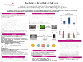
Introduction Results Figure 4
Past studies indicate that the composition of a hydrogel has a great effect on
its biological and mechanical properties.
Dermis derived hydrogels
Pros:
• Similar in structure and composition to extracellular matrix in vivo
• Provides structural support for cellular growth and function1
Controls: 3‐ 15% PEGDA only gels, with a mean swelling ratio of 9.058 and a standard
deviation of 0.346.
•There were two batches of gels made, here labeled as A and B (figure 5).
•Hydrogels were evaluated based on two parameters: the ability to form a gel (figure 4)
and the swelling ratio test (figure 5).
•2% PEGDA concentration solutions were unable to form a gel at both PEGDA molecular
i h
Introduction Results Figure 4
Cons:
• Protein extracted hydrogel degrade very rapidly
•are mechanically weak
Proposed solution: cross‐linking a hydrogel in a way that retains high
biofunctionality and improves the mechanics
•This is of significance in tissue engineering scaffolds, with applications in
chronic diabetic wound healing and tissue regeneration. A biosynthetic
ff
weights.
•3% gels formed at 3.4K da but not at 20K da.
• 3% gels had higher swelling ratios than 4% gels at 3.4K da.
• 20K da PEGDA produced varied data that needs to be repeated.
Figure 5
Swelling Ratio Test
hybrid scaffold made with PEG and a tissue‐derived extract could better
create the complexity of in vivo tissue as compared to an entirely synthetic
scaffold2.
Aim of this study was to introduce polyethelyne di‐acrylate (PEGDA) as a
cross‐linking agent for the dermal extract and to establish a point of gel
formation.
Methods
The dermal extract was obtained by harvesting the tissue of a Long Evans rat
using a technique developed by Uriel et al. as illustrated in figure 2.
Extraction:
•Proteins were extracted from tissue using mechanical and enzymatic means.
•High salt buffer combined with protease inhibitors removed cells and
protected proteins from degradation.
8M dd d t i th l bilit f th bt i d t i
Methods
Discussion
Figure 2
Tissue Derived Hydrogel Technique Image from Chiu Y‐C, et al.
•8M urea was added to increase the solubility of the obtained proteins.
•TNBS assay was used to determine percent PEGylation:
% pegylation = 1‐(ratioPegylated dermal extract /ratioDermal extract )
Hydrogel formation
This study was helpful in establishing a point of gel
formation, which is 3% for 3.4K da PEGDA and 4% for 20K
da PEGDA. Compared to the control group of PEGDA gels
only, 3.4K da PEGDA gels produced consistently higher
swelling ratios, with the lowest concentration of PEGDA
having the highest swelling ratios. This indicates that theFigure 3
Figure 1: Chemical structure PEG and PEGDA
http://homepages.cae.wisc.edu/~bme200/microencapsulation_fall05/
• A larger sample size should be tested to investigate reproducibility
• The gels should be tested for mechanical properties
• The gels should have biofunctional testing to investigate the effects of
pegylation on biological activity
Hydrogel formation:
•Hydrogels were made using PEGylated dermal extract of 1:1 and PEGDA stock
using either 3.4K da or 20K da (figures 1 and 3).
•Stock PEGDA concentrations were made: 2%, 3%, 4%, or 5%.
•Three gels were made per concentration, with a volume of 100 µL per gel.
•Gels were exposed to UV light for 15 minutes, then hydrated for 30 minutes,
and dehydrated in an oven overnight to obtain measurements for a swell ratio
test
g g g
less cross‐linked the gel is, the more water it will be able
to hold.
The data on the effect of the molecular weight of the gel
on its swelling abilities was inconclusive. Additional
testing is necessary to optimize the results at 20K da to
confirm the findings. Moreover, varying molecular weights
of PEGDA will help to test the increase of the point of gel
Future Work
g
Pegylated Dermal Extract
pegylation on biological activity
This research was supported by the National Science Foundation (grant number
0552896). We would like to sincerely thank the members of Dr. Brey’s lab, Jef
Larson and Bin Jiang for their consulting and engineering support during this
project.
1. Uriel S. et al., Tissue Engineering 2009; 15: 309‐321
2. Almany L., Seliktar D., Biomaterials, 2005; 26: 2467‐2477
3. Cheng, M. et al, J.of Biomedical Materials Research, 2010; 92A : 852 ‐ 858
test. p p g
formation where PEGDA length is a true statistical trend.
The obtained data indicated that the point of gel formation for
3.4K da PEGDA occurs at 3% and for 20K PEGDA – at 4%.
Acknowledgements References
Conclusion