Flavin-Containing Dimethylaniline Monooxygenase 5 Drives Malignancies in Hepa...
Poster
1. UofL Design and Print
♦ Abstract
Familial Hypercholesterolemia (FH) is a hereditary
disease resulting in defective Low Density Lipoprotein
Receptor (LDL-R) expression. Due to defective LDL-R,
LDL cholesterol is left to accumulate in the bloodstream,
where it can form atherosclerotic plaques and accelerate
cardiovascular disease. Liver transplant is the only
curative option for FH, but healthy donor livers are in
short supply. Deriving induced pluripotent stem cells
(iPSC) from FH patient fibroblasts and differentiating
them into functional hepatocyte-like cells (HLC) could
offer a significant therapeutic benefit.
We differentiated iPSC into HLC over five stages.
Polymerase Chain Reactions (PCR) performed at the end
of each stage verified the appropriate expression of five
genes (POU5F1, SOX17, HNF4a, AFP, and ALB) used
as markers of cell development and differentiation.
Visualization of PCR products via gel electrophoresis
indicated that the HLC expressed the appropriate makers
for each stage of development.
♦ Introduction & Background
♦ References
Familial Hypercholesterolemia (FH) is an autosomal
dominant disease that results in defective expression of
the Low Density Lipoprotein Receptor (LDL-R). As the
typical FH patient exhibits elevated LDL cholesterol (LDL-
c) levels, cardiovascular diseases (CVD) such as
atherosclerosis or coronary artery disease can result. In
every year since 1900 (except 1918), CVD accounted for
more deaths than any other cause of death in the United
States. It is now the leading cause of death in the world.
Hepatocytes, the parenchymal cells of the liver, have
an especially high concentration of functional LDL-R that
bind circulating LDL-c, which is internalized via receptor-
mediated endocytosis. For this reason, liver transplant is
regarded as the only curative treatment for FH. However,
livers are in short supply, and transplant presents many
challenges (such as immunosuppression, quality of life,
high cost, complication rate, and death).
Induced pluripotent stem cells (iPSC) are embryonic-
like cells that have been reprogrammed from terminally
differentiated adult cells. They express the genes and
factors important for maintaining the defining properties of
embryonic stem cells. Because they are pluripotent (able
to differentiate into the three germ layers – ectoderm,
mesoderm, and endoderm), iPSC can be differentiated
into cells that resemble hepatocytes, termed hepatocyte-
like cells (HLC). We differentiated our iPSC using a
published five-stage protocol (Song, Cell Res, 2009).
♦ Methodology ♦ Conclusions
♦ Acknowledgements
I would like to sincerely thank my mentor Dr. Nolan
Boyd for guiding me through my project and allowing me
to use his lab. I would also like to thank Venkat
Ramakrishnan for supervising me and my progress. I
owe much to my parents, Drs. Huey Tien and Ring Tsai,
for transporting me to and from the lab. Finally, I would
like to acknowledge my science teacher and adult
sponsor, Mr. Robert Baar, who organized the science
fair affairs in my science class.
Development and Characterization of FamilialDevelopment and Characterization of Familial
Hypercholesterolemia Hepatocyte-Like CellsHypercholesterolemia Hepatocyte-Like Cells
Kevin T. Tien
duPont Manual High School, Cardiovascular Innovation Institute, University of Louisville School of Medicine
♦ Purpose
Our lab’s long-term aim is to develop a cell-based
apheresis device that utilizes functionally restored FH
patient-derived HLC (FH-LDLR-HLC). As a first step
towards producing a functional device, the lab
successfully differentiated non-restored FH-iPSC (FH-NT-
iPSC) into HLC (FH-NT-HLC). This experiment sought to
characterize the differentiation of the FH-NT-iPSC into
FH-NT-HLC. The FH-NT-HLC would then serve as a
negative control for functionally restored cells.
♦ Hypothesis
FH-NT-HLC will express the appropriate markers at each
stage of development:
- Stage 0 (undifferentiated): POU5F1
- Stage 1 (early definitive endoderm): SOX17
- Stage 2 (late definitive endoderm/hepatic specification):
HNF4a
- Stage 2 – 5 (hepatic maturation): AFP and ALB
Cells were lysed at the end of each stage of development before being RNA-purified and quantified via QiaShredder
and RNeasy Kits (Qiagen). Isolated RNA was converted into cDNA using SuperScript II Reverse Transcriptase (RT,
Invitrogen). Samples without RT were used as negative controls.
To assess gene expression during HLC development, cell DNA was subjected to Polymerase Chain Reaction (PCR)
at the end of each of five stages. The following genes encode the transcription factors and plasma proteins that were
assessed as markers of HLC development: POU5F1, SOX17, HNF4α, Alpha-fetoprotein (AFP), and Albumin (ALB).
Primers (purchased from Integrated DNA Technologies, IDT) were used to amplify these genes (amplicons). Amplicons
were analyzed via standard gel electrophoresis. GAPDH was used as a loading control.
Gene expression was normalized to GAPDH during densitometry analysis. Densitometry, which indicates the
magnitude of gene expression, was calculated for each band using ImageJ software (NIH). The software measured the
integral of the band for each gene at each stage, allowing us to compare relative expression via a semi-quantitative
means.
We performed these experiments in quadruplicate.
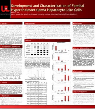
Figure 2: Gene Expression through Various Stages of Hepatocyte-like Cells
OCT4 (POU5F1), a transcription factor, is a marker of pluripotence
that is characteristically expressed in untreated FH-iPSC (Stage 0).
SOX17, another transcription factor, marks the definitive endoderm stage
and is stably expressed at the end of Stage 1, after exposure to
STEMdiff™ Definitive Endoderm media, with decreasing expression
through Stage 5. The HNF4a transcription factor is a late endodermal
marker that is expressed between Stages 2 and 5. Alpha fetoprotein (AFP)
and Albumin are cytoplasmic markers of hepatoblasts/immature
hepatocytes and mature hepatocytes, respectively. They are significantly
up-regulated beginning in Stage 2 and remain stably so through Stage 5.
The GAPDH loading control provided a means for normalization during
densitometry analysis.
Table 1: Densitometry Means of /GAPDH
Table 2: Standard Error Measurements of /GAPDH
Cells exhibited the characteristic genes of
differentiation from iPSC to HLC. Our gel images indicate
that the appropriate genes were expressed at
appropriate times; densitometry measurements
correspond to the expected trends as well.
POU5F1 was expressed during stage 0 as a marker
of pluripotence. SOX17 expression was highest in Stage
1 and gradually decreased in magnitude through Stage
5. HNF4a was first seen in Stage 2 and followed an
expression pattern similar to SOX17. AFP is a marker for
immature hepatocytes while Albumin (ALB) is a marker
associated with mature hepatocytes. Both AFP and ALB
exhibited strong expression starting in Stage 2 (hepatic
specification). AFP expression decreased slightly in
subsequent stages, while ALB was most predominant in
Stage 5 (final stage of differentiation).
The presence of AFP and ALB at Stage 5 indicates that
we (a) have cells that are hepatic in nature and (b) are
potentially of mixed maturity.
♦ Current & Future Directions
The derivation of HLC from FH patient fibroblasts is a
significant milestone in developing a potential therapeutic
for this population. However, as FH is a genetic disease,
it is clear that any cells derived from an FH patient would
be equally as dysfunctional. Therefore, for our iPSC and
HLC to be effective, functional restoration of the LDL-R
activity is warranted. Our lab is working to deliver a
corrective plasmid into iPSC and characterize the
functionally restored HLC.
Further, in order to demonstrate therapeutic potential,
FH-LDLR-HLC will be implanted into FH-model mice and
assessed for their ability to clear LDL cholesterol from
the bloodstream. The ultimate goal would be to utilize
such apheresis devices in human FH patients as a
therapeutic, autologous means of metabolizing their
excess LDL cholesterol and lowering their CVD risk.
Betheseda. (2009). What are Induced Pluripotent Stem Cells? Retrieved October 1, 2013, from National Institutes of
Health: stemcells.nih.gov
Bowen, R. (1998, June 23). Hepatic Histology: Hepatocytes. Retrieved September 26, 2013, from
www.vivo.colostate.edu: http://www.vivo.colostate.edu
Cai, J. (2007). Directed Differentiation of Human Embryonic Stem Cells into Functional Hepatic Cells. Hepatology,
1229-1239.
Cohen, J., Hobbs, H. H., & Rader, D. J. (2003). Monogenic Hypercholesterolemia: New Insights in Pathogenesis
and Treatment. The Journal of Clinical Investigation, 1795-1803.
Hypercholesterolemia. (2007, March). Retrieved September 27, 2013, from Genetics Home Reference:
ghr.nlm.nih.gov
Matsumoto, K., Yoshitomi, H., Rossant, J., & Zaret, K. S. (2001). Liver Organogenesis Promoted by Endothelial
Cells Prior to Vascular Function. www.sciencemag.org, 559-563.
Nunes, S. S., Maijub, J. G., Krishnan, L., Ramakrishnan, V. M., Clayton, L. R., Williams, S. K., . . . Boyd, N. L.
(2013). Generation of a Functional Liver Tissue Mimic Using Adipose Stromal Vascular Fraction Cell-Derived
Vasculatures. Scientific Reports, 1-7.
Song, Z., Cai, J., Liu, Y., Zhao, D., Yong, J., Duo, S., . . . Qin, H. (2009). Efficient Genertation of Hepatocyte-like
Cells from Human Induced Pluripotent Stem Cells. Cell Research.
The University of Utah. (2008). PCR Virtual Lab. Retrieved October 1, 2013, from Learn. Genetics:
learn.genetics.utah.edu
Means S0 S1 S2 S3 S4 S5
POU5F1 0.0715 0.0615 0.0000 0.0000 0.0000 0.0000
SOX17 0.0004 1.4155 0.5915 0.0537 0.1819 0.0247
HNF4A 0.0033 0.1449 0.5626 0.2520 0.4262 0.1932
AFP 0.0064 0.0224 1.6999 1.5441 2.6651 1.2878
ALB 0.0000 0.0260 0.4953 1.3186 2.4401 1.3380
S.E.M. S0 S1 S2 S3 S4 S5
POU5F1 0.0251 0.0341 0.0000 0.0000 0.0000 0.0000
SOX17 0.0004 0.5521 0.1048 0.0146 0.0223 0.0189
HNF4A 0.0033 0.0539 0.1600 0.0965 0.1687 0.0434
AFP 0.0064 0.0087 0.1872 0.1564 1.1704 0.1495
ALB 0.0000 0.0114 0.1329 0.0975 1.0132 0.2479
♦ Results
Figure 4: /GAPDH Means Throughout Differentiation
Day 13730 18 21
iPSC
1. Endoderm
Induction
2. Hepatic
Specification
4. Hepatic
Maturation
3. Hepatoblast
Expansion
5. Mature
HLC
Act A
OSM
Dex
HGF
KGF
FGF4
BMP2
OSM
Dex
N2B27
Figure 1: Flow Chart of Stepwise Differentiation Protocol
Figure 3: Example of Densitometry Curves by ImageJ Software
OCT4 SOX17 HNF4A AFP ALB