SlideShare a Scribd company logo
1 of 1
The regression of Meckel’s cartilage relative to
the development of the canine tympanic bulla.
Keith A. Jarrett1
, J. Michael Mathis2
, & Cathryn K. Stevens-Sparks1
1Department of Biomedical Sciences, School of Veterinary Medicine, Ross University, Bassetterre, St. Kitts, West Indies
2Department of Comparative Biomedical Sciences, School of Veterinary Medicine, Louisiana State University, Baton Rouge, LA 70803
Introduction
Background: Hearing loss in dogs and cats following dental procedures performed under anesthesia
has been documented. Jaw manipulation is a common factor in these cases. The malleomandibular
(MML) ligament has recently been described in the puppy, and represents a connection between the
jaw and the malleus. In the adult dog, it is speculated that the MML becomes entrapped due to the
development and ossification of the tympanic bulla (TB). Fibrous connections of this ligamentous
remnant of Meckel’s cartilage to the tympanic bone have warranted its more appropriate terminology
as the tympanomandibular (TML) ligament in the adult dog. Disruption of the TML may be a possible
cause of acute-onset, conductive deafness in the dog.
Purpose: The goal of this research is to provide a more precise anatomical description of the
relationship of the canine MML and TML to bony skull elements by using micro-computed tomography
and staining techniques to study the peri- and postnatal osseous development of the canine middle
ear, especially in relation to the TB and the rostral process of the malleus.
Results
Discussion
Figure 4 Figure 5 Figure 6 Figure 7
A B
C D
Materials & Methods
A B
C D
A B
C D
A B
C D
Ethanol-Fixed
Specimen
Formalin-Fixed
Specimen
Alcian Blue
Ethanol
Acetic Acid
Alizarin Red
Ethanol
Alizarin Red
Alcian Blue
Ethanol
Potassium Hydroxide
Distilled Water
Sodium Borate
Distilled Water
Figure 1
Figure 3
Figure 2
Fig 2: MML originating from the rostral process of the
malleus and attaching to the medial aspect of the mandible
Fig 3: Tympanic annuli (TA) of 1, 14 & 21 day old specimen
Fig 4
A. Lateral 3D reconstruction of 1 day old canine skull highlighting the TA (green box)
B. Ventral view of 3D reconstruction of 1 day old canine skull highlighting the TA (green box)
C. Manipulated 3D reconstruction matching image D of the TA and surrounding structures
D. Ventral view of the TA with the tympanic membrane removed in order to demonstrate the rostral
process of the malleus (green asterisk) and affiliated MML (green arrow)
Fig 5
A. Lateral 3D reconstruction of 8 day old canine skull with the TA (green box)
B. Ventral view of 3D reconstruction of 8 day old canine skull highlighting the TA (green box)
C. Manipulated 3D reconstruction matching image D of the TA and surrounding structures
D. Ventrolateral view of the TA with the tympanic membrane removed in order to demonstrate the
rostral process of the malleus (green asterisk) and affiliated MML (green arrow)
Fig 6
A. Lateral 3D reconstruction of 14 day old canine skull highlighting the TA (green box)
B. Ventral view of 3D reconstruction of 14 day old canine skull highlighting the TA (green box)
C. Manipulated 3D reconstruction matching image D of the TA and surrounding structures
D. Dorsomedial view of the TA with the tympanic membrane intact in order to demonstrate the
handle of the malleus (green asterisk)
Fig 7
A. Lateral 3D reconstruction of 21 day old canine skull highlighting the TA (green box)
B. Ventral view of 3D reconstruction of 21 day old canine skull highlighting the TA (green box)
C. Manipulated 3D reconstruction matching image D of the TA and surrounding structures
D. Lateral view of the TA with the tympanic membrane removed in order to demonstrate the rostral
process of the malleus (green asterisk) and affiliated MML (green arrow)
Fig 1: Medial view of 8 day old ethanol-fixed specimen
• The MML was visible on all dissected specimen and was observed attaching to the rostral process of the malleus and to the medial aspect of the mandible, directly caudal
to the mandibular foramen. The rostral process of the malleus appeared to become more ossified and was noticeably more concealed by the TA as development
progressed. Also, the ossification of the TB was observed to progress ventrocaudally from the TA while shifting the apex of the TA and associated structures ventrolaterally
from a complete horizontal plane.
• Images obtained using CT have confirmed that the apex of the TA shift ventrolaterally and remain closely affiliated with the mandible. However, further study of specimens
older than 21 days will be required to completely describe the ossification and development of the TB in order to understand its affiliation with the TML.
• It should be noted that while ethanol-fixed specimen and the double-staining technique were used in this study, the two-stage staining technique with formalin-fixed
specimen should be considered in future studies due to a significantly more pronounced uptake of stain.
• This study confirmed that the MML/TML does pass through a foramen located immediately medial to the mandibular fossa of the temporal bone (documented but not
represented here).
*
* *
*
Acknowledgements
We wish to thank Dr. Michel
Vandenplas, Dr. Allison Salmon,
Dr. Martha Littlefield, Dr.
Margaret McNulty, and various
support staff and faculty of Ross
University SVM and LSU SVM.
This research project was funded and
supported by Ross University School
of Veterinary Medicine, St. Kitts.

More Related Content

Similar to SS15PosterFinal

Anchorage condition during canine retraction using transpalatal arch with con...
Anchorage condition during canine retraction using transpalatal arch with con...Anchorage condition during canine retraction using transpalatal arch with con...
Anchorage condition during canine retraction using transpalatal arch with con...Shahriar Tomal
 
parasite poster for linkin
parasite poster for linkinparasite poster for linkin
parasite poster for linkinRachael Swedberg
 
Deep Learning-based Histological Segmentation Differentiates Cavitation Patte...
Deep Learning-based Histological SegmentationDifferentiates Cavitation Patte...Deep Learning-based Histological SegmentationDifferentiates Cavitation Patte...
Deep Learning-based Histological Segmentation Differentiates Cavitation Patte...Wookjin Choi
 
Frequency of Sella Turcica Bridge and Clinoid Enlargement in Lateral Cephalom...
Frequency of Sella Turcica Bridge and Clinoid Enlargement in Lateral Cephalom...Frequency of Sella Turcica Bridge and Clinoid Enlargement in Lateral Cephalom...
Frequency of Sella Turcica Bridge and Clinoid Enlargement in Lateral Cephalom...Iván E Pérez
 
congenital vertical talus by DR.Girish motwani
congenital vertical talus by DR.Girish motwanicongenital vertical talus by DR.Girish motwani
congenital vertical talus by DR.Girish motwaniGirish Motwani
 
Clinical Approach of a Tooth with Radix Entomolaris and Five Root Canals
Clinical Approach of a Tooth with Radix Entomolaris and Five Root CanalsClinical Approach of a Tooth with Radix Entomolaris and Five Root Canals
Clinical Approach of a Tooth with Radix Entomolaris and Five Root CanalsAbu-Hussein Muhamad
 
Transplantation of decidous teeth pulp.pptx
Transplantation of decidous teeth pulp.pptxTransplantation of decidous teeth pulp.pptx
Transplantation of decidous teeth pulp.pptxAshokKp4
 
,Dana info=www.blackwell synergy.com+j.1601-1546.2005.00129
,Dana info=www.blackwell synergy.com+j.1601-1546.2005.00129,Dana info=www.blackwell synergy.com+j.1601-1546.2005.00129
,Dana info=www.blackwell synergy.com+j.1601-1546.2005.00129ibugesic1234
 
Treatment-of-tooth-discoloration-after-the-use-of-white-mineral-trioxide-aggr...
Treatment-of-tooth-discoloration-after-the-use-of-white-mineral-trioxide-aggr...Treatment-of-tooth-discoloration-after-the-use-of-white-mineral-trioxide-aggr...
Treatment-of-tooth-discoloration-after-the-use-of-white-mineral-trioxide-aggr...Ilya Belobrov
 
Intact navigation skills after bilateral loss of striate cortex
Intact navigation skills after bilateral loss of striate cortexIntact navigation skills after bilateral loss of striate cortex
Intact navigation skills after bilateral loss of striate cortexJosé Luis Moreno Garvayo
 
Hero Robles 2019 MRC Symposium
Hero Robles 2019 MRC SymposiumHero Robles 2019 MRC Symposium
Hero Robles 2019 MRC SymposiumHero Robles
 
Is epigenetic variation the key to success in seagrass clones?
Is epigenetic variation the key to success in seagrass clones?Is epigenetic variation the key to success in seagrass clones?
Is epigenetic variation the key to success in seagrass clones?Alexander Jueterbock
 
Senior Seminar composite scaffold Poster-Template-42x60
Senior Seminar composite scaffold Poster-Template-42x60Senior Seminar composite scaffold Poster-Template-42x60
Senior Seminar composite scaffold Poster-Template-42x60Eric Queen
 
Primer-directed enzymatic amplification of DNA with a thermostable DNA polyme...
Primer-directed enzymatic amplification of DNA with a thermostable DNA polyme...Primer-directed enzymatic amplification of DNA with a thermostable DNA polyme...
Primer-directed enzymatic amplification of DNA with a thermostable DNA polyme...José Luis Moreno Garvayo
 

Similar to SS15PosterFinal (20)

Anchorage condition during canine retraction using transpalatal arch with con...
Anchorage condition during canine retraction using transpalatal arch with con...Anchorage condition during canine retraction using transpalatal arch with con...
Anchorage condition during canine retraction using transpalatal arch with con...
 
5)inactive squamousal com
5)inactive squamousal com5)inactive squamousal com
5)inactive squamousal com
 
BCUR Poster
BCUR PosterBCUR Poster
BCUR Poster
 
parasite poster for linkin
parasite poster for linkinparasite poster for linkin
parasite poster for linkin
 
Deep Learning-based Histological Segmentation Differentiates Cavitation Patte...
Deep Learning-based Histological SegmentationDifferentiates Cavitation Patte...Deep Learning-based Histological SegmentationDifferentiates Cavitation Patte...
Deep Learning-based Histological Segmentation Differentiates Cavitation Patte...
 
Kimm
KimmKimm
Kimm
 
Frequency of Sella Turcica Bridge and Clinoid Enlargement in Lateral Cephalom...
Frequency of Sella Turcica Bridge and Clinoid Enlargement in Lateral Cephalom...Frequency of Sella Turcica Bridge and Clinoid Enlargement in Lateral Cephalom...
Frequency of Sella Turcica Bridge and Clinoid Enlargement in Lateral Cephalom...
 
congenital vertical talus by DR.Girish motwani
congenital vertical talus by DR.Girish motwanicongenital vertical talus by DR.Girish motwani
congenital vertical talus by DR.Girish motwani
 
Clinical Approach of a Tooth with Radix Entomolaris and Five Root Canals
Clinical Approach of a Tooth with Radix Entomolaris and Five Root CanalsClinical Approach of a Tooth with Radix Entomolaris and Five Root Canals
Clinical Approach of a Tooth with Radix Entomolaris and Five Root Canals
 
Transplantation of decidous teeth pulp.pptx
Transplantation of decidous teeth pulp.pptxTransplantation of decidous teeth pulp.pptx
Transplantation of decidous teeth pulp.pptx
 
Articulo
ArticuloArticulo
Articulo
 
,Dana info=www.blackwell synergy.com+j.1601-1546.2005.00129
,Dana info=www.blackwell synergy.com+j.1601-1546.2005.00129,Dana info=www.blackwell synergy.com+j.1601-1546.2005.00129
,Dana info=www.blackwell synergy.com+j.1601-1546.2005.00129
 
Treatment-of-tooth-discoloration-after-the-use-of-white-mineral-trioxide-aggr...
Treatment-of-tooth-discoloration-after-the-use-of-white-mineral-trioxide-aggr...Treatment-of-tooth-discoloration-after-the-use-of-white-mineral-trioxide-aggr...
Treatment-of-tooth-discoloration-after-the-use-of-white-mineral-trioxide-aggr...
 
Monique Mai - Final Poster
Monique Mai - Final PosterMonique Mai - Final Poster
Monique Mai - Final Poster
 
Intact navigation skills after bilateral loss of striate cortex
Intact navigation skills after bilateral loss of striate cortexIntact navigation skills after bilateral loss of striate cortex
Intact navigation skills after bilateral loss of striate cortex
 
Hero Robles 2019 MRC Symposium
Hero Robles 2019 MRC SymposiumHero Robles 2019 MRC Symposium
Hero Robles 2019 MRC Symposium
 
Is epigenetic variation the key to success in seagrass clones?
Is epigenetic variation the key to success in seagrass clones?Is epigenetic variation the key to success in seagrass clones?
Is epigenetic variation the key to success in seagrass clones?
 
Senior Seminar composite scaffold Poster-Template-42x60
Senior Seminar composite scaffold Poster-Template-42x60Senior Seminar composite scaffold Poster-Template-42x60
Senior Seminar composite scaffold Poster-Template-42x60
 
81st publication sjodr- 6th name
81st publication  sjodr- 6th name81st publication  sjodr- 6th name
81st publication sjodr- 6th name
 
Primer-directed enzymatic amplification of DNA with a thermostable DNA polyme...
Primer-directed enzymatic amplification of DNA with a thermostable DNA polyme...Primer-directed enzymatic amplification of DNA with a thermostable DNA polyme...
Primer-directed enzymatic amplification of DNA with a thermostable DNA polyme...
 

SS15PosterFinal

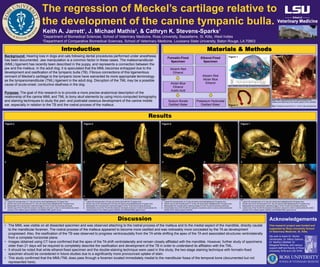
  • 1. The regression of Meckel’s cartilage relative to the development of the canine tympanic bulla. Keith A. Jarrett1 , J. Michael Mathis2 , & Cathryn K. Stevens-Sparks1 1Department of Biomedical Sciences, School of Veterinary Medicine, Ross University, Bassetterre, St. Kitts, West Indies 2Department of Comparative Biomedical Sciences, School of Veterinary Medicine, Louisiana State University, Baton Rouge, LA 70803 Introduction Background: Hearing loss in dogs and cats following dental procedures performed under anesthesia has been documented. Jaw manipulation is a common factor in these cases. The malleomandibular (MML) ligament has recently been described in the puppy, and represents a connection between the jaw and the malleus. In the adult dog, it is speculated that the MML becomes entrapped due to the development and ossification of the tympanic bulla (TB). Fibrous connections of this ligamentous remnant of Meckel’s cartilage to the tympanic bone have warranted its more appropriate terminology as the tympanomandibular (TML) ligament in the adult dog. Disruption of the TML may be a possible cause of acute-onset, conductive deafness in the dog. Purpose: The goal of this research is to provide a more precise anatomical description of the relationship of the canine MML and TML to bony skull elements by using micro-computed tomography and staining techniques to study the peri- and postnatal osseous development of the canine middle ear, especially in relation to the TB and the rostral process of the malleus. Results Discussion Figure 4 Figure 5 Figure 6 Figure 7 A B C D Materials & Methods A B C D A B C D A B C D Ethanol-Fixed Specimen Formalin-Fixed Specimen Alcian Blue Ethanol Acetic Acid Alizarin Red Ethanol Alizarin Red Alcian Blue Ethanol Potassium Hydroxide Distilled Water Sodium Borate Distilled Water Figure 1 Figure 3 Figure 2 Fig 2: MML originating from the rostral process of the malleus and attaching to the medial aspect of the mandible Fig 3: Tympanic annuli (TA) of 1, 14 & 21 day old specimen Fig 4 A. Lateral 3D reconstruction of 1 day old canine skull highlighting the TA (green box) B. Ventral view of 3D reconstruction of 1 day old canine skull highlighting the TA (green box) C. Manipulated 3D reconstruction matching image D of the TA and surrounding structures D. Ventral view of the TA with the tympanic membrane removed in order to demonstrate the rostral process of the malleus (green asterisk) and affiliated MML (green arrow) Fig 5 A. Lateral 3D reconstruction of 8 day old canine skull with the TA (green box) B. Ventral view of 3D reconstruction of 8 day old canine skull highlighting the TA (green box) C. Manipulated 3D reconstruction matching image D of the TA and surrounding structures D. Ventrolateral view of the TA with the tympanic membrane removed in order to demonstrate the rostral process of the malleus (green asterisk) and affiliated MML (green arrow) Fig 6 A. Lateral 3D reconstruction of 14 day old canine skull highlighting the TA (green box) B. Ventral view of 3D reconstruction of 14 day old canine skull highlighting the TA (green box) C. Manipulated 3D reconstruction matching image D of the TA and surrounding structures D. Dorsomedial view of the TA with the tympanic membrane intact in order to demonstrate the handle of the malleus (green asterisk) Fig 7 A. Lateral 3D reconstruction of 21 day old canine skull highlighting the TA (green box) B. Ventral view of 3D reconstruction of 21 day old canine skull highlighting the TA (green box) C. Manipulated 3D reconstruction matching image D of the TA and surrounding structures D. Lateral view of the TA with the tympanic membrane removed in order to demonstrate the rostral process of the malleus (green asterisk) and affiliated MML (green arrow) Fig 1: Medial view of 8 day old ethanol-fixed specimen • The MML was visible on all dissected specimen and was observed attaching to the rostral process of the malleus and to the medial aspect of the mandible, directly caudal to the mandibular foramen. The rostral process of the malleus appeared to become more ossified and was noticeably more concealed by the TA as development progressed. Also, the ossification of the TB was observed to progress ventrocaudally from the TA while shifting the apex of the TA and associated structures ventrolaterally from a complete horizontal plane. • Images obtained using CT have confirmed that the apex of the TA shift ventrolaterally and remain closely affiliated with the mandible. However, further study of specimens older than 21 days will be required to completely describe the ossification and development of the TB in order to understand its affiliation with the TML. • It should be noted that while ethanol-fixed specimen and the double-staining technique were used in this study, the two-stage staining technique with formalin-fixed specimen should be considered in future studies due to a significantly more pronounced uptake of stain. • This study confirmed that the MML/TML does pass through a foramen located immediately medial to the mandibular fossa of the temporal bone (documented but not represented here). * * * * Acknowledgements We wish to thank Dr. Michel Vandenplas, Dr. Allison Salmon, Dr. Martha Littlefield, Dr. Margaret McNulty, and various support staff and faculty of Ross University SVM and LSU SVM. This research project was funded and supported by Ross University School of Veterinary Medicine, St. Kitts.