SlideShare a Scribd company logo
1 of 4
Download to read offline
Treatment of Tooth Discoloration after the Use
of White Mineral Trioxide Aggregate
Ilya Belobrov, BDSc, and Peter Parashos, BDSc, MDSc, PhD
Abstract
Introduction: A number of vital pulp therapy tech-
niques have been recommended to preserve pulp vitality
in teeth with complicated crown fractures, especially in
young patients. Calcium hydroxide has been the gold
standard as a pulp capping material, but recently
mineral trioxide aggregate (MTA) has been recommen-
ded. This case report describes the treatment of tooth
discoloration caused by white MTA (WMTA) used for
the management of a complicated crown fracture.
Methods: A partial pulpotomy was performed with
the use of WMTA after a complicated crown fracture
of the upper right central incisor. Seventeen months
later, the WMTA was removed because of tooth discol-
oration, and internal bleaching was performed. Results:
Upon access, the WMTA was completely discolored.
After it was removed, a significant color change was
observed in the tooth crown, which was further
improved with internal bleaching. The tooth remained
vital, and a dentin bridge was confirmed clinically and
radiographically. Conclusions: The recommendation
to use WMTA for vital pulp therapy in the esthetic
zone may need to be reconsidered. Should discoloration
occur with the use of WMTA, the technique described
may be used to improve the esthetics. (J Endod
2011;37:1017–1020)
Key Words
Complicated crown fracture, mineral trioxide aggregate,
partial pulpotomy, tooth discoloration
Acomplicated crown fracture involves enamel, dentin, and the pulp (1). It is a rela-
tively common type of dental trauma with a reported prevalence in the permanent
dentition of 14% to 21% (2, 3).
A number of procedures have been recommended for the treatment of traumat-
ically exposed pulps in young patients. The aim of treatment is to preserve pulp vitality to
allow for continued physiological root development, including closure of the root apex
anddevelopment oflateralrootdentin. Thistreatment aimsto prevent cervicalrootfrac-
tures caused by thin dentinal walls (4). Vital pulp therapy in older patients is less
predictable but can still be successful (5). However, the decision whether to retain
the pulp is usually governed by the amount of remaining tooth structure rather than
the age of the patient.
Direct pulp capping is a procedure in which the capping agent is placed on the
tissue that has been exposed to microorganisms and inflammation is present (6).
Partial pulpotomy, as described by Cvek (7), has an excellent prognosis and consists
of the aseptic, surgical removal of the exposed pulp and dentin surrounding the expo-
sure to a depth of 1.5 to 2.0 mm followed by sealing of the exposed pulp with a suitable
material. Unlike direct pulp capping, the partial pulpotomy procedure creates space for
the placement and retention of the pulp capping material. Calcium hydroxide has been
the gold standard for vital pulp therapy since the 1930s (8). However, the dentin bridge
formed is porous, with multiple tunnel defects (9). In some cases, it is associated with
a chronic inflammatory response associated with the use of Ca(OH)2 (10). Subse-
quently, many materials have been shown to be biologically compatible with exposed
pulps and permit an environment that is conductive to dentin bridge formation (11).
The healing of dental pulp exposures is not dependent on the type of pulp capping mate-
rial but is related to the capacity of these materials to prevent bacterial leakage (11).
Mineral trioxide aggregate (MTA) has been shown to prevent dye and bacterial
leakage and has a high level of biocompatibility (12, 13). Based on animal and
human studies, MTA is considered a suitable pulp capping material (5, 14–17). Its
use has been recommended for the treatment of complicated crown fractures (18).
One of the potential drawbacks of using gray MTA for vital pulp therapy in anterior
teeth is the subsequent development of crown discoloration (19). For this reason, white
MTA (WMTA) was developed. Although it has been shown to be comparable to gray
MTA as a pulp capping agent (20), reports show discoloration of WMTA in vitro
and that WMTA discolors primary teeth in vivo after pulpotomy (21, 22). The
following case report describes the treatment of a complicated crown fracture in
a permanent anterior tooth with WMTA and the favorable outcome of treatment for
the resulting crown discoloration.
Case Report
A 12-year-old girl reported to the Emergency Department of the Royal Dental
Hospital of Melbourne 4 hours after fracturing a tooth on the back of a chair. The
From Melbourne Dental School, University of Melbourne, Melbourne, Australia.
Address requests for reprints to Associate Professor Peter Parashos, Melbourne Dental School, University of Melbourne, 720 Swanston Street, Victoria, 3010,
Australia. E-mail address: parashos@unimelb.edu.au
0099-2399/$ - see front matter
Copyright ª 2011 American Association of Endodontists.
doi:10.1016/j.joen.2011.04.003
Case Report/Clinical Techniques
JOE — Volume 37, Number 7, July 2011 Tooth Discoloration by MTA 1017
intraoral examination revealed a complicated crown fracture of the
maxillary right central incisor (tooth #8). The fractured coronal frag-
ment had been placed into a glass of milk. All teeth in the anterior
segment of maxillary and mandibular arches responded to CO2 pulp
testing at the time of presentation. Radiographic examination
(Fig. 1A) confirmed the clinical findings that tooth #8 had substantial
coronal tooth structure remaining for restoration by reattachment of
the coronal fragment or by a direct bonded resin composite restoration.
After the administration of local anesthesia, a partial pulpotomy
was performed under rubber dam on tooth #8 using a high-speed
size 2 round diamond bur with copious water coolant. The coronal
pulp stump was rinsed with 1% sodium hypochlorite for 2 minutes until
hemostasis was achieved. ProRootWMTA (DentsplyTulsaDental,Tulsa,
OK) was placed onto the pulp followed by Vitrebond (3M Dental Prod-
ucts Division, St Paul, MN), etching, Adper Single Bond Plus Adhesive
(3M Espe, St Paul, MN), and flowable composite (Revolution Formula
2; Kerr, Orange, CA) to bond the coronal tooth fragment. The enamel
surface at the fracture line was beveled, and a direct resin composite
restoration (Tetric N-Ceram; Ivoclar Vivadent, Schaan, Liechtenstein)
was placed to reinforce the two fragments (Fig. 1B). The patient was
subsequently reviewed in the Endodontics Unit of the Royal Dental
Hospital of Melbourne and did not report any immediate postoperative
discomfort.
The patient was reviewed at 1, 5, and 17 months. Throughout the
follow-up period, the maxillary anterior teeth remained asymptomatic
and responsive to CO2 pulp testing. At 1 month, a slight gray discolor-
ation couldbe seen justapical to the fracture line (Fig. 2A). At 5 months,
there was further evidence of crown discoloration (Fig. 2B).
At 17 months, the patient and her parents were concerned with the
crown discoloration (Fig. 2C). The tooth responded to CO2 pulp testing,
and a hard tissue bridge and continued apical root maturation were
Figure 1. (A) A periapical radiograph of tooth #8 upon presentation. (B) The
immediate postoperative radiograph showing the placement of WMTA and the
reattachment of the coronal fragment. (C) A radiograph at 17 months showing
the formation of hard-tissue bridge and root maturation. (D) A radiograph
post-bleaching and restoration of the access cavity.
Figure 2. (A) An intraoral photograph at the 1-month follow-up. Note the slight gray discoloration apical to the fracture line. (B) Further crown discoloration at
the 5-month recall. (C) Distinctly noticeable crown discoloration 17 months after partial pulpotomy with WMTA. (D) The removal of discolored WMTA.
Case Report/Clinical Techniques
1018 Belobrov and Parashos JOE — Volume 37, Number 7, July 2011
radiographically evident (Fig. 1C). After discussion with the parents and
the patient concerning internal bleaching, a local anesthetic was admin-
istered. Under a rubber dam, the pulp chamber was reaccessed. Using
the operating microscope, a high-speed size 2 round stainless steel bur
with water coolant was used tocompletelyremovethe discolored WMTA
(Fig. 2D), exposing the hard-tissue bridge (Fig. 3A). The removal of
WMTA was attempted using ultrasonics but was proved unsuccessful
because of cutting inefficiency; therefore, the use of a high-speed bur
with water cooling under a microscope was used, which was a quicker
and just as conservative an option.
Considerable improvement in the color of the tooth was seen
immediately after the removal of the discolored WMTA (Fig. 3B). At
the same appointment, a mixture of sodium perborate and saline was
placed in the access cavity to internally bleach the crown. No adverse
pulpal effects were anticipated because of the lack of porosity in the
dentine bridge formed after the use of MTA (10). Then, the access
opening was sealed with Cavit W (3M, Dental Products Division, St
Paul, MN).
The patient returned 1 week later asymptomatic, and the tooth was
still responding to CO2 pulp testing. Under rubber dam isolation, the
tooth was reaccessed, and the internal bleaching paste was removed
(Fig. 3C). The access cavity was subsequently restored with a polycar-
boxylate cement (Durelon, 3M ESPE) and resin composite restoration
(Tetric N-Ceram). The labial composite was also replaced with a trans-
lucent (4 Seasons medium enamel value, Ivoclar Vivadent) composite
resin restoration to match the patients improved esthetics (Figs. 1D and
3D). Because there was no noticeable improvement in the patient’s oral
hygiene, further oral hygiene instructions were also provided. The
patient was recalled 1 month later, and there was no change in the color
of the crown; the tooth continued to respond to CO2 pulp testing.
Discussion
Thiscaseconfirmedthatset WMTAwasresponsible forthe gradual
discoloration of tooth #8. The intraoral photograph in Figure 2D clearly
distinguishes between the discolored WMTA and the surrounding
dentin. The careful removal of WMTA under the operating microscope
ensured that no additional tooth structure was removed. Although
internal bleaching was used in this case, most of the discoloration
was within the WMTA (Figs. 2D and 3A) and not in the dentin. Consider-
able improvement in crown color was achieved after the removal of the
WMTA (Figs. 2C and 3B). Only a minor improvement was seen in the
color of the dentin internally (Fig. 3A and C) after internal bleaching.
Therefore, internal bleaching may not be required.
Although the biologic and the esthetic goals of treatment were
achieved in this case, it may be reasonable to reconsider using
WMTA for vital pulp therapy in the esthetic zone. Should discoloration
occur after the use of WMTA in the esthetic zone, careful removal of set
WMTA may be attempted after confirmation of dentin bridge formation.
Reattachment of the coronal fragment provides several advantages
including exact restoration of crown morphology, reduced chair time,
excellent esthetics, similar wear rate to opposing teeth, and a positive
emotional response from the patient (23). When simple reattachment
(ie, no preparation) is used, the use of resin composite in the adhesive
Figure 3. (A) All of the discolored WMTA was removed until a hard-tissue bridge was reached. (B) A noticeable improvement in the color of the crown could be
seen immediately after the removal of discolored WMTA. (C) The dentin color after 1 week of internal bleaching. (D) The excellent esthetic result after the removal
of discolored WMTA and internal bleaching.
Case Report/Clinical Techniques
JOE — Volume 37, Number 7, July 2011 Tooth Discoloration by MTA 1019
interface significantly increases the fracture strength compared with
bonding agents only (24). Beveling of the margins further increases
the fracture resistance and has been shown to have a better retention
and esthetic prognosis than direct composite resin restoration (25).
MTA rather than calcium hydroxide was used for pulp capping
after the partial pulpotomy. The main advantages of MTA are that it
provides a good protective barrier against bacterial penetration (26)
and is biocompatible (27). Furthermore, the presence of blood has
little impact on the degree of leakage (12). It has also been shown
that the bioactive property of MTA is superior (when compared with
Ca[OH]2 and other materials) in dentin bridge formation after pulp
capping and pulpotomies (10, 17). Hence, the use of MTA has been
recommended as the material of choice for vital pulp therapy (28, 29).
Because of the potential staining of tooth structure in an area in
which esthetics is important, a white powder form of MTA was manu-
factured (30). However, in vitro studies have reported a gray discolor-
ation of WMTA used in single-rooted human teeth and plastic blocks
after the setting reaction (21, 31). In both studies, the material was
affected in depth, which led the authors to speculate that a chemical
reaction was responsible for the discoloration.
Histologically, MTA forms a superior dentin bridge with less
underlying inflammation than Ca(OH)2 (10, 14). It also has greater
success for direct pulp capping carious exposures when there is
bacterial presence (32). However, no clinical study has compared
the use of MTA and Ca(OH)2 after traumatic exposure of the pulp.
Cvek (7) reported 96% success for partial pulpotomies using
Ca(OH)2. Therefore, the use of WMTA in the esthetic zone after trau-
matic exposure of the pulp, with its potential for discoloration of tooth
structure, must be questioned.
Acknowledgments
The authors deny any conflicts of interest related to this study.
References
1. Glendor U, Marcenes W, Jo A. Classification, epidemiology and Etiology. In:
Andreasen J, Andreasen F, Andersson L, eds. Textbook and Color Atlas of Traumatic
Injuries to the Teeth. 4th ed. Oxford: Blackwell Munksgaard; 2007:217–54.
2. Davis GT, Knott SC. Dental trauma in Australia. Aust Dent J 1984;29:217–21.
3. Onetto JE, Flores MT, Garbarino ML. Dental trauma in children and adolescents in
Valparaiso, Chile. Endod Dent Traumatol 1994;10:223–7.
4. Cvek M. Prognosis of luxated non-vital maxillary incisors treated with calcium
hydroxide and filled with gutta-percha. A retrospective clinical study. Endod Dent
Traumatol 1992;8:45–55.
5. Bogen G, Kim JS, Bakland LK. Direct pulp capping with mineral trioxide aggregate:
an observational study. J Am Dent Assoc 2008;139:305–15.
6. Cvek M, Cleaton-Jones PE, Austin JC, Andreasen JO. Pulp reactions to exposure after
experimental crown fractures or grinding in adult monkeys. J Endod 1982;8:391–7.
7. Cvek M. A clinical report on partial pulpotomy and capping with calcium hydroxide
in permanent incisors with complicated crown fracture. J Endod 1978;4:232–7.
8. Cox CF, Hafez AA. Biocomposition and reaction of pulp tissues to restorative treat-
ments. Dent Clin North Am 2001;45:31–48.
9. Cox CF, Subay RK, Suzuki S, Suzuki SH, Ostro E. Biocompatibility of various dental
materials: pulp healing with a surface seal. Int J Periodontics Restorative Dent 1996;
16:240–51.
10. Nair PN, Duncan HF, Pitt Ford TR, Luder HU. Histological, ultrastructural and quan-
titative investigations on the response of healthy human pulps to experimental
capping with mineral trioxide aggregate: a randomized controlled trial. Int Endod
J 2008;41:128–50.
11. Cox CF, Keall CL, Keall HJ, Ostro E, Bergenholtz G. Biocompatibility of surface-sealed
dental materials against exposed pulps. J Prosthet Dent 1987;57:1–8.
12. Torabinejad M, Higa RK, McKendry DJ, Pitt Ford TR. Dye leakage of four root end
filling materials: effects of blood contamination. J Endod 1994;20:159–63.
13. Torabinejad M, Parirokh M. Mineral trioxide aggregate: a comprehensive literature
review—part II: leakage and biocompatibility investigations. J Endod 2010;36:
190–202.
14. Aeinehchi M, Eslami B, Ghanbariha M, Saffar AS. Mineral trioxide aggregate (MTA)
and calcium hydroxide as pulp-capping agents in human teeth: a preliminary report.
Int Endod J 2003;36:225–31.
15. Ramachandran Nair PNR, Duncan HF, Pitt Ford TRP, Luder HU. Histological, ultra-
structural and quantitative investigations on the response of healthy human pulps to
experimental capping with mineral trioxide aggregate: a randomized controlled
trial. Int Endod J 2008;41:128–50.
16. Accorinte MLR, Loguercio AD, Reis A, et al. Evaluation of two mineral trioxide aggre-
gate compounds as pulp-capping agents in human teeth. Int Endod J 2009;42:
122–8.
17. Pitt Ford T, Torabinejad M, Abedi H, Bakland L, Kariyawasam S. Using mineral
trioxide aggregate as a pulp-capping material. J Am Dent Assoc 1996;127:1491.
18. Flores MT, Andersson L, Andreasen JO, et al. Guidelines for the management of trau-
matic dental injuries. I. Fractures and luxations of permanent teeth. Dent Traumatol
2007;23:66–71.
19. Karabucak B, Li D, Lim J, Iqbal M. Vital pulp therapy with mineral trioxide aggregate.
Dent Traumatol 2005;21:240–3.
20. Parirokh M, Asgary S, Eghbal MJ, et al. A comparative study of white and grey
mineral trioxide aggregate as pulp capping agents in dog’s teeth. Dent Traumatol
2005;21:150–4.
21. Boutsioukis C, Noula G, Lambrianidis T. Ex vivo study of the efficiency of two tech-
niques for the removal of mineral trioxide aggregate used as a root canal filling
material. J Endod 2008;34:1239–42.
22. Maroto M, Barberıa E, Planells P, Garcıa Godoy F. Dentin bridge formation after
mineral trioxide aggregate (MTA) pulpotomies in primary teeth. Am J Dent 2005;
18:151–4.
23. Reis A, Loguercio AD, Kraul A, Matson E. Reattachment of fractured teeth: a review of
literature regarding techniques and materials. Oper Dent 2004;29:226–33.
24. Pusman E, Cehreli ZC, Altay N, Unver B, Saracbasi O, Ozgun G. Fracture resistance of
tooth fragment reattachment: effects of different preparation techniques and adhe-
sive materials. Dent Traumatol 2010;26:9–15.
25. Andreasen FM, Noren JG, Andreasen JO, Engelhardtsen S, Lindh-Stromberg U. Long-
term survival of fragment bonding in the treatment of fractured crowns: a multicenter
clinical study. Quintessence Int 1995;26:669–81.
26. Torabinejad M, Rastegar AF, Kettering JD, Pitt Ford TR. Bacterial leakage of mineral
trioxide aggregate as a root-end filling material. J Endod 1995;21:109–12.
27. Torabinejad M, Hong CU, Pitt Ford TR, Kettering JD. Cytotoxicity of four root end
filling materials. J Endod 1995;21:489–92.
28. Parirokh M, Torabinejad M. Mineral trioxide aggregate: a comprehensive literature
review—part III: clinical applications, drawbacks, and mechanism of action.
J Endod 2010;36:400–13.
29. Witherspoon DE. Vital pulp therapy with new materials: new directions and treat-
ment perspectives—permanent teeth. J Endod 2008;34:S25–8.
30. Glickman GN, Koch KA. 21st-century endodontics. J Am Dent Assoc 2000;
131(suppl):39S–46S.
31. Watts JD, Holt DM, Beeson TJ, Kirkpatrick TC, Rutledge RE. Effects of pH and mixing
agents on the temporal setting of tooth-colored and gray mineral trioxide aggregate.
J Endod 2007;33:970–3.
32. Mente J, Geletneky B, Ohle M, et al. Mineral trioxide aggregate or calcium hydroxide
direct pulp capping: an analysis of the clinical treatment outcome. J Endod 2010;36:
806–13.
Case Report/Clinical Techniques
1020 Belobrov and Parashos JOE — Volume 37, Number 7, July 2011

More Related Content

What's hot

Surgery of Labially Impacted Canine & Orthodontic Management – A Case Report
Surgery of Labially Impacted Canine & Orthodontic Management – A Case ReportSurgery of Labially Impacted Canine & Orthodontic Management – A Case Report
Surgery of Labially Impacted Canine & Orthodontic Management – A Case ReportAbu-Hussein Muhamad
 
Single step apexification with mineral trioxide aggregate
Single step apexification with mineral trioxide aggregateSingle step apexification with mineral trioxide aggregate
Single step apexification with mineral trioxide aggregateAbu-Hussein Muhamad
 
JOURNAL CLUB: “Comparative Evaluation of Postoperative Pain and Success Rate ...
JOURNAL CLUB: “Comparative Evaluation of Postoperative Pain and Success Rate ...JOURNAL CLUB: “Comparative Evaluation of Postoperative Pain and Success Rate ...
JOURNAL CLUB: “Comparative Evaluation of Postoperative Pain and Success Rate ...Urvashi Sodvadiya
 
MANAGEMENT OF OPEN APEX IN PERMANENT TEETH WITH CALCIUM HYDROXIDE PASTE
MANAGEMENT OF OPEN APEX IN PERMANENT TEETH WITH CALCIUM HYDROXIDE PASTEMANAGEMENT OF OPEN APEX IN PERMANENT TEETH WITH CALCIUM HYDROXIDE PASTE
MANAGEMENT OF OPEN APEX IN PERMANENT TEETH WITH CALCIUM HYDROXIDE PASTEAbu-Hussein Muhamad
 
Repair of teeth with cracks in crowns and roots: An observational clinical study
Repair of teeth with cracks in crowns and roots: An observational clinical studyRepair of teeth with cracks in crowns and roots: An observational clinical study
Repair of teeth with cracks in crowns and roots: An observational clinical studyDR.AJAY BABU GUTTI M.D.S
 
JOURNAL CLUB: Dilaceration: Review of an Endodontic Challenge
JOURNAL CLUB: Dilaceration: Review of an Endodontic ChallengeJOURNAL CLUB: Dilaceration: Review of an Endodontic Challenge
JOURNAL CLUB: Dilaceration: Review of an Endodontic ChallengeUrvashi Sodvadiya
 
Prosthodontics ( inhibition of denture plaque)
Prosthodontics ( inhibition of denture plaque)Prosthodontics ( inhibition of denture plaque)
Prosthodontics ( inhibition of denture plaque)DHANANJAYSHETH1
 
Management of Open Apex in Permanent Teeth with Biodentine
Management of Open Apex in Permanent Teeth with BiodentineManagement of Open Apex in Permanent Teeth with Biodentine
Management of Open Apex in Permanent Teeth with BiodentineAbu-Hussein Muhamad
 
Regenerative endodontics / endodontics courses
Regenerative endodontics / endodontics coursesRegenerative endodontics / endodontics courses
Regenerative endodontics / endodontics coursesIndian dental academy
 
Bio hybrid the next gen implant
Bio hybrid the next gen implantBio hybrid the next gen implant
Bio hybrid the next gen implantIJARIIT
 
JOURNAL CLUB: Stability of Bonded Resin Composite Restorations to Enamel afte...
JOURNAL CLUB: Stability of Bonded Resin Composite Restorations toEnamel afte...JOURNAL CLUB: Stability of Bonded Resin Composite Restorations toEnamel afte...
JOURNAL CLUB: Stability of Bonded Resin Composite Restorations to Enamel afte...Urvashi Sodvadiya
 
Pulp therapy in pediatric dentistry revised 2
Pulp therapy in pediatric dentistry revised 2Pulp therapy in pediatric dentistry revised 2
Pulp therapy in pediatric dentistry revised 2alaa Mohamed
 
JOURNAL CLUB: Impact of Access Cavity Design and Root Canal Taper on Fracture...
JOURNAL CLUB: Impact of Access Cavity Design and Root Canal Taper on Fracture...JOURNAL CLUB: Impact of Access Cavity Design and Root Canal Taper on Fracture...
JOURNAL CLUB: Impact of Access Cavity Design and Root Canal Taper on Fracture...Urvashi Sodvadiya
 
complete denture after a stroke ( Prosthodontics)
complete denture after a stroke ( Prosthodontics)complete denture after a stroke ( Prosthodontics)
complete denture after a stroke ( Prosthodontics)DHANANJAYSHETH1
 
Clinical endodontic management during the covid 19 pandemic
Clinical endodontic management during the covid 19 pandemicClinical endodontic management during the covid 19 pandemic
Clinical endodontic management during the covid 19 pandemicDR.AJAY BABU GUTTI M.D.S
 

What's hot (20)

Surgery of Labially Impacted Canine & Orthodontic Management – A Case Report
Surgery of Labially Impacted Canine & Orthodontic Management – A Case ReportSurgery of Labially Impacted Canine & Orthodontic Management – A Case Report
Surgery of Labially Impacted Canine & Orthodontic Management – A Case Report
 
Single step apexification with mineral trioxide aggregate
Single step apexification with mineral trioxide aggregateSingle step apexification with mineral trioxide aggregate
Single step apexification with mineral trioxide aggregate
 
JOURNAL CLUB: “Comparative Evaluation of Postoperative Pain and Success Rate ...
JOURNAL CLUB: “Comparative Evaluation of Postoperative Pain and Success Rate ...JOURNAL CLUB: “Comparative Evaluation of Postoperative Pain and Success Rate ...
JOURNAL CLUB: “Comparative Evaluation of Postoperative Pain and Success Rate ...
 
Vital Pulp Therapy
Vital Pulp TherapyVital Pulp Therapy
Vital Pulp Therapy
 
MANAGEMENT OF OPEN APEX IN PERMANENT TEETH WITH CALCIUM HYDROXIDE PASTE
MANAGEMENT OF OPEN APEX IN PERMANENT TEETH WITH CALCIUM HYDROXIDE PASTEMANAGEMENT OF OPEN APEX IN PERMANENT TEETH WITH CALCIUM HYDROXIDE PASTE
MANAGEMENT OF OPEN APEX IN PERMANENT TEETH WITH CALCIUM HYDROXIDE PASTE
 
Repair of teeth with cracks in crowns and roots: An observational clinical study
Repair of teeth with cracks in crowns and roots: An observational clinical studyRepair of teeth with cracks in crowns and roots: An observational clinical study
Repair of teeth with cracks in crowns and roots: An observational clinical study
 
JOURNAL CLUB: Dilaceration: Review of an Endodontic Challenge
JOURNAL CLUB: Dilaceration: Review of an Endodontic ChallengeJOURNAL CLUB: Dilaceration: Review of an Endodontic Challenge
JOURNAL CLUB: Dilaceration: Review of an Endodontic Challenge
 
Prosthodontics ( inhibition of denture plaque)
Prosthodontics ( inhibition of denture plaque)Prosthodontics ( inhibition of denture plaque)
Prosthodontics ( inhibition of denture plaque)
 
Pediatric endodontics
Pediatric endodonticsPediatric endodontics
Pediatric endodontics
 
Vital Pulp Therapy
Vital Pulp TherapyVital Pulp Therapy
Vital Pulp Therapy
 
Management of Open Apex in Permanent Teeth with Biodentine
Management of Open Apex in Permanent Teeth with BiodentineManagement of Open Apex in Permanent Teeth with Biodentine
Management of Open Apex in Permanent Teeth with Biodentine
 
Regenerative endodontics / endodontics courses
Regenerative endodontics / endodontics coursesRegenerative endodontics / endodontics courses
Regenerative endodontics / endodontics courses
 
Bio hybrid the next gen implant
Bio hybrid the next gen implantBio hybrid the next gen implant
Bio hybrid the next gen implant
 
JOURNAL CLUB: Stability of Bonded Resin Composite Restorations to Enamel afte...
JOURNAL CLUB: Stability of Bonded Resin Composite Restorations toEnamel afte...JOURNAL CLUB: Stability of Bonded Resin Composite Restorations toEnamel afte...
JOURNAL CLUB: Stability of Bonded Resin Composite Restorations to Enamel afte...
 
Pulp therapy in pediatric dentistry revised 2
Pulp therapy in pediatric dentistry revised 2Pulp therapy in pediatric dentistry revised 2
Pulp therapy in pediatric dentistry revised 2
 
JOURNAL CLUB: Impact of Access Cavity Design and Root Canal Taper on Fracture...
JOURNAL CLUB: Impact of Access Cavity Design and Root Canal Taper on Fracture...JOURNAL CLUB: Impact of Access Cavity Design and Root Canal Taper on Fracture...
JOURNAL CLUB: Impact of Access Cavity Design and Root Canal Taper on Fracture...
 
complete denture after a stroke ( Prosthodontics)
complete denture after a stroke ( Prosthodontics)complete denture after a stroke ( Prosthodontics)
complete denture after a stroke ( Prosthodontics)
 
Mutilated Occlusion Fixed-Removable Approach- A Case Report
Mutilated Occlusion Fixed-Removable Approach- A Case ReportMutilated Occlusion Fixed-Removable Approach- A Case Report
Mutilated Occlusion Fixed-Removable Approach- A Case Report
 
revascularisation
revascularisationrevascularisation
revascularisation
 
Clinical endodontic management during the covid 19 pandemic
Clinical endodontic management during the covid 19 pandemicClinical endodontic management during the covid 19 pandemic
Clinical endodontic management during the covid 19 pandemic
 

Similar to Treatment-of-tooth-discoloration-after-the-use-of-white-mineral-trioxide-aggregate

Single-Step Apexification with Mineral Trioxide Aggregate (MTA) –Case Reports
Single-Step Apexification with Mineral Trioxide Aggregate (MTA) –Case ReportsSingle-Step Apexification with Mineral Trioxide Aggregate (MTA) –Case Reports
Single-Step Apexification with Mineral Trioxide Aggregate (MTA) –Case ReportsAbu-Hussein Muhamad
 
Reaction of the pulp to various capping materials 2003
Reaction of the pulp to various capping materials 2003Reaction of the pulp to various capping materials 2003
Reaction of the pulp to various capping materials 2003Asmaa Ali
 
Autogenous tooth fragment reattachment ; a 12 years follow-up
Autogenous  tooth  fragment  reattachment ; a 12  years follow-upAutogenous  tooth  fragment  reattachment ; a 12  years follow-up
Autogenous tooth fragment reattachment ; a 12 years follow-upAbu-Hussein Muhamad
 
Zirconium Dental Implants And Crown for Congenitally Missing Maxillary Latera...
Zirconium Dental Implants And Crown for Congenitally Missing Maxillary Latera...Zirconium Dental Implants And Crown for Congenitally Missing Maxillary Latera...
Zirconium Dental Implants And Crown for Congenitally Missing Maxillary Latera...Abu-Hussein Muhamad
 
ORTHODONTIC TREATMENT OF PERIODONTALLY DAMAGED TEETH - AN INTERDISCIPLINARY A...
ORTHODONTIC TREATMENT OF PERIODONTALLY DAMAGED TEETH - AN INTERDISCIPLINARY A...ORTHODONTIC TREATMENT OF PERIODONTALLY DAMAGED TEETH - AN INTERDISCIPLINARY A...
ORTHODONTIC TREATMENT OF PERIODONTALLY DAMAGED TEETH - AN INTERDISCIPLINARY A...Abu-Hussein Muhamad
 
Crestal bone loss around dental implants after implantation of Tricalcium pho...
Crestal bone loss around dental implants after implantation of Tricalcium pho...Crestal bone loss around dental implants after implantation of Tricalcium pho...
Crestal bone loss around dental implants after implantation of Tricalcium pho...Dr. Anuj S Parihar
 
vital pulp therapy.pptx
vital pulp therapy.pptxvital pulp therapy.pptx
vital pulp therapy.pptxengahmedfathy5
 
PULP REVASCULARIZATION OF A NECROTIC INFECTED IMMATURE PERMANENT TOOTH: A CAS...
PULP REVASCULARIZATION OF A NECROTIC INFECTED IMMATURE PERMANENT TOOTH: A CAS...PULP REVASCULARIZATION OF A NECROTIC INFECTED IMMATURE PERMANENT TOOTH: A CAS...
PULP REVASCULARIZATION OF A NECROTIC INFECTED IMMATURE PERMANENT TOOTH: A CAS...Abu-Hussein Muhamad
 
tooth aulsion.docx
tooth aulsion.docxtooth aulsion.docx
tooth aulsion.docxwrite5
 
BIOLOGICAL POSTS AND CROWN AN ESTHETIC RECOVERY OF SMILE
BIOLOGICAL POSTS AND CROWN AN ESTHETIC RECOVERY OF SMILEBIOLOGICAL POSTS AND CROWN AN ESTHETIC RECOVERY OF SMILE
BIOLOGICAL POSTS AND CROWN AN ESTHETIC RECOVERY OF SMILEPLikitha
 
traumatic injuries management.ppt
traumatic injuries management.ppttraumatic injuries management.ppt
traumatic injuries management.pptAsheeshSawhny1
 
Restoration of endodontically treated teeth review & treatment recomendations
Restoration of endodontically treated teeth review & treatment recomendationsRestoration of endodontically treated teeth review & treatment recomendations
Restoration of endodontically treated teeth review & treatment recomendationsJinny Shaw
 
Modern Treatment for Congenitally Missing Teeth : A Multidisciplinary Appro...
Modern Treatment for Congenitally Missing Teeth   : A Multidisciplinary Appro...Modern Treatment for Congenitally Missing Teeth   : A Multidisciplinary Appro...
Modern Treatment for Congenitally Missing Teeth : A Multidisciplinary Appro...Abu-Hussein Muhamad
 
zanardi2015-3 copy.pdf
zanardi2015-3 copy.pdfzanardi2015-3 copy.pdf
zanardi2015-3 copy.pdfMhandika1
 
Direct and Indirect pulp capping
Direct and Indirect pulp cappingDirect and Indirect pulp capping
Direct and Indirect pulp cappingAnju Thomas
 
Split denture prosthesis for xerostomia- Journal Club
Split denture prosthesis for xerostomia- Journal Club Split denture prosthesis for xerostomia- Journal Club
Split denture prosthesis for xerostomia- Journal Club Dr. Prathamesh Fulsundar
 

Similar to Treatment-of-tooth-discoloration-after-the-use-of-white-mineral-trioxide-aggregate (20)

Single-Step Apexification with Mineral Trioxide Aggregate (MTA) –Case Reports
Single-Step Apexification with Mineral Trioxide Aggregate (MTA) –Case ReportsSingle-Step Apexification with Mineral Trioxide Aggregate (MTA) –Case Reports
Single-Step Apexification with Mineral Trioxide Aggregate (MTA) –Case Reports
 
Reaction of the pulp to various capping materials 2003
Reaction of the pulp to various capping materials 2003Reaction of the pulp to various capping materials 2003
Reaction of the pulp to various capping materials 2003
 
Autogenous tooth fragment reattachment ; a 12 years follow-up
Autogenous  tooth  fragment  reattachment ; a 12  years follow-upAutogenous  tooth  fragment  reattachment ; a 12  years follow-up
Autogenous tooth fragment reattachment ; a 12 years follow-up
 
ToothSurfaceLoss_Part2
ToothSurfaceLoss_Part2ToothSurfaceLoss_Part2
ToothSurfaceLoss_Part2
 
Zirconium Dental Implants And Crown for Congenitally Missing Maxillary Latera...
Zirconium Dental Implants And Crown for Congenitally Missing Maxillary Latera...Zirconium Dental Implants And Crown for Congenitally Missing Maxillary Latera...
Zirconium Dental Implants And Crown for Congenitally Missing Maxillary Latera...
 
ORTHODONTIC TREATMENT OF PERIODONTALLY DAMAGED TEETH - AN INTERDISCIPLINARY A...
ORTHODONTIC TREATMENT OF PERIODONTALLY DAMAGED TEETH - AN INTERDISCIPLINARY A...ORTHODONTIC TREATMENT OF PERIODONTALLY DAMAGED TEETH - AN INTERDISCIPLINARY A...
ORTHODONTIC TREATMENT OF PERIODONTALLY DAMAGED TEETH - AN INTERDISCIPLINARY A...
 
Crestal bone loss around dental implants after implantation of Tricalcium pho...
Crestal bone loss around dental implants after implantation of Tricalcium pho...Crestal bone loss around dental implants after implantation of Tricalcium pho...
Crestal bone loss around dental implants after implantation of Tricalcium pho...
 
vital pulp therapy.pptx
vital pulp therapy.pptxvital pulp therapy.pptx
vital pulp therapy.pptx
 
PULP REVASCULARIZATION OF A NECROTIC INFECTED IMMATURE PERMANENT TOOTH: A CAS...
PULP REVASCULARIZATION OF A NECROTIC INFECTED IMMATURE PERMANENT TOOTH: A CAS...PULP REVASCULARIZATION OF A NECROTIC INFECTED IMMATURE PERMANENT TOOTH: A CAS...
PULP REVASCULARIZATION OF A NECROTIC INFECTED IMMATURE PERMANENT TOOTH: A CAS...
 
tooth aulsion.docx
tooth aulsion.docxtooth aulsion.docx
tooth aulsion.docx
 
BIOLOGICAL POSTS AND CROWN AN ESTHETIC RECOVERY OF SMILE
BIOLOGICAL POSTS AND CROWN AN ESTHETIC RECOVERY OF SMILEBIOLOGICAL POSTS AND CROWN AN ESTHETIC RECOVERY OF SMILE
BIOLOGICAL POSTS AND CROWN AN ESTHETIC RECOVERY OF SMILE
 
traumatic injuries management.ppt
traumatic injuries management.ppttraumatic injuries management.ppt
traumatic injuries management.ppt
 
Restoration of endodontically treated teeth review & treatment recomendations
Restoration of endodontically treated teeth review & treatment recomendationsRestoration of endodontically treated teeth review & treatment recomendations
Restoration of endodontically treated teeth review & treatment recomendations
 
Modern Treatment for Congenitally Missing Teeth : A Multidisciplinary Appro...
Modern Treatment for Congenitally Missing Teeth   : A Multidisciplinary Appro...Modern Treatment for Congenitally Missing Teeth   : A Multidisciplinary Appro...
Modern Treatment for Congenitally Missing Teeth : A Multidisciplinary Appro...
 
597563.pdf
597563.pdf597563.pdf
597563.pdf
 
Untitled 2 2
Untitled 2 2Untitled 2 2
Untitled 2 2
 
Oral surgery and orthodontic for orthodontists by Almuzian
Oral surgery and orthodontic for orthodontists by AlmuzianOral surgery and orthodontic for orthodontists by Almuzian
Oral surgery and orthodontic for orthodontists by Almuzian
 
zanardi2015-3 copy.pdf
zanardi2015-3 copy.pdfzanardi2015-3 copy.pdf
zanardi2015-3 copy.pdf
 
Direct and Indirect pulp capping
Direct and Indirect pulp cappingDirect and Indirect pulp capping
Direct and Indirect pulp capping
 
Split denture prosthesis for xerostomia- Journal Club
Split denture prosthesis for xerostomia- Journal Club Split denture prosthesis for xerostomia- Journal Club
Split denture prosthesis for xerostomia- Journal Club
 

Treatment-of-tooth-discoloration-after-the-use-of-white-mineral-trioxide-aggregate

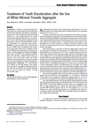
  • 1. Treatment of Tooth Discoloration after the Use of White Mineral Trioxide Aggregate Ilya Belobrov, BDSc, and Peter Parashos, BDSc, MDSc, PhD Abstract Introduction: A number of vital pulp therapy tech- niques have been recommended to preserve pulp vitality in teeth with complicated crown fractures, especially in young patients. Calcium hydroxide has been the gold standard as a pulp capping material, but recently mineral trioxide aggregate (MTA) has been recommen- ded. This case report describes the treatment of tooth discoloration caused by white MTA (WMTA) used for the management of a complicated crown fracture. Methods: A partial pulpotomy was performed with the use of WMTA after a complicated crown fracture of the upper right central incisor. Seventeen months later, the WMTA was removed because of tooth discol- oration, and internal bleaching was performed. Results: Upon access, the WMTA was completely discolored. After it was removed, a significant color change was observed in the tooth crown, which was further improved with internal bleaching. The tooth remained vital, and a dentin bridge was confirmed clinically and radiographically. Conclusions: The recommendation to use WMTA for vital pulp therapy in the esthetic zone may need to be reconsidered. Should discoloration occur with the use of WMTA, the technique described may be used to improve the esthetics. (J Endod 2011;37:1017–1020) Key Words Complicated crown fracture, mineral trioxide aggregate, partial pulpotomy, tooth discoloration Acomplicated crown fracture involves enamel, dentin, and the pulp (1). It is a rela- tively common type of dental trauma with a reported prevalence in the permanent dentition of 14% to 21% (2, 3). A number of procedures have been recommended for the treatment of traumat- ically exposed pulps in young patients. The aim of treatment is to preserve pulp vitality to allow for continued physiological root development, including closure of the root apex anddevelopment oflateralrootdentin. Thistreatment aimsto prevent cervicalrootfrac- tures caused by thin dentinal walls (4). Vital pulp therapy in older patients is less predictable but can still be successful (5). However, the decision whether to retain the pulp is usually governed by the amount of remaining tooth structure rather than the age of the patient. Direct pulp capping is a procedure in which the capping agent is placed on the tissue that has been exposed to microorganisms and inflammation is present (6). Partial pulpotomy, as described by Cvek (7), has an excellent prognosis and consists of the aseptic, surgical removal of the exposed pulp and dentin surrounding the expo- sure to a depth of 1.5 to 2.0 mm followed by sealing of the exposed pulp with a suitable material. Unlike direct pulp capping, the partial pulpotomy procedure creates space for the placement and retention of the pulp capping material. Calcium hydroxide has been the gold standard for vital pulp therapy since the 1930s (8). However, the dentin bridge formed is porous, with multiple tunnel defects (9). In some cases, it is associated with a chronic inflammatory response associated with the use of Ca(OH)2 (10). Subse- quently, many materials have been shown to be biologically compatible with exposed pulps and permit an environment that is conductive to dentin bridge formation (11). The healing of dental pulp exposures is not dependent on the type of pulp capping mate- rial but is related to the capacity of these materials to prevent bacterial leakage (11). Mineral trioxide aggregate (MTA) has been shown to prevent dye and bacterial leakage and has a high level of biocompatibility (12, 13). Based on animal and human studies, MTA is considered a suitable pulp capping material (5, 14–17). Its use has been recommended for the treatment of complicated crown fractures (18). One of the potential drawbacks of using gray MTA for vital pulp therapy in anterior teeth is the subsequent development of crown discoloration (19). For this reason, white MTA (WMTA) was developed. Although it has been shown to be comparable to gray MTA as a pulp capping agent (20), reports show discoloration of WMTA in vitro and that WMTA discolors primary teeth in vivo after pulpotomy (21, 22). The following case report describes the treatment of a complicated crown fracture in a permanent anterior tooth with WMTA and the favorable outcome of treatment for the resulting crown discoloration. Case Report A 12-year-old girl reported to the Emergency Department of the Royal Dental Hospital of Melbourne 4 hours after fracturing a tooth on the back of a chair. The From Melbourne Dental School, University of Melbourne, Melbourne, Australia. Address requests for reprints to Associate Professor Peter Parashos, Melbourne Dental School, University of Melbourne, 720 Swanston Street, Victoria, 3010, Australia. E-mail address: parashos@unimelb.edu.au 0099-2399/$ - see front matter Copyright ª 2011 American Association of Endodontists. doi:10.1016/j.joen.2011.04.003 Case Report/Clinical Techniques JOE — Volume 37, Number 7, July 2011 Tooth Discoloration by MTA 1017
  • 2. intraoral examination revealed a complicated crown fracture of the maxillary right central incisor (tooth #8). The fractured coronal frag- ment had been placed into a glass of milk. All teeth in the anterior segment of maxillary and mandibular arches responded to CO2 pulp testing at the time of presentation. Radiographic examination (Fig. 1A) confirmed the clinical findings that tooth #8 had substantial coronal tooth structure remaining for restoration by reattachment of the coronal fragment or by a direct bonded resin composite restoration. After the administration of local anesthesia, a partial pulpotomy was performed under rubber dam on tooth #8 using a high-speed size 2 round diamond bur with copious water coolant. The coronal pulp stump was rinsed with 1% sodium hypochlorite for 2 minutes until hemostasis was achieved. ProRootWMTA (DentsplyTulsaDental,Tulsa, OK) was placed onto the pulp followed by Vitrebond (3M Dental Prod- ucts Division, St Paul, MN), etching, Adper Single Bond Plus Adhesive (3M Espe, St Paul, MN), and flowable composite (Revolution Formula 2; Kerr, Orange, CA) to bond the coronal tooth fragment. The enamel surface at the fracture line was beveled, and a direct resin composite restoration (Tetric N-Ceram; Ivoclar Vivadent, Schaan, Liechtenstein) was placed to reinforce the two fragments (Fig. 1B). The patient was subsequently reviewed in the Endodontics Unit of the Royal Dental Hospital of Melbourne and did not report any immediate postoperative discomfort. The patient was reviewed at 1, 5, and 17 months. Throughout the follow-up period, the maxillary anterior teeth remained asymptomatic and responsive to CO2 pulp testing. At 1 month, a slight gray discolor- ation couldbe seen justapical to the fracture line (Fig. 2A). At 5 months, there was further evidence of crown discoloration (Fig. 2B). At 17 months, the patient and her parents were concerned with the crown discoloration (Fig. 2C). The tooth responded to CO2 pulp testing, and a hard tissue bridge and continued apical root maturation were Figure 1. (A) A periapical radiograph of tooth #8 upon presentation. (B) The immediate postoperative radiograph showing the placement of WMTA and the reattachment of the coronal fragment. (C) A radiograph at 17 months showing the formation of hard-tissue bridge and root maturation. (D) A radiograph post-bleaching and restoration of the access cavity. Figure 2. (A) An intraoral photograph at the 1-month follow-up. Note the slight gray discoloration apical to the fracture line. (B) Further crown discoloration at the 5-month recall. (C) Distinctly noticeable crown discoloration 17 months after partial pulpotomy with WMTA. (D) The removal of discolored WMTA. Case Report/Clinical Techniques 1018 Belobrov and Parashos JOE — Volume 37, Number 7, July 2011
  • 3. radiographically evident (Fig. 1C). After discussion with the parents and the patient concerning internal bleaching, a local anesthetic was admin- istered. Under a rubber dam, the pulp chamber was reaccessed. Using the operating microscope, a high-speed size 2 round stainless steel bur with water coolant was used tocompletelyremovethe discolored WMTA (Fig. 2D), exposing the hard-tissue bridge (Fig. 3A). The removal of WMTA was attempted using ultrasonics but was proved unsuccessful because of cutting inefficiency; therefore, the use of a high-speed bur with water cooling under a microscope was used, which was a quicker and just as conservative an option. Considerable improvement in the color of the tooth was seen immediately after the removal of the discolored WMTA (Fig. 3B). At the same appointment, a mixture of sodium perborate and saline was placed in the access cavity to internally bleach the crown. No adverse pulpal effects were anticipated because of the lack of porosity in the dentine bridge formed after the use of MTA (10). Then, the access opening was sealed with Cavit W (3M, Dental Products Division, St Paul, MN). The patient returned 1 week later asymptomatic, and the tooth was still responding to CO2 pulp testing. Under rubber dam isolation, the tooth was reaccessed, and the internal bleaching paste was removed (Fig. 3C). The access cavity was subsequently restored with a polycar- boxylate cement (Durelon, 3M ESPE) and resin composite restoration (Tetric N-Ceram). The labial composite was also replaced with a trans- lucent (4 Seasons medium enamel value, Ivoclar Vivadent) composite resin restoration to match the patients improved esthetics (Figs. 1D and 3D). Because there was no noticeable improvement in the patient’s oral hygiene, further oral hygiene instructions were also provided. The patient was recalled 1 month later, and there was no change in the color of the crown; the tooth continued to respond to CO2 pulp testing. Discussion Thiscaseconfirmedthatset WMTAwasresponsible forthe gradual discoloration of tooth #8. The intraoral photograph in Figure 2D clearly distinguishes between the discolored WMTA and the surrounding dentin. The careful removal of WMTA under the operating microscope ensured that no additional tooth structure was removed. Although internal bleaching was used in this case, most of the discoloration was within the WMTA (Figs. 2D and 3A) and not in the dentin. Consider- able improvement in crown color was achieved after the removal of the WMTA (Figs. 2C and 3B). Only a minor improvement was seen in the color of the dentin internally (Fig. 3A and C) after internal bleaching. Therefore, internal bleaching may not be required. Although the biologic and the esthetic goals of treatment were achieved in this case, it may be reasonable to reconsider using WMTA for vital pulp therapy in the esthetic zone. Should discoloration occur after the use of WMTA in the esthetic zone, careful removal of set WMTA may be attempted after confirmation of dentin bridge formation. Reattachment of the coronal fragment provides several advantages including exact restoration of crown morphology, reduced chair time, excellent esthetics, similar wear rate to opposing teeth, and a positive emotional response from the patient (23). When simple reattachment (ie, no preparation) is used, the use of resin composite in the adhesive Figure 3. (A) All of the discolored WMTA was removed until a hard-tissue bridge was reached. (B) A noticeable improvement in the color of the crown could be seen immediately after the removal of discolored WMTA. (C) The dentin color after 1 week of internal bleaching. (D) The excellent esthetic result after the removal of discolored WMTA and internal bleaching. Case Report/Clinical Techniques JOE — Volume 37, Number 7, July 2011 Tooth Discoloration by MTA 1019
  • 4. interface significantly increases the fracture strength compared with bonding agents only (24). Beveling of the margins further increases the fracture resistance and has been shown to have a better retention and esthetic prognosis than direct composite resin restoration (25). MTA rather than calcium hydroxide was used for pulp capping after the partial pulpotomy. The main advantages of MTA are that it provides a good protective barrier against bacterial penetration (26) and is biocompatible (27). Furthermore, the presence of blood has little impact on the degree of leakage (12). It has also been shown that the bioactive property of MTA is superior (when compared with Ca[OH]2 and other materials) in dentin bridge formation after pulp capping and pulpotomies (10, 17). Hence, the use of MTA has been recommended as the material of choice for vital pulp therapy (28, 29). Because of the potential staining of tooth structure in an area in which esthetics is important, a white powder form of MTA was manu- factured (30). However, in vitro studies have reported a gray discolor- ation of WMTA used in single-rooted human teeth and plastic blocks after the setting reaction (21, 31). In both studies, the material was affected in depth, which led the authors to speculate that a chemical reaction was responsible for the discoloration. Histologically, MTA forms a superior dentin bridge with less underlying inflammation than Ca(OH)2 (10, 14). It also has greater success for direct pulp capping carious exposures when there is bacterial presence (32). However, no clinical study has compared the use of MTA and Ca(OH)2 after traumatic exposure of the pulp. Cvek (7) reported 96% success for partial pulpotomies using Ca(OH)2. Therefore, the use of WMTA in the esthetic zone after trau- matic exposure of the pulp, with its potential for discoloration of tooth structure, must be questioned. Acknowledgments The authors deny any conflicts of interest related to this study. References 1. Glendor U, Marcenes W, Jo A. Classification, epidemiology and Etiology. In: Andreasen J, Andreasen F, Andersson L, eds. Textbook and Color Atlas of Traumatic Injuries to the Teeth. 4th ed. Oxford: Blackwell Munksgaard; 2007:217–54. 2. Davis GT, Knott SC. Dental trauma in Australia. Aust Dent J 1984;29:217–21. 3. Onetto JE, Flores MT, Garbarino ML. Dental trauma in children and adolescents in Valparaiso, Chile. Endod Dent Traumatol 1994;10:223–7. 4. Cvek M. Prognosis of luxated non-vital maxillary incisors treated with calcium hydroxide and filled with gutta-percha. A retrospective clinical study. Endod Dent Traumatol 1992;8:45–55. 5. Bogen G, Kim JS, Bakland LK. Direct pulp capping with mineral trioxide aggregate: an observational study. J Am Dent Assoc 2008;139:305–15. 6. Cvek M, Cleaton-Jones PE, Austin JC, Andreasen JO. Pulp reactions to exposure after experimental crown fractures or grinding in adult monkeys. J Endod 1982;8:391–7. 7. Cvek M. A clinical report on partial pulpotomy and capping with calcium hydroxide in permanent incisors with complicated crown fracture. J Endod 1978;4:232–7. 8. Cox CF, Hafez AA. Biocomposition and reaction of pulp tissues to restorative treat- ments. Dent Clin North Am 2001;45:31–48. 9. Cox CF, Subay RK, Suzuki S, Suzuki SH, Ostro E. Biocompatibility of various dental materials: pulp healing with a surface seal. Int J Periodontics Restorative Dent 1996; 16:240–51. 10. Nair PN, Duncan HF, Pitt Ford TR, Luder HU. Histological, ultrastructural and quan- titative investigations on the response of healthy human pulps to experimental capping with mineral trioxide aggregate: a randomized controlled trial. Int Endod J 2008;41:128–50. 11. Cox CF, Keall CL, Keall HJ, Ostro E, Bergenholtz G. Biocompatibility of surface-sealed dental materials against exposed pulps. J Prosthet Dent 1987;57:1–8. 12. Torabinejad M, Higa RK, McKendry DJ, Pitt Ford TR. Dye leakage of four root end filling materials: effects of blood contamination. J Endod 1994;20:159–63. 13. Torabinejad M, Parirokh M. Mineral trioxide aggregate: a comprehensive literature review—part II: leakage and biocompatibility investigations. J Endod 2010;36: 190–202. 14. Aeinehchi M, Eslami B, Ghanbariha M, Saffar AS. Mineral trioxide aggregate (MTA) and calcium hydroxide as pulp-capping agents in human teeth: a preliminary report. Int Endod J 2003;36:225–31. 15. Ramachandran Nair PNR, Duncan HF, Pitt Ford TRP, Luder HU. Histological, ultra- structural and quantitative investigations on the response of healthy human pulps to experimental capping with mineral trioxide aggregate: a randomized controlled trial. Int Endod J 2008;41:128–50. 16. Accorinte MLR, Loguercio AD, Reis A, et al. Evaluation of two mineral trioxide aggre- gate compounds as pulp-capping agents in human teeth. Int Endod J 2009;42: 122–8. 17. Pitt Ford T, Torabinejad M, Abedi H, Bakland L, Kariyawasam S. Using mineral trioxide aggregate as a pulp-capping material. J Am Dent Assoc 1996;127:1491. 18. Flores MT, Andersson L, Andreasen JO, et al. Guidelines for the management of trau- matic dental injuries. I. Fractures and luxations of permanent teeth. Dent Traumatol 2007;23:66–71. 19. Karabucak B, Li D, Lim J, Iqbal M. Vital pulp therapy with mineral trioxide aggregate. Dent Traumatol 2005;21:240–3. 20. Parirokh M, Asgary S, Eghbal MJ, et al. A comparative study of white and grey mineral trioxide aggregate as pulp capping agents in dog’s teeth. Dent Traumatol 2005;21:150–4. 21. Boutsioukis C, Noula G, Lambrianidis T. Ex vivo study of the efficiency of two tech- niques for the removal of mineral trioxide aggregate used as a root canal filling material. J Endod 2008;34:1239–42. 22. Maroto M, Barberıa E, Planells P, Garcıa Godoy F. Dentin bridge formation after mineral trioxide aggregate (MTA) pulpotomies in primary teeth. Am J Dent 2005; 18:151–4. 23. Reis A, Loguercio AD, Kraul A, Matson E. Reattachment of fractured teeth: a review of literature regarding techniques and materials. Oper Dent 2004;29:226–33. 24. Pusman E, Cehreli ZC, Altay N, Unver B, Saracbasi O, Ozgun G. Fracture resistance of tooth fragment reattachment: effects of different preparation techniques and adhe- sive materials. Dent Traumatol 2010;26:9–15. 25. Andreasen FM, Noren JG, Andreasen JO, Engelhardtsen S, Lindh-Stromberg U. Long- term survival of fragment bonding in the treatment of fractured crowns: a multicenter clinical study. Quintessence Int 1995;26:669–81. 26. Torabinejad M, Rastegar AF, Kettering JD, Pitt Ford TR. Bacterial leakage of mineral trioxide aggregate as a root-end filling material. J Endod 1995;21:109–12. 27. Torabinejad M, Hong CU, Pitt Ford TR, Kettering JD. Cytotoxicity of four root end filling materials. J Endod 1995;21:489–92. 28. Parirokh M, Torabinejad M. Mineral trioxide aggregate: a comprehensive literature review—part III: clinical applications, drawbacks, and mechanism of action. J Endod 2010;36:400–13. 29. Witherspoon DE. Vital pulp therapy with new materials: new directions and treat- ment perspectives—permanent teeth. J Endod 2008;34:S25–8. 30. Glickman GN, Koch KA. 21st-century endodontics. J Am Dent Assoc 2000; 131(suppl):39S–46S. 31. Watts JD, Holt DM, Beeson TJ, Kirkpatrick TC, Rutledge RE. Effects of pH and mixing agents on the temporal setting of tooth-colored and gray mineral trioxide aggregate. J Endod 2007;33:970–3. 32. Mente J, Geletneky B, Ohle M, et al. Mineral trioxide aggregate or calcium hydroxide direct pulp capping: an analysis of the clinical treatment outcome. J Endod 2010;36: 806–13. Case Report/Clinical Techniques 1020 Belobrov and Parashos JOE — Volume 37, Number 7, July 2011