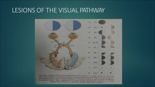
2. Optic nerve lesions (A,B)
Causes : Optic atrophy, traumatic avulsion, acute optic neuritis etc.
1.Distal optic nerve lesion (A)
• Complete blindness of affected side
• Abolition of direct light reflex on affected side
• Accommodation reflex intact
3. 2. Proximal optic
nerve lesion (B)
• Blindness on affected side
• Contralateral hemianopia
• Aboli8on of direct light reflex on affected side
• Accommoda8on reflex intact
5. Causes :
I. Intrinsic causes – Lesions which produce thickening of
chiasma. Eg. Gliomas, multiple sclerosis
II. Extrinsic causes – Compressive lesions. Eg. Pitutary
adenoma, meningioma
III. Other causes – Include metabolic, toxic and
inflammatory syndromes. Eg. Lymphoid
hypophysitis, sarcoidosis
6. Optic tract lesions (E)
I. Intrinsic causes – Demyelinating diseases and infarction.
II. Extrinsic causes – Compressive lesions. Eg. Pitutary adenomas,
tumours of optic thalamus
III. Other causes – syphilitic meningitis, tubercular meningitis
Causes :
7. Optic tract lesions
• Incongruous homonymous hemianopia
• Contralateral hemianopic pupillary responses (Wernicke’s
reaction)
• Optic disc changes – Descending type of partial optic
atrophy is produced characterized by temporal pallor on
the side of the lesion and bow tie atrophy on the
contralateral side.
• Visual acuity is intact
9. Lateral geniculate nucleus lesions(E)
• Incongruous homonymous hemianopia
• Pupillary reflexes are normal as the fibres go to pretectal nucleus and
not the LGN
• Optic disc pallor may occur due to partial descending atrophy
10. Lesions of optic radiations (F,G)
Common lesions include :
• Vascular occlusions
• Tumours
• Trauma
• Temporal lobectomy for seizures
11. Lesions of optic radiations
• Superior quadrantic hemianopia(F) – Pie in the sky lesions.
It is explained by the fact that inferior fibres of optic
radiations contain fibres from ipsilateral lower temporal
retina and contralateral lower nasal retina.(part of optic
radiations in temporal lobe)
• Inferior quadrantic hemianopia(G) – Pie on the floor
lesions. This is the same as above. Difference being the
superior fibres are affected. (part of optic radiations in
parietal lobe)
12. • Complete homonymous hemianopia(H) – produced when
all fibres of op8c radia8ons are involved some8mes sparing
the macular fibres as they lie centrally.
• Pupillary reflexes are spared
• Op8c disc atrophy does not occur
13. Visual cortex lesions (I,J,K)
• Congruous homonymous hemianopia – macular field of vision is
spared. It is a feature of occlusion of posterior cerebral artery.
• Congruous homonymous macular defects – occurs in lesions at the tip
of occipital cortex following head injuries or gun shot injuries
14. • Bilateral homonymous macular defects – presenting like
bilateral central scotoma occur in bilateral lesions of
occipital cortex
• Pupillary light reflexes are normal
• Optic atrophy doesn’t occur.
Other manifestations of occipital lobe lesions include :
• Cortical blindness
• Dyschromatopsia
15. • Visual hallucinations
• Palinopsia – Persistent perception of visual image
• Visual anesthesia – transposition of visual stimulus from
one hemifield to another
• Polyopsia – multiple images of single object which do not
disappear on closing the eye.