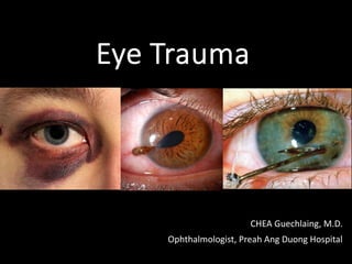
2. Introduction
• The eye is protected from direct injury by lids,
eyelashes and the projecting margins of the orbit.
Nevertheless, it can be injured in a variety of ways; by
chemicals, heat, radiation and mechanical trauma.
14. • Mechanism:
blunt trauma (impacting obj > ⦶ orbital aperture) > ↑ IOP > blow out
at weakest point along post. medial part of floor (maxillary bone)
• Diagnosis:
• History: orbital aperture struck by obj > its diameter (ball, fist…)
• Physical examination:
_ Eyelid signs: ecchymosis, edema
_ Diplopia with limitation of upgaze/downgaze/both + pain in inf orbit
_ Enophthalmos + ptosis of globe
_ Hypoesthesia of infraorbital nerve
_ Emphysema of orbit and eyelid
• Orbital imaging: CT orbit (coronal, axial, sagittal views)
2. Orbit – Orbital floor fractures
15. • Mechanism:
Naso-orbital-ethmoidal (NOE) fractures < faces striking a solid surface.
• Diagnosis:
• Characteristic: depressed bridge of nose + traumatic telecanthus.
• NOE divides into 3 categories:
1/ Type I: central fragment of bone attached to canthal tendon
2/ Type II: comminuted fractures of the central fragment
3/ Type III: comminuted tendon attachment or an avulsed tendon
• Complications: cerebral + ocular damage, severe epistaxis, CSF
rhinorrhea, damage to lacrimal drainage system…
2. Orbit – Medial Orbital Fractures
16. • Mechanism:
OCS result from ↑↑↑orbital pressure from hemorrhage
occur in: trauma, surgery, retrobulbar or peribulbar injections,
or pre-existing orbital disease
• Diagnosis:
• ↓vision + afferent pupillary defect + ↑IOP
• + tight orbit + limited EOM + proptosis
• Management:
Lateral canthotomy + inferior cantholysis
2. Orbit – Orbital Compartment Syndrome
18. 3. Trauma to Globe – Blunt Trauma
• blunt force > peripheral volume displacement with ↑ pressure >
damage to area of least resistance along lens, iris root, TM
• Sheering of vessels causes hyphema and force > scleral ruptures
posterior to muscle insertion and limbus
20. 3. Blunt Trauma – Cornea
Corneal abrasion: breach of epithelium
+ stains well with florescien
Symptoms: pain, FB sensation, tearing, discomfort blinking
Treatment: patching/bandage contact lens > pain relief
topical antibiotics prophylaxis
heals within 24-48h
21. 3. Blunt Trauma – Anterior Chamber
Hyphema: blood in AC due to injury to iris/ant. ciliary Vx
Medical management:
• Min. rebleeding:
_ restric physical activity
_ elevate bed head
• Control inflammation
• Control IOP
23. 3. Blunt Trauma – Anterior Uvea
• Traumatic mydriasis: tear of iris sphincter
• Iridodialysis: separation of iris root from ciliary body
• Cyclodialysis: separation of ciliary body from its
attachment to the scleral spur resulting a cleft
24. • Angle recession: tear between the longitudinal and circular muscle
fibers of ciliary body, characterized by deepening of ant chamber
and widening of ciliary body on gonioscopy and leads to glaucoma.
Closed Globe Injuries
3. Blunt Trauma – Anterior Uvea
25. 3. Blunt Trauma – Lens
• Cataract: rosette formation
• Subluxation: secondary to partial tearing of suspensory
ligament but lens is present in the pupillary area
• Dislocation: 360º rupture of zonular fibres
29. 3. Blunt Trauma – Optic nerve
Traumatic optic neuropathy
• follows ocular, orbital or head trauma as sudden visual loss that cannot
be explained by other ocular pathology
• Classification:
_ direct: due to blunt or sharp optic nerve damage from agents
_ indirect: secondary transmitted force
• Presentation:
_ Poor vision + afferent pupillary defect +
optic nerve head: normal >> developing pallor