1. H/D Exchange uses the difference in mass between Hydrogen and Deuterium to
better understand which surface regions of the protein make contact with the
siRNA. The more a part of the protein is exposed the more exchange will occur
and a greater difference in mass will be seen in mass spectrometry data. If the
structure of the protein has areas that are folded and less exposed or protected by
other interactions, these areas will have less exchange and therefore the mass
will be relatively the same. Data for both p19 and p14 was collected last summer
using the following procedure. Protein samples were prepared in a HEPES buffer
solution for the H/D exchange reactions. The reaction was performed by mixing
p19 or p14 protein sample in deuterium oxide to create a mostly deuterated
solution. Deuterium was allowed to exchange with hydrogen for 30 seconds, 300
seconds, or 3000 seconds at which point the reaction was quenched by adding
4% formic acid and freezing in liquid nitrogen. Three trials of each time point were
created with and without
siRNA added to the protein
solution. Control samples
were made exactly the
same except for water was
added instead of deuterium
oxide. Before data
collection each replicate
was defrosted, urea was
added, and the protein was
digested with Protease XIII
to create multiple peptides.
Figure 1 from Hamuro et al.
diagrams the process of
H/D Exchange as used in this experiment. The data for each trial was collected
on Thermo Scientific’s Accela HPLC and LTQ Orbitrap XL Discovery mass
spectrometer. The data was analyzed using the software HDX Workbench, which
calculated the percent deuteration (%D) values for each peptide that was
identified for every replicate. These %D values were then compared to the values
for p19 to find similarities, keeping in mind the three dimensional structure of p19.
Structure and Binding Specificity of PoLV
p14 in comparison to CIRV p19 using H/D
Exchange and Fluorescence Polarization
Julia L. Meier and Jeffrey M. Vargason
Department of Chemistry, George Fox University, Newberg, OR 97132
Viruses have developed a defense system to an organism’s viral silencing
mechanism by creating proteins that bind to viral small interfering RNA (siRNA)
and inhibits the cleavage pathway from cleaving the viral RNA. This study centers
on the viral suppressor of RNA silencing p14 from the Pothos Latent Virus. The
structure and binding preferences of p14 are unknown, but it is known that PoLV
p14 has a 20% amino acid sequence similarity to another viral suppressor CIRV
p19, and that p14 and p19 have been found to bind similarly. The structure of p19
has been found using x-ray crystallography. Using the technique of Hydrogen-
Deuterium Exchange (H/D), the two proteins were compared to find structural
features of p14.
Methods
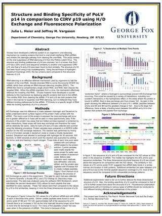
“protection factor” where a Hydrogen’s surroundings prevent H/D Exchange from
occurring. This could be caused by a variety of interactions, including non-
covalent interactions or the hydrophobic effect. In the areas where the protein is
bound to siRNA, there is less exchange and thus a lower %D. As seen in the
graph showing the difference between p14 and p14 + siRNA, peptides between
amino acid 65 and 88 had the greatest change in %D. Due to the large %D
difference, there is likely a binding event between the siRNA and p14 in this
section that protects the peptide from exchange.
Background
RNA silencing is an effective defense mechanism used by organisms to halt the
invasion of the viral RNA. Double stranded RNA is cut by the enzyme DICER into
siRNA, which then activate an RNA-induced silencing complex (RISC). The
siRNA then bind to complimentary single strand RNA, and RISC then cleaves the
targeted RNA. When the siRNA originates from a virus, the mechanism effectively
destroys the invading viral RNA. Viruses however have developed a counter
defense to this mechanism by deploying specific proteins that bind to the siRNA.
This inhibits RISC from cleaving the siRNA. P14 and p19 are both proteins that
work in this manner to suppress viral RNA silencing, but it is believed they have
different binding preferences for the siRNA. P19 binds to a specific length of RNA
while the binding specificity of p14 is unknown.
Results
Abstract
Future Directions
Additional experiments need to be run to further deduce the three dimensional
structure of p14. Also, the binding specificity of p14 could be compared to that of
p19 to further reveal more about the structure of p14. Fluorescence polarization
experiments with p14 binding to various types of siRNA should be conducted to
find different binding affinities and preferences of the protein.
Acknowledgements
This project was funded by The Paul K. Richter Memorial Fund and the Evalyn
E.C. Richter Memorial Fund
Figure 3: Differential H/D Exchange
The graphs in Figure 2 show the %D values for p14 without siRNA and with siRNA
at each of the three time points. Eleven usable peptides were found to cover
amino acids 47-88 and 110-120, approximately half of the sequence for p14.
Overall there is an increase in %D as the time intervals became longer since there
is more time for exchange to occur. For many of the peptides a lower %D was
seen when in the presence of siRNA. This trend is because of the
In figure 3, the sequence of p14 and p19 are compared for the %D values at 300s
when in the presence of siRNA. The middle row highlights the 20% amino acid
sequence similarities. The three dimensional structural features of p19 are shown
above the sequence for comparison with the %D values. The similarity in %D
values at p14’s amino acids 74-81 suggests that siRNA likely binds in a similar
manner and location in both proteins. Additional similarities are seen between p14
and p19 but due to the minimal peptide sequence coverage in p14, most of the
sequence cannot be compared to find comparable exchange patterns.
Sources:
Hamuro Y, Coales SJ, Southern MR, Nemeth-Cawley JF, Stranz DD, Griffin PR. (2003). Rapid analysis of
protein structure and dynamics by hydrogen/deuterium exchange mass spectrometry. J. Biomol. Tech. 14,
171–182
Figure 1: H/D Exchange Process
Figure 2: % Deuteration at Multiple Time Points
CIRV p19 MERAIQGN DTREQANGERWDGGSGGITSPFKLPDESPSWTEWRLYNDETNSNQDNPLGFK
+
PLV p14 MENSQTGVLCPNRCQVCSHTTYIR
CIRV P19 ESWGFGKVVFKRYLRYDRTEASLHRVLG----SWTGDSVNYAASRFLGANQVGCTYSIRFR
ES G G R+ R+ T+ + G SW D L + G + +IR
PLV P14 ESSGQGGRQACRFTRFV-TQPRVVSEQGIQYRSWLSDRGFPIT--LLSTSG-GLSTTIRGH
CIRV P19 GVSVTISGGSRTLQHLCEMAIRSKQELLQLTPVEV ESNVSRGCPEGIETFK KESE
GV++TI G S++L + C +A V S
PLV P14 GVAITIQGDSKSLLNFCRVAYDVFHHPVVQSEVCH GSGPATSDEITTKF
20% 30% 40% 50% 60% 70% 80% 90%
α1 α2
β1 β2 α3 α4 β3
β4 α5
10 20
30 40 50 60 70 80
90 100 110 120 130