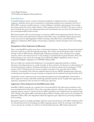
1. Austin Wright-Pettibone
151218: Ribozyme Regulated Silencing Devices
Introduction:
Controlled regulatory activity is crucial to functional complexity in engineered systems. Among living
organisms, metabolic activity can be constitutively or dynamically regulated, and is mediated at the level of
DNA, RNA, or protein assembly processes. A major challenge in metabolic engineering has been creating
fast-acting controls that can be readily induced to affect targeted cell activity (Na 2013). This quest, in part to
divert metabolic flux away from undesired products and toward desired ones, has led to the development of
post-transcriptional RNA-based controls.
RNA-based controls offer several advantages over protein or DNA-based engineering methods. The more
limited set of bases and conformational structures makes RNA easier to predictably engineer than proteins.
At the same time, the rapid degradation of RNA transcripts makes RNA more useful than DNA in
application. Furthermore, RNA displays a broad range of regulatory activities through its host of noncoding
RNAs.
Upregulation of Gene Expression by Ribozymes
These noncoding RNAs include many classes of biochemical regulators. Among them, hammerhead and glmS
ribozymes stand out as small self-catalyzing transcripts, able to induce functional changes within a cell after
cleavage of their 5’-PPP terminus. Mechanistically, following transcription, these ribozyme form a hairpin
structure, which is then cleaved near the 5’-end, resulting in a terminal hydroxyl group (D’Amare 2010). The
hydroxyl group, in turn, causes the transcript to be catabolized by RppH- degradation, which is slow as
compared with RppH+ degradation of 5’-PPP-RNA (Deana 2008).
Previous studies have exploited the idempotence of contemporary engineering methods to combine
ribozymes and coding sequences in a single transcript. In these constructs, the ribozyme is coded upstream of
the desired expression sequence. Following transcription, the ribozyme subunit cleaves, producing a 5’-OH
terminus. This 5’-OH group has been shown to impede association of the transcript with the RNase
machinery. Consequently, these ribozyme-regulated expression devices (rREDs) upregulate gene expression
by allowing more translation events per transcript as compared with the unaltered transcript (Carothers 2011).
Upregulation of gene expression by post-transcriptional regulation has broad applicability. Functionally, it
allows engineers to increase yield while minimizing undesired catabolic activity. Coupled with suppression of
expression, upregulation forms the basis for tunable gene expression.
Suppression of Gene Expression by Small Ribonucleic Acids
Small RNA (sRNA) molecules are a separate class of noncoding RNAs that induce gene knockdown at the
post-transcriptional level (Na 2013). These short, noncoding sequences, fold into stem-loop structures with a
targeting sequence near the 5’ end that promotes mRNA recognition and complementary base pairing (Vogel
2011). Pairing of the sRNA-mRNA transcripts facilitates RNase E binding, which activates the transcripts for
degradation (Masse 2003). This degradation effectively suppresses gene expression by preventing translation
from occurring.
Recognition sequences are highly specifiable within the conserved architecture of the sRNA scaffold. In 2013,
Yoo described protocols for exchanging the mRNA targeting sequence within the seed regio of the sRNA as
a way to increase the versatility of a small set of well-described structures. sRNA binds upstream the
2. ribosome binding site through
antisense pairing. By
specifying this transcript-
dependent sequence, engineers
can guide their designed
regulator to virtually any gene
of interest (Yoo 2013).
Binding to the transcript is
then assisted by a hexameric
chaperone protein, Hfq. Hfq
mediates the annealing of
sRNA to its cognate mRNA
through a brief catalytic
encounter, lowering the
activation energy for sRNA-
mRNA interaction (Vogel
2011). This activity depends
on the roughly equal local
concentrations of mRNA and
sRNA. At highly skewed concentrations of sRNA or mRNA, there is a greater probability of sRNA-sRNA or
mRNA-mRNA interactions than mRNA-sRNA ones. (Soper 2010). This corresponds to reduced repression
of gene expression: fewer interactions on the protein’s surface will be successful mRNA-sRNA binding
events, and so more transcripts will be translated by the ribosomes.
Ribozyme-Regulated Silencing Device: Towards Tunable RNA-based Gene Suppression?
Combining ribozyme-regulated expression devices with sRNA scaffolds, we set out to create a novel
ribozyme-regulated silencing device (rRSD) that induced increased repression of metabolic activity by
increasing the half-life of sRNA. Our design strategy bound the ribozyme, sRNA, and genetic targeting
sequence inside a single transcript. The genetic targeting sequence was complimentary to RFP, resulting in
complex formation between the rRSD and mRNA transcripts. Following transcription of the rRSD, we
hypothesized that the ribozyme subunit would cleave, producing a scar sequence with a 5’-OH terminus that
would be slowly degraded as compared to an analogous transcript with a 5’-PPP head. This, in turn, would
result in a lower fluorescent reading during the test phase, as each rRSD interacted with more mRNA
transcripts during its lifetime than analogous transcripts with a 5’-PPP head. Consequently, validation of our
hypothesis would occur if the rRSD exhibited lower fluorescent activity at lower concentrations than an
analogous device with a 5’-PPP head.
Results and Discussion:
To test our hypothesis, we constructed three sets of devices. First, we built an sRNA control. Against this we
tested three scar controls, which contained the sRNA along with a short upstream flanking sequence. This
sequence matched the post-cleavage scar of our rRSDs, with a 5’-PPP group substituted in place of the
rRSDs 5’-OH terminus (see Figure 1b,c).
We expected the sRNA control and the scar control to perform analogously, and then to see increased
suppression when the 5’-OH group was added. Unexpectedly, fluorescence assays of these two groups
revealed appreciable differences between the scar control and the sRNA control (see Figure 2). At low levels
of induction, the scar controls appeared more able to repress RFP activity, suggesting upstream structure has
Figure 1: Transcript structure and theoretical function for (A) rRSD construct, (B) scar control, and
(C) sRNA control. Induction causes each device and control to transcribe an sRNA, which then is
targeted to RFP, suppressing activity. The presence of the 5’-OH terminus was hypothesized to
increase transcript half-life in the rRSD, resulting in greater suppression at lower levels of induction.
The scar construct and sRNA control were hypothesized to have analogous functions if the scar
sequence 5’ the sRNA had no effect.
3. an appreciable effect on device
performance. This effect is variable,
however: between our three scar
controls, two of them performed
significantly better than our sRNA
control, while the third performed
only moderately better, and
performed slightly worse at the
lowest levels of induction. While
structural analysis offered no
immediate clues, it is possible
secondary structure around the
targeting sequence in our scar
control interrupts the association of
the construct with the mRNA.
At higher levels of induction, any
advantage that the scar control
displayed at low concentrations
disappeared. Between 60 and 80uM of induction (termed medium to high), all three devices began
performing worse than the sRNA control. We hypothesized this was due to saturation by the scar controls,
but at present have not identified the precise mechanism by which this saturation appears to occur. Given the
shift in performance between low and medium to high levels of induction, it is possible the upstream
structure does partially slow degradation, but that this slower degradation, in turn, saturates the system at
higher levels. At that point, the rate of sRNA degradation could overtake the rate of complexing between the
sRNA and mRNA (Vogel 2011).
Taking these observations into the test phase for our rRSDs we sought to understand whether analogous
behavior could be seen between the scar control and the ribozyme regulated silence device. Further, we
hoped to validate our original hypothesis that the 5’-OH terminus would confer the greatest suppression.
Each rRSD contained a ribozyme attached upstream the sRNA sequence, bounded by a left and right
flanking sequence (see Figure 1a). Following transcription, the ribozyme would cleave, generating a device
analogous to the previously discussed scar control, albeit with a 5’-OH, rather than a 5’-PPP, terminus.
Fluorescence assays revealed a wide range of behaviors among the devices. Of the four rRSDs successfully
constructed, two failed to perform better than the sRNA control at any level of induction, and only one
(device 3) performed better than the scar control under a wide range of conditions (see Figure 3a-c). As such,
this appeared to invalidate our original hypothesis, while raising new questions around device and scar
behavior.
Notably, both the devices that outperformed the sRNA control at low levels of induction exhibited similar
saturation effects as the scar control. Following initial periods of increasing suppression, devices 1 and 3 both
saturated at medium to high levels of induction. Device 4 did this to an extent as well, but was consistently
outperformed by both the sRNA control and the scar control.
Interestingly, devices 3 and 4 were designed to have the same structure upon cleavage. Consequently, based
on the similar performance of device 3 and its corresponding scar control (see Figure 3c), the varied
performance between devices 3 and 4 indicates cleavage events did not occur in at least one devices, most
likely device 4. It would appear, then, device 3 interacted with cellular components prior to cleavage. This
Figure 2: Scar Control vs sRNA Control Performance. Saturation is observed to occur at
low levels of induction in two of the three scar controls. The sRNA control (shown in blue)
represses more fully at higher levels of induction.
0
1000
2000
3000
4000
5000
6000
7000
8000
0 20 40 60 80 100Fluorescence
[IPTG]
sRNA Control
Scar Control 1
Scar Control 2
Scar Control 3
4. might also explain its poor performance and
suggest that ribonucleotides 5’ the sRNA may
impede the transcript’s targeting ability.
Meanwhile, device 2 evidenced a linear response
curve, rather than a characteristic sigmoidal
response. This indicates low levels of activity even
at high levels of induction. Without further
evidence, all that can be conclusively stated
regarding device 2 is that while we know
transcription occurred due to the negative slope, it
does not appear the device was particularly high
functioning. Rather, the upstream structure
appears to have impeded device functionality,
which is an interesting result suggesting we can
impede sRNA functionality by 5’ nucleotide
addition. With reference back to sRNA
biochemistry, the 5’ end is associated with binding
to the Hfq (Vogel 2011). If this binding is
interrupted due to extensive sequence structure 5’
the sRNA itself, then the frequency of sRNA-
mRNA complex events may be reduced, and
repression of gene expression would decrease.
From device 3 we also saw signs that a cleaved
ribozyme attached to an sRNA subunit can
perform as well as its corresponding scar control.
Future constructions could implement this useful
result by incorporating the ribozymes self-catalytic
ability to act as a separator element between two
subunits in a larger transcript. Effectively, a large
transcript could be controlled by a single
promoter. Then, within that transcript a regulation
scheme could be carried out such that one part of
the transcript regulates the activity of another.
Following transcription, the ribozyme would
cleave, and then the regulator element of the
transcript would then be free to target either an
element within the transcript or some other
targeted cell activity. If targeted to the transcript
itself, this would create a Type I Incoherent
Feedforward Pathway (see Figure 4a). If targeted
to another cell activity, we could use it to knock
out a competing cell process, creating a switching
pathway: gene expression for our desired activity
would be increased as the competing one was
commensurately decreased (see Figure 4b). For example, a cell expressing Yellow Fluorescent Protein (YFP)
could be switched to express Red Fluorescent Protein (RFP) by creating a transcript containing a ribozyme
separating RFP from an sRNA targeted to YFP. Transcription would then result in the ribozyme cleaving; the
A: Device 1 and its corresponding Scar Control 1. Both had analogous right flanking
sequences. Device 1 had nominally better performance than the scar control at the
lowest levels of induction. Beyond that, it was outperformed by both the scar and
sRNA control.
B: Device 2 and its corresponding Scar Control 2. Both had analogous right flanking
sequences. Device 2 was outperformed at every level. Its linear response curve
suggests it never achieved saturating activity, which might be due to fast degradation
of the device by cellular machinery.
C: Devices 3 and 4 had analogous right flanking sequences as their corresponding
Scar Control 3. The varied performance between devices 3 and 4 suggesting at least
one device is interacting prior to cleavage. Further, device 3’s performance suggests
it is possible to achieve similar expression using a 5’-OH terminus as with a 5’-PPP
terminus.
0
1000
2000
3000
4000
5000
6000
7000
8000
0 20 40 60 80 100
Fluorescence
[IPTG] (uM)
Scar-Control
Device 1
sRNA-Control
0
1000
2000
3000
4000
5000
6000
7000
8000
0 20 40 60 80 100
Fluorescence
[IPTG] (uM)
Scar-Control
Device 2
sRNA-Control
0
1000
2000
3000
4000
5000
6000
7000
8000
0 20 40 60 80 100
Fluorescence
[IPTG] (uM)
Scar-Control
Device 3
Device 4
sRNA-Control
5. sRNA targeting the YFP, decreasing yellow
fluorescence; and the RFP being translated
to create green fluorescence. Such a
switching mechanism could be advantageous
over models that tie multiple promoters to
the same inducer by eliminating the
competition for inducer molecules to release
the repressor molecule that prevents
transcription. Thus, lower levels of induction
could yield higher levels of complex
expression.
Open Questions and
Conclusions:
While these experiments did not yield
confirmation of our original hypothesis, they
raised many new questions surrounding
sRNA-ribozyme interactions and in vitro device functionality. Beyond the questions previously posed, several
others stand out as important obstacles to be addressed before moving forward. While there are nearly
endless possible flanking sequences within the combinatorial library, we have selected only a handful for
testing. These were optimized for favorable cleavage and transcript stability, but it is evident other factors
have influenced the functionality of our devices. Understanding the mechanisms of ribozyme activity prior to
cleavage and incorporating these results into our selection process may promote orthogonality of parts while
also ensuring device functionality under a range of conditions. Relatedly, it is currently unclear what level of
interaction our devices are having with Hfq and whether their 5’ structures are impacting the targeting or
complexing of the sRNA with either the Hfq or the mRNA. Saturation may be occurring due to steric of
chemical hindrance of sRNA dissociation from Hfq.
Conclusively, we have shown that sequences 5’ of the sRNA can improve device performance at low
expression levels. At minimum, the hydroxyl group does not appear to damage the functionality of our
devices. Rather, the left flanking sequence appears to have more of an effect, as exemplified by the
performance of devices 3 and 4. This flanking sequence may confer additional secondary structure that
encourages device interaction prior to cleavage or that slows the rate of cleavage sufficiently so that device
interaction becomes a more timely event than cleavage. We have concurrently shown that cleaved devices
with a 5’-OH terminus can deliver equivalent activity as scarred constructs with a 5’-PPP terminus. This
potentially useful result could aid in the construction of Type I Incoherent Feedforward Pathways, or in
switching mechanisms for more complex regulation. Further testing should aim to elucidate the effects
surrounding ribozyme interactions with sRNA and its associated partners.
Materials and Methods:
In Silico design of Plasmids:
I did not work on plasmid design during these experiments.
Plasmid Construction:
Plasmids were constructed by polymerase chain reaction and Gibson assembly. All rRSD constructs were
built off a plasmid control containing the MicC scaffold sRNA downstream from an RFP targeting sequence.
By linearizing the plasmid upstream from the RFP targeting sequence and downstream the scaffold, we
Figure 4: Theoretical applications for rRSD mediated pathways. (a) A Type I
Incoherent Feedforward Pathway, wherein ribozyme cleavage separates a silencing
sRNA from a coding sequence. The sRNA is targeted to suppress the coding
sequence following ribozyme cleavage. (b) A Switching Pathway. The coding sequence
and sRNA are separated by a ribozyme, which cleaves upon transcription. The sRNA
targets a sequence off the transcript with a competing phenotype as the coding
sequence on the transcript. Meanwhile, transcription of the coding sequence switches
the expressed phenotype.
6. obtained backbone fragments for our test constructs. Our inserts were built off a gblock ordered from
Operon containing a 222nt sequence housing the ribozyme, left and right flanking sequences, and the RFP
targeting sequence. Using Gibson Assembly, we then fused the insert and backbone, circularizing the
completed plasmid.
To increase the variety of our rRSDs, we exchanged the flanking sequences along either side of the ribozyme.
Primers covering both flanking sequences and the corresponding overhang regions were ordered from
Operon and fused using primer extension. With the first plasmid constructed using a gblock, subsequent
iterations could be built off the construct with minimal changes using the ordered primers stitched together in
the form of a small, 136nt, insert. This yielded a cost-effective method for high volume assembly of our insert
regions. These could then be circularized using Gibson Assembly through combination with a longer static
backbone sequence cloned off the construct.
To test our hypothesis that increased suppression resulted from the 5’-OH group, rather than from the scar
sequence remaining post-cleavage, we constructed plasmids containing the post-cleavage scar with a 5’-PPP
terminus, along with the sRNA and RFP targeting sequence. These were assembled alongside another set of
control plasmids containing only the functional sRNA. Transcription was repressed by LacI, until induction
by IPTG. All plasmids were built using Gibson Assembly to construct a template, which could be used as a
guide for subsequent modification carried out through the aforementioned primer extension method.
Following plasmid construction, we chemically transformed the constructs into mg1655, allowing cells to
grow on antibiotic plates and in liquid culture before sending them out for sequence verification.
Testing
We proceeded to test the effectiveness of our rRSD constructs at suppressing RFP expression in E. coli. We
used standard plasmids containing RFP as the targets of experimentation. Our constructed rRSD and scar
control plasmids produced transcripts that bound to the RFP mRNA, preventing translation of the RFP to
varying degrees.
Using electroporation we transformed our constructs along with the RFP plasmid into BL21 strain E. coli
cells. From there, we prepared liquid cultures of LB, carbenicillin, and kanamycin, and allowed the cells to
grow at 37 degrees C until they reached stationary phase. To test device functionality, we prepared 96-well
plates over a range of settings for each device. We induced cultures at 0, 20, 40, 60, 80, 100, 150, and 200uM
IPTG. We also conducted binary tests in the absence (0uM) and presence (100uM) of arabinose to confirm
results were independent of induction of the RFP plasmid. Measurements were conducted using fluorescence
assays, measuring absorbance at 340nm and 600nm and fluorescence in the red emission wavelengths. For
later iterations, we limited the range of testing to 0, 20, 40, 60, 80, and 100uM IPTG after noting device
performance was readily determined by the lower concentrations.
Citations:
1. Carothers, J. et al. Model Driven Engineering of RNA Devices to Quantitatively Program Gene
Expression. Science 334, 1716-1719 (2011).
2. D’Amare. A., Scott, W. Small Self-cleaving Ribozymes. CSH Perspectives in Biology 2, 1-10 (2010).
3. Deana, A., Celesnik H., Belasco, JG. The bacterial enzyme RppH triggers messenger RNA
degradation by 5’ pyrophosphate removal. Nature 451, 355-358 (2008).
4. Massé, E., Escorcia, F. E. & Gottesman, S. Coupled degradation of a small regulatory RNA and its
mRNA targets in Escherichia coli. Genes Dev. 17, 2374–2383 (2003)
5. Morita, T., Maki, K. & Aiba, H. RNase E-based ribonucleoprotein complexes: mechanical basis of
mRNA destabilization mediated by bacterial noncoding RNAs. Genes Dev. 19, 2176–2186 (2005).
7. 6. Na, D. et al. Metabolic engineering of Escherichia coli using synthetic small regulatory RNAs. Nat.
Biotechnol. 31, 170-174 (2013).
7. Soper, T., Mandin, P., Majdalani, N., Gottesman, S. & Woodson, S. A. Positive regulation by small RNAs
and the role of Hfq. Proc. Natl Acad. Sci. USA 107, 9602–9607 (2010).
8. Vogel, J., Luisi, BF. Hfq and its Constellation of RNA. Nat. Microbio. 9, 578-589 (2011).
9. Yoo, SM., Na, D., Lee, SY. Design and use of synthetic regulatory small RNAs to control gene
expression in Escherichia coli. Nat. prot 8, 1694-1707 (2013).